![]()
![]()
![]()
Use LEFT and RIGHT arrow keys to navigate between flashcards;
Use UP and DOWN arrow keys to flip the card;
H to show hint;
A reads text to speech;
49 Cards in this Set
- Front
- Back
- 3rd side (hint)
|
Axial Skeleton |
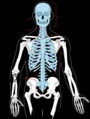
Bones of the cranium, neck and trunk |
|
|
|
Appendicular Skeleton |
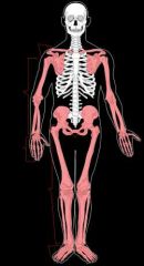
Bones of the limbs and their girdles |
|
|
|
Cartilage |
A resilient, semi-rigid form of connective tissue that forms part of te skeleton where more flexibility is required |
|
|
|
Bone |
Living tissue that provides support for the body, protection of its vital structures, mechanical basis, storage of salts, and continuous suppy of new blood cells. |
|
|
|
A fibrous connective tissue enveloping the bones except at the surfaces of the joints. |
Periosteum. |
|
|
|
the fibrous connective tissue that envelops cartilage where it is not at a joint. |
Perochondrium |
|
|
|
2 types of bone |
Compact and Spongy. |
|
|
|
What is another word for spongy bone |
Trabecular or cancellous |
|
|
|
Where are blood cells and blood platelets formed? |
In the medullary cavity of adult bones and between the trabeculae of spongy bone. |
|
|
|
Sesamoid bones |
Develop in certain tendons and are found where tendons cross the ends of long bones in limbs; (ie the patella in the knee) |
|
|
|
What do sesamoid bones do? |
They protect the tendon from excessive wear and often change the angle of the tendons as they pass to their attachments (ie the patella in the knee) |
|
|
|
Small, rounded, articular head is what bone marking |
Capitulum |
(ie radius) |
|
|
In bone markings, what is a rounded, knuckle-like, articular area, usually occuring in pairs |
Condyle |
|
|
|
Crest is what in bone marking? |
Ridge of bone. |
|
|
|
Epicondyle |
Eminence superior to a condyle |
|
|
|
Facet |
Smooth, flat area that isnusually covered with cartilage, where a bone articulates with another bone |
|
|
|
Foramen |
Passage through a bone |
(ie foramen magnum) |
|
|
Fossa |
Hollow or depressed area |
Infraspinous fossa of scapula |
|
|
Groove |
Elongated depression of humerus |
Radial groove of humerus |
|
|
Line |
Linear elevation |
(ie superior nuchal line) |
|
|
Malleolus |
Rounded process |
(ie lateral and medial malleolus aka ankle) |
|
|
Notch |
Indentation at the edge of a bone |
|
|
|
Protuberance |
Projection of bone |
(ie external occipital protuberance) |
|
|
Spine |
Thorn-like process |
|
|
|
Spinous process |
Projecting spine-like part |
(ie C7 spinous process) |
|
|
Trochanter |
Large blunt elevation |
(ie greater trochanter) |
|
|
Trochlea |
Spool-like articular process or a process that acts as a pulley |
|
|
|
Tubercle |
Small raised eminance |
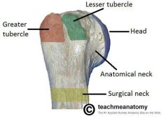
(ie greater tubercle of the humerus) |
|
|
Tuberosity |
Large rounded elevation |

(ie deltoid tuberosity of humerus in upper arm) |
|
|
Diaphysis |
Shaft of a bone ossified from the primary ossification center |
|
|
|
Epiphyses |
End of bone. Secondary ossification centres that appear in other parts of developing bone |
|
|
|
Epiphyseal line |
The seam formed during the fusion process where the diaphysis forms with the epiphyses and bone growth ceases |
|
|
|
Synovial joints have three classifications |
Articular Capsule, articular cavity, and articular cartilage |
|
|
|
What are the types of joints? |
Synovial, fibrous, cartilaginous |
|
|
|
Synovial joints |
Provide free movements between the bones they join. Usually reinforced by accessory ligaments that are either extrinsic or are a thickening of a portion of the articular capsule (intrinsic) |
|
|
|
Fibrous joints |
Are articulating bones of a fibrous joint and are united bu fibrous tissue |
|
|
|
3 types of fibrous joints |
Sutures: bones are close togetherinterlocking along a line or overlapping (ie lines along the cranium) Syndesmosis: unites bones with a sheet of fibrous tissue, either a ligament or a fibrous membrane (ie interosseous membrane in the forearm that joins the radius and ulna) Gomphosis: a peg like process that fits into a socket articulation (ie teeth into maxillae and mandible) |
|
|
|
Cartilaginous joints |
Are articulating structures of cartilaginous joints that are united by hyaline cartilageor fibrocartilage |
|
|
|
2 types of Cartilaginous joints |
Primary Cartilaginous Joints (Synchondroses): bones are united by hyaline cartilage which permits slight bending during early life (ie those present during development of long bone) Secondary Cartilaginous Joints (Symphisis): strong, slightly movable joints united fibrocartilage (ie fibrocartilaginous intervertebral discs) |
|
|
|
6 types of Synovial joints |
1. Plane (acriomioclavicular joint) 2. Hinge (humerus articulates with the radius and ulna) 3. Saddle (base of the thumb) 4. Condyloid (metacarpophalangeal joint) 5. Ball and Socket (glenohumeral joint) 6. Pivot (atlantoaxial joint) |
|
|
|
Plane joints |
Permits gliding or sliding movements in the plane of the articular surfaces. |
|
|
|
Hinge joints |
Uniaxial: flexion and extension only |
|
|
|
Saddle joints |
Biaxial: permit ABduction, Adduction, Flexion, and Extension |
|
|
|
Condyloid joint |
Biaxial: permit flexion, extension, ABduction, and Adduction |
|
|
|
Ball and socket joints |
Multi-axial: flexion, extension, ABduction, Adduction, medial and lateral rotation, circumduction. Highly mobile joints |
|
|
|
Pivot joints |
Uniaxial: permit rotation around a central axis |
|
|
|
Hilton's law |
States that nerves supplying a joint also supply the muscles moving the joint or the skin covering their distal attachments |
|
|
|
Aponeurosis |
Flat shedts formed by tendons of some muscles. They anchor the muscle to the skeleton, deep fascia, or other aponeurosis |
|
|
|
Functions of muscles |
Agonists: prime movers; the main muscles responsible for producing a specific movement Fixators: they steady proximal partsnof a limb while movements are occuring in the distal parts Synergist: complements the action of the agonist Antagonist: muscles whose actions are opposite to the agonist |
|