Reading...
![]()
Play button
![]()
Play button
![]()
Use LEFT and RIGHT arrow keys to navigate between flashcards;
Use UP and DOWN arrow keys to flip the card;
H to show hint;
A reads text to speech;
21 Cards in this Set
- Front
- Back
|
The Lumbar Plexus
|
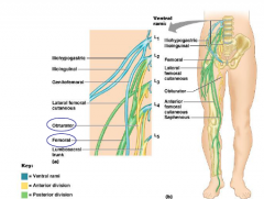
- arises from the 1st four lumbar spin nerves: L1-L4
- located within the psoas major muscle - some of its minor branches innervate the abdominal wall, but its main branches descend to innervate the anterior thigh, via the femoral and obturator nerves |
|
|
The Sacral Plexus
|
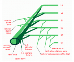
- originates from spinal nerves L4-S4 and is found directly behind the lumbar plexus
- about ½ of the branches of the sacral plexus serve the buttocks and lower limb but the most important branch for the lower limb is the 'sciatic' nerve, which divides into the tibial nerve and common fibular nerves |
|
|
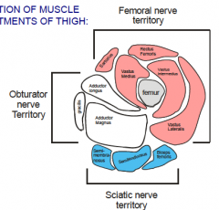
Innervation of Muscle Compartments of Thigh
|
|
|
|
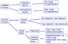
Innervation of Lower Limb Diagram
|
|
|
|
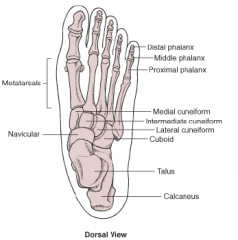
Bones of the Foot
|
|
|
|
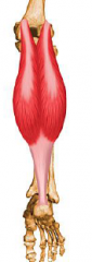
Muscles of the Posterior Compartment of the Leg
|
Superficial:
- gastrocnemius - plantaris - soleus Deep: - popliteus - FDL - flexor digitorum longus -FHL - flexor hallicus longus - tibialis posterior Cross knew and flex leg = gastrocnemius, plantar is, popliteus Invert foot = FDL, FHL, tibialis posterior |
|
|
Gastrocnemius
|
Superficial
O: via 2 heads from the medial and lateral condyles of the femur In: posterior calcareous bone, via the calcaneal tendon A: plantar flexes foot when knee is extended, but can also flex the knee since it crosses the knee joint I: tibial nerve |
|
|
Soleus
|
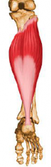
Superficial
O: an extensive origin from superior tibia, fibula and interosseus membrane In: posterior calcareous bone, via the calcaneal tendon A: plantar flexes foot, impotent postural and locomoto muscle during running and walking I: tibial nerve |
|
|
Plantaris
|
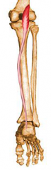
Superficial
O: posterior femur above the lateral condyle In: via a long, thin tendon into the calcareous bone A: assists in knee flexion and plants flexion of foot I: tibial nerve |
|
|
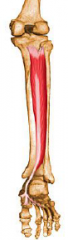
Flexor Digitorum longus (FDL)
|
Deep:
O: extensive origin on the posterior tibia In: tendon runs behind medial malleolus and splits into distal phalanges of toes 2-5 A: plantar flexes and inverts foot, flexes the toes, helps the foot 'grip' the ground I: tibial nerve |
|
|
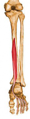
Flexor Hallucis Longus
|
Deep
O: middle part of the shaft of fibula, interosseous membrane In: tendon runs under foot to the distal phalanx of big toe A: plantar flexes and inverts foot, flexes the big toe at all joints, serves as a push off muscle during walking I: tibial nerve |
|
|
Popliteus
|
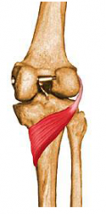
Deep
O: lateral condyle of the femur and lateral meniscus of the knee In: proximal tibia A: flexes and rotate leg medially to 'unlock' the knee from full extension when flexion begins I: tibial nerve |
|
|
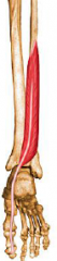
Tibialis Posterior
|
Deep
O: superior tibia and fibula and also interosseous membrane In: tendon passes behind medial malleolus and under arch of foot to insert into several tarsals and metatarsals 2-4 A: prime mover of foot inversion, plantar flexes the foot I: tibial nerve |
|
|
3 tendons of the 3 posterior compartment flexor muscles along the medial side of the food
|
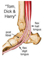
"Tom, Dick and Harry"
- post. tibial, flex hall longs, flex, digit longus |
|
|
Muscles of the Lateral Compartment of the Leg
|
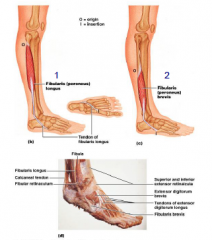
1. peroneus longus (fibularis longus) *
2. peroneus brevis (fibularis brevis) * *plantar flexion and eversion of foot |
|
|
Peroneus Longus
|
O: head and upper portion of the lateral side of the fibula
In: by a long tendon that curves under the foot t othe first metatarsal and medial cuneiform A: plantar flexes and everts the foot I: superficial fibular nerve |
|
|
Peroneus Brevis
|
O: distal shaft of fibula
In: via a tendon running behind lateral malleolus to insert on proximal end of 5th metatarsal A: plantar flexes and everts foot I: superficial fibular nerve |
|
|
Muscles of the Anterior Compartment of the Leg
|
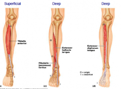
1. Tibialis Anterior (TA)
2. Extensor Digitorum Longus (EDL) 3. Extensor Hallicus Longus (EHL) * all dorsiflex the foot at the ankle |
|
|
Tibialis Anterior
|
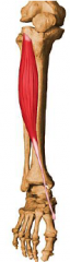
Superficial
O: lateral condyle and upper 2/3 of tibial shaft, interosseous membrane In: via a tendon into the medial cuneiform and first metatarsal A: prime mover of foot dorsiflexion, inverts the foot I: deep fibular nerve |
|
|
Extensor Digitorum Longus
|
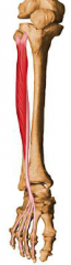
Deep
O: lateral condyle of the tibia, proximal 1/3 of fibula and interosseous membrane In: middle and distal phalanged of toes 2-5 A: prime mover of toe extension and dorsiflexes the foot I: deep fibular nerve |
|
|
Extensor Hallucis Longus
|
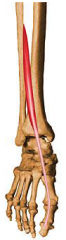
Deep
O: shaft of the fibula and interosseous membrane In: tendon inserts into distal phalanx of the big toe A: extends the big toe, dorsiflexes the foot I: deep fibular nerve |