![]()
![]()
![]()
Use LEFT and RIGHT arrow keys to navigate between flashcards;
Use UP and DOWN arrow keys to flip the card;
H to show hint;
A reads text to speech;
118 Cards in this Set
- Front
- Back
|
Alimentary Canal (Mouth, pharynx, esophagus, stomach, small intestine, large intestine)
Accessory (Teeth, tongue, gallbladder, salivary glands, liver, pancreas) |
2 Main Groups of the Digestive System and name the Organs (6,6) |
|
|
Gastrointestinal tract Gut Digestive Tract |
3 Synonyms for the Alimentary Canal |
|
|
Ingestion |
- Taking food into the digestive tract (eating) |
|
|
Propulsion |
- Moves food through the alimentary canal includes swallowing which is initiated voluntarily and peristalsis, involves alternating waves of contraction and relaxation of muscles in the organ walls |
|
|
Swallowing |
- Voluntary propulsion of food through the alimentary canal |
|
|
Peristalsis |
- Major means of propulsion which involves alternating waves of contraction and relaxation of muscles in the organ walls - Main effect is to squeeze food along the tract, but some mixing occurs as well |
|
|
- Digests and absorbs foods - Route that food takes |
What is the 2 main functions of the alimentary canal aka gastrointestinal tract? |
|
|
Food does not pass through the accessory organs |
Although usually important, why are the accessory organs of the digestive system named so? |
|
|
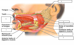
Modified skeletal muscle originating from the hyoid bone |
What muscle is the tongue made of? Where is its origin? |
|
|
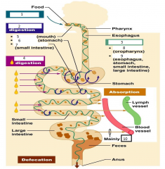
Ingestion Propulsion Mechanical Digestion Chemical Digestion Absorption Defecation |
What are the six essential activities of the "disassembly" line o the GI tract to make nutrients more available to the body? |
|
|
Carbohydrate: 4Kcal per gram - Quick energy source Protein: 4Kcal per gram - Structure and regulation Lipid: 9Kcal per gram - Energy storage, cushion, insulation |
Name the 3 Food Macromolecules; the Kcal per gram, and their function(s) |
|
|
Vitamins - Coenzymes Minerals - Electrolytes Water - Hydrolysis and dilution of waste |
Name the 3 other essential nutrients besides carbohydrates, proteins and lipids and their main function(s) |
|
|
Chewing (Teeth in mouth) Mixing (Tongue in mouth) Churning (stomach) Segmentation (small intestine) |
Give 4 examples of Mechanical Digestion and where they occur |
|
|
Swallowing (oropharynx) Peristalsis (esophagus, stomach, intestines) |
Give 2 examples of Propulsion and which area(s) they occur |
|
|
Mouth Salivary Amylase Blood Lymph |
Chemical digestion begins in the __________ by the enzyme ____________ and occurs all throughout the small intestines. Absorption occurs in all the intestines to take nutrients to the ____________ and ______________ vessels, although the capillaries don't really absorb fats. |
|
|
Lacteals - Lymph Vessels - Nodes - Trunk - Duct - Heart - Hepatic Portal Artery - Liver |
- Lymph vessels that absorb fats - Fats take very long to process, so not used as much although bigger Kcal/gram Trace the route from these vessels to the liver. (7) |
|
|
Large Intestines |
Anything remaining in the ______________________ after absorption is very toxic, especially whatever is stored in the rectum. Influential to colon cancer, the most common being the sigmoid. |
|
|
Bolus |
A ball of food formed by the mouth with mucus to make it sticky |
|
|
Segmentation |
- Rhythmic local constrictions of the small intestine that mixes foods with digestive juices and makes absorption more efficient by repeatedly moving different parts of the food mass over the intestinal wall |
|
|
Digestion |
- Series of catabolic steps in which enzymes secreted into the lumen of the alimentary canal break down complex food molecules to their chemical building blocks |
|
|
Absorption |
- Passage of digested end products (plus vitamins, minerals and water) from the lumen of the GI tract through the mucosal cells by active or passive transport into the blood or lymph |
|
|
Defecation |
- Eliminate indigestible substances from the body via the anus in the form of feces |
|
|
Chemical Digestion |
- Catabolic breakdown of food by hydrolysis (Decomposition Reactions) Ex: Amylase breaks down starch, pepsin breaks down proteins |
|
|
- Cleanses the mouth - Dissolves food chemicals so they can be tasted - Moistens food and helps compact it into a bolus - Contains the enzyme amylase that begins the digestion of starchy foods |
Saliva Functions (4) (ABCD)
|
|
|
- Parotid (Anterior to ear) - Submandibular Glands (Medial aspect of mandibular) - Sublingual Glands (Under tongue) |
3 Salivary Glands and their Locations |
|
|
- Serous cells: Watery secretion containing enzymes, ions and mucin - Mucous cells: Mucus |
2 Types of Secretory Cells of the Salivary Glands and their secretions |
|
|
- Water: 97-99.5% - Electrolytes: Na+, K+, Cl-, PO4-2, HCO3- - Proteins: Amylase, Mucin, lysozyme, defensions, IgA - Waste Products: Urea & uric acid |
4 Components of Saliva and examples |
|
|
- Ingestion - Mechanical Digestion: Mastication - Propulsion: Swallowing - Chemical Digestion: Amylase |
4 Digestive processes of the Mouth |
|
|
Cardiac Sphincter Cardia, Gastroesophageal sphincter, Gastroesophageal junction |
- Physiological sphincter surrounded by diaphragm that helps keep it closed when food is not being swallowed - Mucus cells help protect the esophagus form reflux of stomach acid - Also called? (3) |
|
|
- Mucosa: Nonkeratinized stratifed squamous epithelium - Submucosa: Esophageal glands - Muscularis externa: Skeletal and smooth muscle - Fibrous adventitia: Connective Tissue |
4 Layers of Esophagus and the tissue (Slightly different from alimentary canal layers) |
|
|
Peritoneum |
- Of the abdominopelvic cavity, most extensive of the serous membranes - Between the visceral and parietal layers is its cavity, containing serous fluid secreted by serous cells to prevent friction of mobile digestive organs |
|
|
Mesentery |
- Double layer of peritoneum, a sheet of two serous membranes fused back to back that extends to the digestive organs from the body wall - Provides routes for blood vessels, lymphatics, and nerves to reach the digestive viscera - Holds organs in place - Stores fat |
|
|
Retroperitoneal Intraperitoneal |
- Term referring to how some organs are not suspended by a mesentery and instead adhere to the dorsal abdominal wall - Ex: Kidneys, pancreas, duodenum *And those that remain in the peritoneal cavity and keep their mesentery are called? |
|
|
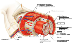
Mucosa Submucosa Muscularis Externa Serosa |
4 Tunics (Layers) of the alimentary canal |
|
|
Mucosa Lining epithelium Lamina Propria Muscularis mucosae |
- Layer of the GI tract that secretes mucus, digestive enzymes and hormones - Absorbs the digested products into the blood - Protects against infectious disease - Stratified squamous or simple columnar with goblet cells * Has 3 Sublayers, what are they? |
|
|
Lining Epithelium of Mucosa |
- Sublayer of Mucosa layer that contains simple columnar epithelium (Or stratified squamous in the mouth, esophagus and anus) - Protects certain digestive organs from being digested by enzymes working within cavities - Can contain both enzyme-synthesizing or hormone-secreting cells |
|
|
Lamina Propria of the Mucosa |
- Sublayer of the mucosa that is loose areolar tissue - Its capillaries nourish the epithelium and absorb digested nutrients - May contain isolated lymphoid follicles, part of MALT |
|
|
Muscularis Mucosae of Mucosa |
- Sublayer of the mucosa that produces local movements of the mucosa and can enhance absorption and secretion |
|
|
Submucosa |
- Layer of dense connective tissue with elastic fibers containing rich supply of blood and lymphatic vessels, lymphoid follicles and nerve fibers which supply the surrounding tissues of the GI tract |
|
|
Muscularis Externa |
- Smooth muscle layer that is responsible for segmentation and peristalsis - Has inner circular layer and outer longitudinal layer |
|
|
Serosa Esophagus Adventitia |
- Outermost layer of the intraperitoneal organs, is the visceral peritoneum - Areolar connective tissue covered with mesothelium, which is a simple squamous epithelium - This layer is not present in what part of the body? Instead there is a/an? |
|
|
Splanchnic Circulation |
- Includes those arteries that branch off the abdominal aorta to serve the digestive organs and the hepatic portal circulation - The arterial supply = Celiac trunk [Hepatic - Liver; splenic - spleen; gastric - stomach; mesenteric - intestines] - Hepatic portal circulation collects nutrient rich venous blood draining from the digestive viscera and delivers to the liver |
|
|
- Mechanical and Chemical stimuli by local receptors - Extrinsic Nervous control by CNS centers - Intrinsic nervous control by local centers |
3 ways to regulate digestion |
|
|
- Oral - Thoracic (Pleural) - Abdominal (Peritoneal) |
3 Cavities of the Digestive System |
|
|
Hepatic Portal Vein (From small intestines to liver) Hepatic Vein (From liver to inferior vena cava) |
Which vein has the highest blood sugar right after eating? Which vein has the highest blood sugar after fasting to regulate glucose levels? |
|
|
Hepatic Portal Circulation |
- Includes hepatic portal vein and hepatic portal venules - Collects nutrients-rich venous blood from the small intestine to deliver to the liver for metabolic processing and storage |
|
|
Liver |
What is considered the chemical capitol of the body? |
|
|
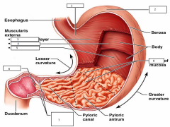
Stomach |
- Chemical breakdown of proteins begin here by digestive enzyme pepsin and food bolus is converted to chyme |
|
|
Lesser Omentum Greater Omentum |
- Mesentery that runs from the liver to the lesser curvature of the stomach - Mesentery that drapes inferiorly from greater curvature of stomach to small intestine |
|
|
- Longitudinal - Circular - Oblique |
3 Layers of the Stomach that allows it to churn and physically mix food, by breaking it down? The esophagus and intestines have 2. |
|
|
Rugae |
- Folds in the stomach wall that enables the stomach to expand and increases surface area |
|
|
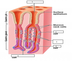
- Simple columnar epithelium - Goblet cells that produce a coat of alkaline mucus - Villi - Increase surface area - Gastric Pits |
4 Structural Characteristics of the Stomach? |
|
|
Mucous cells - Mucus Parietal Cells - HCl Serous acinar "Chief" Cells - Pepsinogen |
Gastric pits contain what gastric glands with 3 types of cells. What are they and their secretions? |
|
|
Zymogens |
Enzymes secreted in inactive state Ex: Trypsinogen, procarboxypeptidase |
|
|
- Holds ingested food - Breaks down food physically and chemically - Delivers chime to the small intestine - Digests proteins with the enzyme pepsin - Secretes intrinsic Factor |
5 Major Functions of the Stomach (HBDDS) |
|
|
Rennin |
Chemical in babies that acts like Hydrochloric acid to curdle milk |
|
|
Hydrochloric Acid |
- Pepsinogen secreted by the chief cells are activated to pepsin to break down proteins by the presence of _____________ secreted the parietal cells since pepsin it a zymogen (inactive protein). |
|
|
- Gastrin (Hormone) - Histamine (Neurotransmitter) - Endorphins (Hormone) - Serotonin (Neurotransmitter) - Cholecystokinin CCK (Hormone) - Somatostatin (Hormone) - Secretin |
What are 7 examples of Hormones that the Enteroendocrine organ cells secrete? (SSS C E H G) |
|
|
Gastrin |
- Increases HCl secretion and stimulates gastric emptying - Produced by stomach mucosa gastric cells |
|
|
Histamine |
- Activates parietal cells to release HCl (Anti-inflammatory stimulus by basophils/mast cells In other areas of the body) - Secreted by stomach mucosa |
|
|
Endorphins |
- Brain chemicals that are natural pain-killers to alleviate spicy foods |
|
|
Serotonin |
- Causes contraction in stomach muscle - produced by stomach mucosa - Neurotransmitter that makes you feel good |
|
|
Cholecystokinin (CCK) |
-Tropic hormone, produced by duodenal mucosa stimulated by presence of acidic, fatty chyme - Targets stomach, liver, pancreas, gallbladder, hepatopancreatic sphincter - Results in entry of pancreatic juice and bile to stomach (by causing gall bladder to contract) |
|
|
Somatostatin |
- Produced by duodenal mucosa and stomach mucosa, inhibits secretion from stomach, pancrease, gallbladder and liver |
|
|
1. Gastric Pit 2. Parietal Cells 3. Gastric Clands 4. Chief Cells 5. Enteroendocrine cells |
|
|
|
Absorption of vitamin B12, which is important for mature erythrocytes
|
What is intrinsic factor required for?
|
|
|
Duodenum - Workhouse Jejunum - Moves food Ileum - Compacts food |
3 Subdivisions of the Small Intestines and their main function
|
|
|
Carboxylases - Carbohydrates (Ex: Amylase) Proteases - Proteins (Ex: Trypsin) Lipases - Fats Nucleases - Nucleic acids |
4 digestive enzymes present in the duodenum by the pancreas and what they break down (And Examples if applicable)
|
|
|
Liver - Common Hepatic Duct - Cystic Duct - Gallbladder
Common Hepatic Duct and Cystic Duct
|
Trace the flow of bile from liver to the gall bladder (4)
The two ducts that fuse to form the bile duct?
|
|
|
Bile
|
Chemical substance that emulsifies fats, NOT DIGESTION, the purpose is to increase surface area
|
|
|
- Plicae circulares - Villi - Microvilli |
3 Structural Modifications of the Small Intestine that increases surface area?
|
|
|
Plicae circulares
|
- Deep circular folds of the mucosa and submucosa that project into the lumen of the small intestine
|
|
|
Villi |
Fingerlike extension of the mucosa
|
|
|
Microvilli
|
Tiny projections of membranes of simple columnar epithelial cells
|
|
|
Brunner's Glands = Duodenal Glands
|
- Glands that make copious amounts of alkaline mucus from the submucosa in the duodenum, lubricate and neutralize acidic chyme
|
|
|
Intestinal Crypts
|
- Site of enzyme production in the small intestines mucosa , collectively called intestinal juice |
|
|
Duodenum
|
- Break up of fats starts and ends at?
|
|
|
Peyer's Pathces
|
- Found in the submucosa of the ileum - Contains mucous associated lymphoid tissues (MALT) |
|
|
Liver Right, left, caudate, quadrant |
- What is the largest organ in the body? - Name its four lobes |
|
|
1. Cholecystokinin 2. Secretin 3. Bile Salts 4. Bile 5. Vagus nerve |
Acidic, fatty chime entering duodenum causes release of _______1_____ and ____2______ from duodenal wall enteroendocrine cells. These enter the blood stream. ______3______ and _____2_____ transported by bloodstream stimulate liver to produce ___4___ more rapidly. ______5_____ causes weak contractions of gallbladder. ______1_____ from bloodstream causes gallbladder to contract and ____4____ enters duodenum. ____3____ reabsorbed into blood |
|
|
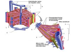
Lobules
|
- Structural and functional units of the liver - Composed of hepatocyte plates radiating outward from a central vein - Portal triads found at the corners |
|
|
1. Bile Duct 2. Portal Venule 3. Portal arteriole 4. Portal triad 5. Hepatocyte plates 6. Hepatic Portal Vein 7. Liver Sinusoids 8. Central Vein 9. Kupffer cells |
|
|
|
- Bile duct: Carries bile from gallbladder to duodenum - Hepatic Arteriole: Supplies oxygen rich blood to the liver - Hepatic Portal Venule: Carries venous blood with nutrients from small intestine |
What do the portal triads consist of, and what are their functions? (3)
|
|
|
Liver Sinusoids
|
- Enlarged leaky capillaries located between hepatic plates in the lobules of the liver |
|
|
Kupffer cells
|
- Hepatic macrophages found in liver sinusoids
|
|
|
- Produces bile - Processes nutrients - Stores fat-soluble vitamins - Detoxification |
4 Functions of Hepatocytes in the Liver (BNVD)
|
|
|
- Bile Salts - Bile pigments: bilirubin - Cholesterol - Neutral fats - Phospholipids - Electrolytes |
Composition of Bile (6) (SBCFPE)
|
|
|
- Emulsifies fat - Facilitates fat and cholesterol absorption - Helps solubilize cholesterol |
3 Functions of Bile Salts
|
|
|
Gallbladder
|
- Organ of the body that stores and concentrates bile, releases bile through the cystic duct which flows into the "common" bile duct
|
|
|
- Secretin and bile salts: Causes liver to produce bile - Cholecystokini: Causes gall bladder to contract - Vagus Nerve: Causes weak contractions of gallbladder |
The presence of acidic, fatty chime in the duodenum triggers - Release of hormones (2) - Stimulation of a brain nerve (1) What are they and what do they cause? |
|
|
Serous Acinar cells: Pancreatic juice with enzymes for all macromolecules secreted Alpha Cells: Glucagon Beta cells: Insulin |
What cells are responsible for the exocrine functions and endocrine functions of the pancreas? What do they secrete? (3)
|
|
|
- Enzymes - Electrolytes - Water |
3 Main Components of Pancreatic Juice |
|
|
Secretin
|
- Produced by duodenal mucosa - Stimulated by presence of acidic chyme - Target stomach, pancreas (secrete bicarbonate-rich pancreatic juice) and liver (produce bile) |
|
|
Segmentation Jejunum, Ileum |
What is the most common motion of the small intestine? What parts of the small intestine are the nutrients absorbed? |
|
|
Cecum Vermiform Appendix Ascending, Transverse, Descending, Sigmoid Colon Rectum Anal Canal |
8 Subdivisions of the Large Intestine and
|
|
|
Teniae coli Haustra Epiploic appendages |
3 Unique Features of the Large Intestine/Colon
|
|
|
Teniae coli
|
- 3 Bands of longitudinal smooth muscle in muscularis externa found in the layers of the large intestine
|
|
|
Haustra
|
- Pocket like sacs caused by the tone of the teniae coli in the large intestine
|
|
|
Epiploic appendages
|
- Fat-filled pouches of visceral peritoneum within the large intestine
|
|
|
Internal: Smooth Muscle External: Skeletal Muscle |
The anus has two anal sphincters are closed except during defecation. What type of muscle can be found in these sphincters?
|
|
|
- Colonize the colon - Ferment indigestible carbohydrates - Release irritating acids and gases - Synthesize B and K vitamins |
4 Functions of the Intestinal Bacterial Flora
|
|
|
- Necessary for normal blood clotting proteins produced by the liver
|
What is the function of Vitamin K synthesized by intestinal microbiota?
|
|
|
- Vitamin, water, electrolyte absorption - Propulsion of fecal material toward the anus |
2 Major Functions of the Large Intestine
|
|
|
Nitrogen
|
- An essential element that is present in proteins and nucleic acids but not in carbohydrates or lipids - Can be inhaled through the atmosphere |
|
|
- Mouth - Small Intestines - Stomach - Small Intestine - Small Intestine - Small Intestine |
Where does carbohydrate digestion begin and end? Where does protein digestion begin and end? Where does fat digestion begin and end? Where does nucleic acid digestion begin and end? |
|
|
Gastric Glands - Chief Cells - Pepsin
Intestinal Crypts - Pancreas- Amylase, Trypsin, Lipase, Nuclease |
What structures of the stomach secrete digestive enzymes? Which specific cells? What is the digestive enzyme?
What structures of the duodenum secrete digestive enzymes? Made by which organ? What are the digestive enzymes?
|
|
|
- Salivary, pancreatic, small intestine brush border |
Name the 3 types of amylase. |
|
|
Stomach - Pepsin @ Stomach Pancreas - Trypsin, chymotrypsin, carboxypeptidase @ Small Intestine Brush Border - Aminopeptidases, carboxypeptidases, dipeptidases @ Small Intestine |
Name the digestive enzymes used for protein digestion by which organs they are being secreted, and where it is being digested. (3 - 1,3,3) |
|
|
- Co-Transport with Na+ - Facilitated Diffusion |
Describe the 2 ways of absorption (from the small intestines) of carbohydrates and proteins
|
|
|
1. Emulsification 2. Bile Salts 3. Digestion 4. Pancreatic Lipase 5. Micelles 6. Diffusion 7. Chylomicrons 8. Exocytosis 9. Lacteals |
The absorption of fats starts by ___1___ by ____2____ to increase the surface area available for ____3____ by enzymes such as ___4___. ____5____ formed in intestinal lumen deposit fatty elements to enter epithelial cells by ____6____, and the fatty elements combine with proteins to create ____7____ by the Smooth ER and Golgi. These are transported by vesicles to be extruded from the epithelial cells by ___8____, where they enter ___9___ to be transported to the systemic circulation by lymph. |
|
|
Bile Salts Pancreatic Lipase |
2 Enzymes/Chemicals used to Chemically Digest Fats |
|
|
Micelles |
Collections of fatty elements clustered together with bile salts in such a way that the polar ends of the molecules face the water and the nonpolar portions form the core - Fatty acids, monoglycerides and bile salts |
|
|
Pancreatic ribonucleases, pancreatic deoxyribonucleases @ Small Intestine Active Transport |
What are the 2 digestive enzymes for nucleic acids and where does digestion occur? Describe the method of absorption. |
|
|
1. Small Intestines 2. Osmosis 3. Large Intestine 4. Stomach |
95% of water is absorbed in the ____1____ by ___2___. The rest of water absorption occurs in the ___3___ and only a small amount is absorbed by the ___4___. |
|
|
1. Ingestion 2. Mechanical 3. Propulsion 4. Chemical 5. Chewing 6. Churning 7. Segmentation 8. Swallowing 9. Peristalsis 10. H2O |
|
|
|
1. Mucosa 2. Lining Epithelium 3. Lamina Propria 4. Muscularis Mucosae 5. Submucosa 6. Muscularis Externa 7. Longitudinal 8. Circular 9. Serosa |
|
|
|
1. Parotid Gland 2. Parotid Duct 3. Masseter Muscle 4. Submandibular Duct 5. Submandibular Gland 6. Sublingual Ducts 7. Sublingual Gland |
|
|
|
1. Cardia 2. Fundus 3. Longitudinal 4. Circular 5. Oblique 6. Pylorus 7. Pyloric Sphincter 8. Rugae |
|