Reading...
![]()
Play button
![]()
Play button
![]()
Use LEFT and RIGHT arrow keys to navigate between flashcards;
Use UP and DOWN arrow keys to flip the card;
H to show hint;
A reads text to speech;
10 Cards in this Set
- Front
- Back

Plate 216:
Identify 1, 3, 4, 5 (name region of hip), 6, 7 (name artery), 8, 9, 11 (name sheath), 14, 15, 16 (name ligament) |
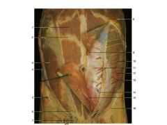
1. Rectus abdominis muscle
3. Internal abdominal oblique m 4. External abdominal oblique m 5. Anterior superior iliac spine 6. Ilio-inguinal n 7. Spermatic cord 8. Costal margin 9. Superior epigastric a 11. Posterior layer of rectus sheath 14. Arcuate line 15. Inferior Epigastric a 16. Inguinal ligament |
|
|
Describe Arcuate line.
|
It is a horizontal line that demarcates the lower limit of the posterior layer of the rectus sheath. It is also where the inferior epigastric vessels perforate the rectus abdominus.
|
|

Plate 207
Identify 33, 35, 36 |
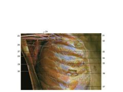
33. External intercostal m
35. Internal intercostal m 36. Anterior intercostal a., v., n. |
|
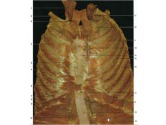
Plate 206
Identify: 2, 4, 5, 6 (name a & n), 7, 8, 10, 13, 15 (name m), 16 (name a & v), 17, 18, 19 |

2. Clavicle
4. internal intercostal m 5. transversus thoracic m 6. intercostal a and n 7. musculophrenic a 8. superior epigastric a and v 10. rectus abdominis m 13. internal thoracic a and v 15. Innermost intercostal m 16. intercostal a and v 17. xiphoid process 18. linea alba and posterior layer of rectus sheath 19. transversus abdominis m |
|
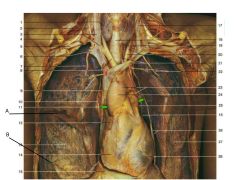
Plate 269
Identify A, B, 2 (ignore a), 3 (not how it runs with jugular), 7, 15, 26 (there's a n, a, and v here) |

A. Horizonal fissure
B. Oblique fissure 2. Phrenic n (and scalenus anterior m 3. Vagus n (and internal jugular v) 7. Internal thoracic a 15. Cut edge of pericardium 26. Left phrenic n and left cardacophrenic a and v |
|
|
Describe the difference between perietal pleura and perietal peritoneum.
|
Perietal pleura lines the thoracic cavity. Perietal peritoneum lines the abdominal cavity.
|
|
|
What is the pubic symphysis?
|
It is the midline cartilaginous joint (secondary cartilaginous) uniting the superior rami of the left and right pubic bones
|
|
|
What is the pyramidalis m.
|
It is a small triangular muscle anterior to the rectus abdominis and within the rectus sheath.
|
|
|
What is the root of lung?
|
A little above the middle of the mediastinal surface of each lung, and nearer its posterior than its anterior border, is its root, by which the lung is connected to the heart and the trachea.
|
|
|
Review the boundaries of the inguinal canal (p43 lab man.) - work on post and sup - the rest you got.
|
Deep: deep inguinal ring
Superficial: superficial inguinal ring Anterior: aponeurosis of the external oblique muscle Posterior: transversalis fascia, reinforced medially by the conjoint tendon. Inferior (floor): Inguinal ligament Superior (root): the arching fibers of the internal oblique and the transversus abdominis muscles |