Reading...
![]()
Play button
![]()
Play button
![]()
Use LEFT and RIGHT arrow keys to navigate between flashcards;
Use UP and DOWN arrow keys to flip the card;
H to show hint;
A reads text to speech;
58 Cards in this Set
- Front
- Back
|
The embryonic period is weeks __________ of human development.
|
3-8
|
|
|
The 3 germ layers (ecto, meso and endoderm) become recognizably human in the _________________ period of development.
|
embryonic
|
|
|
The period during which the conceptus is most likely to develop a major congenital malformation is the _______________ period.
|
embryonic
|
|
|
Name the 6 things the ectodermal germ layer gives rise to.
|
1. nervous system (central and peripheral)
2. sensory epithelium 3. epidermis 4. subcutaneous glands 5. pituitary gland 6. tooth enamel |
|
|
The formation of the CNS begins with the development of a pear-shaped thickening of the ectoderm called the ________________.
|
neural plate
|
|
|
The lateral edges of the neural plate, the __________, fuse to form the neural tube.
|
neural folds
|
|
|
The cranial and caudal neuropores finally fuse at days _____ and _____, respectively.
|
25 and 27
|
|
|
spinal (posterior root) ganglia and sensory ganglia of cranial nerves V, VII, IX, and X; automonic ganglia; the adrenal medulla; Schwann cells; connective tissues of the anterior part of the skull and meninges; melanocytes; C cells of the thyroid gland; and the conotruncal septum of the heart are all derivatives of the ___________.
|
neural crest
|
|
|
As part of the formation of the face and anterior neck, neural crest cells migrate into ______________ and form connective tissues
|
pharyngeal arches
|
|
|
The 3 sections of the mesodermal germ layer, medial to lateral, are:
|
1. paraxial
2. intermediate 3. lateral plate |
|
|
The paraxial mesoderm becomes segmented into 42-44 blocks of tissue called __________.
|
somites
|
|
|
The age of the embryo is expressed by the number of ________.
|
somites (20 days is 1 to 4 somites, 25 days is 17 to 20 somites, 30 days is 34-35 somites)
|
|
|
Cartilage, bones of the axial skeleton (including vertebral column) are formed by the ________________ division of the somite.
|
ventromedial sclerotome
|
|
|
Dorsomedial muscle forming region of the somite is also called the (epimeric/hypomeric) region.
|
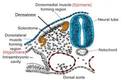
epimeric
|
|
|
Dorsolateral muscle forming region of the somite is also called the (epimeric/hypomeric) region.
|

hypomeric
|
|
|
Each somite has a _______________, which forms the dermis of the skin for its segment of the body.
|
dorsal dermatome
|
|
|
The intermediate mesoderm differentiates into the ___________.
|
urogenital structures (kidney, gonads)
|
|
|
The lateral plate mesoderm divides into 2 layers separated by the _____________ cavity.
|
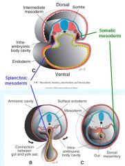
somatic/parietal layer
|
|
|
The _________________ division of the lateral plate mesoderm joins the overlying ectoderm to form the ventral and lateral body walls.
|
somatic/parietal
|
|
|
The _________________ division of the lateral plate mesoderm joins the underlying endoderm to form the wall of the gut.
|
splanchnic/visceral
|
|
|
The intraembryonic cavity is lined with ___________________ for the _________, __________, and _________ cavities of the adult.
|
the serous membranes that will later secrete serous fluid
pericardial, pleural, peritoneal |
|
|
Blood vessel formation first occurs in the _______________ and later in the ___________.
|
-extraembryonic mesoderm surrounding yolk sac
-lateral plate mesoderm (**later--lateral mesoderm) |
|
|
The ____________ becomes the major hematopoietic organ of the fetus by week 6.
|
liver
|
|
|
The liver sends stem cells to colonize the ____________, the definitive blood-forming organ after the seventh month of gestation.
|
bone marrow
|
|
|
The main organ system derivative of the endoderm is the...
|
GI tract
|
|
|
The GI tract forms as a result of ___________ and ___________ folding of the trilaminar germ disc.
|
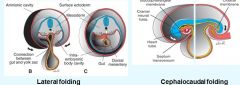
-cephalocaudal
-lateral |
|
|
Cephalocaudal folding causes the formation of a ____________ and _______ fold in the embryo, causing the "fetal position."
|
-head fold
-tail fold |
|
|
The endoderm-derived gastrointestinal tract is comprised of the __________, the _________, and the ___________ between the first two.
|
foregut, hindgut, and midgut between the foregut and hindgut
|
|
|
The midgut is temporarily connected to the yolk sac by the _____________.
|
vitelline drainage
|
|
|
The respiratory system appears as an outgrowth of the ventral wall of the foregut called the _______________.
|
respiratory diverticulum (lung bud)
|
|
|
The ________________ germ layer contributes to the urinary system, thyroid/parathyroid, liver/pancreas, tonsils and thymus and inner ear.
|
endodermal
|
|
|
The _________ period is from week 9 of gestation until birth.
|
fetal
|
|
|
The fetal period begins ____ weeks after fertilization and _____ weeks after the first day of the last normal menstrual period.
|
38
40 |
|
|
The __________ period is the time of growth and functional maturation of tissues and organs.
|
fetal
|
|
|
Primary ossification centers are present in all long bones and the skull by ________ weeks of gestation.
|
12
|
|
|
During the 4th and 5th months of gestation, the head of the fetus grows (more slowly/faster) than the rest of the body.
|
more slowly
|
|
|
At birth, the circumference of the baby's ________ is greater than any other part of the body.
|
skull
|
|
|
The inner cell mass of the blastocyst, the __________, forms the ___________.
|
embryoblast
forms embryo |
|
|
The outer cell mass of the blastocyst, the __________, forms the __________.
|
trophoblast
fetal portion of the PLACENTA |
|
|
During the 2nd week of development, the trophoblast differentiates into the outer _____________ and the inner ______________.
|
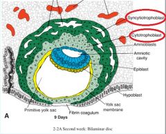
-syncytiotrophoblast
-cytotrophoblast |
|
|
2nd week of development: The endometrium of the uterus undergoes the ___________________, in which endometrial cells around the conceptus become loaded with glycogen and lipids and the tissue becomes edematous.
|
decidua reaction
|
|
|
2nd week of development: Large spaces called ___________ appear in the syncytiotrophoblast as it is invaded by dilated capillaries of the endometrium called ____________. This establishes the the _______________ circulation.
|
-lacunae
-sinusoids -uteroplacental circulation |
|
|
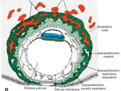
2nd week of development: The ___________ is pinched off from the primary yolk sac during the formation of the _________________ cavity.
|
secondary yolk sac
chorionic |
|
|
2nd week of development: The amnion amnion and yolk sac remain attached to the chorion (extraembryonic mesoderm plus the two layers of trophoblast) across the chorionic cavity by the ______________, which later becomes the umbilical cord.
|
connecting stalk
|
|
|
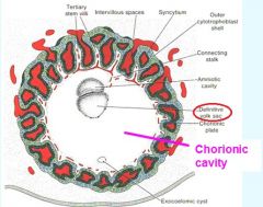
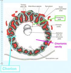
3rd week of development: the ________________ entirely surrounds the trophoblast and attaches it to the endometrium.
|
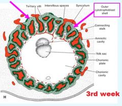
outer cytotrophoblast shell
|
|
|
__________________ can traverse the placental barrier freely.
|
Drugs and viruses
|
|
|
The 4 placental functions include:
|
1. exchange of gasses
2. exchange of nutrients/electrolytes 3. transmission of maternal antibodies (maternal immunoglobulin G/IgG) 4. production of hormones |
|
|
During the first two months the syncytiotrophoblast secretes __________________ to maintain the corpus luteum.
|
human chorionic gonadotropin (hCG)
|
|
|
Early pregnancy tests test the hormone _________.
|
hCG
|
|
|
By the end of the fourth month, the placenta produces enough _________________ to maintain pregnancy if the corpus luteum were removed
|
progesterone
|
|
|
By the end of the fourth month, the placenta produces ____________ to stimulate uterine growth and mammary gland development.
|
estrogens
|
|
|
The umbilical cord forms from the ___________ and the __________.
|
connecting stalk
vitelline duct |
|
|
The _____________ contains the allantois and umbilical vessels.
|
connecting stalk
|
|
|
Amniotic fluid is derived mainly from _____________.
|
maternal blood
|
|
|
The four functions of amniotic fluid are:
|
1. shock absorption
2. preventing embryonic adhesion to amnion 3. allowing fetal movements 4. allowing fetal growth |
|
|
In the fifth month, __________ is swallowed by the fetus, contributing to hypotonic urine.
|
amniotic fluid
|
|
|
In anencephaly or intestinal atresia, amniotic fluid is present in __________. This is called ___________.
|
-excess
-polyhydramnios |
|
|
Amniotic fluid may be deficient in amount (______________), e.g., due to renal agenesis or amnion rupture, resulting in clubfoot or lung hypoplasia.
|
-oligohydramnios
|