![]()
![]()
![]()
Use LEFT and RIGHT arrow keys to navigate between flashcards;
Use UP and DOWN arrow keys to flip the card;
H to show hint;
A reads text to speech;
33 Cards in this Set
- Front
- Back
|
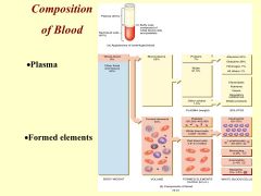
Compositionof blood: formed elements, plasma |
|
|
|
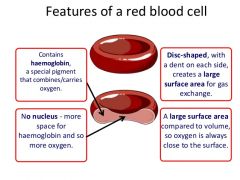
Features of RBCs (red blood cells) |
|
|
|
The process of RBCs production. |
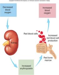
Erythropoiesis (from Greek 'erythro' meaning "red" and 'poiesis' meaning "to make") is the process which produces red blood cells (erythrocytes). It is stimulated by decreased O2 in circulation, which is detected by the kidneys, which then secrete the hormone erythropoietin. Erythropoiesis•Format rate of 3 million/sec•Takes12-15 days•Regulatedby hormone erythropoietin–Producedby the kidney–Stimulatesred cell production–Worksthrough negative feedback loop |
|
|
Normal Lifespan of RBCs. |
•LifeCycle •RBCslive only 120 days–wearout from bending to fit through capillaries–norepair possible due to lack of organelles •Wornout cells removed by fixed macrophages in spleen & liver •Breakdownproducts are recycled |
|
|
The process/steps in blood clotting or coagulation. |
INTRINSIC PATHWAY Takesminutes 1.Bloodvessel endotheliumruptures 2.Collagen fibers are exposed 3.Platelets cling and attract+ release •Plateletfactors •Ca++ 4.Many steps to formationof Clottingfactor X BASICSTEPS-COMMON PATHWAY 1.FactorX >>>>>> Prothrombinase2.Prothrombin>>(prothrombinase)>>> Thrombin 3. Fibrinogen>>(Thrombin)>> Fibrin threads •Threadsentrap red cells, platelets, and plasma in clot 4. Clot shrinks •Pullsvessel together |
|
|
Types of Leucocytes.(white blood cells) |
Granular •Neutrophils •Basolphils •Eosinophils Agranular •Lymphocytes •Monocytes |
|
|
Plasma proteins, types and functions. |
Albimins Globulins Fibrinogen |
|
|
Coverings of the heart. |
OUTER LAYER COVERINGS: Fibrous Pericardium Parietal Layer of the serous pericardium Epicardium (Visceral layer of the Serous pericardium) INNER LAYER COVERINGS: Endocardium |
|
|
Layers of the Heart |
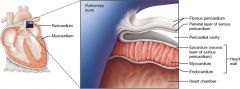
Layers of the Heart Wall. The heart wall is composed of three layers: the epicardium, myocardium, and endocardium. The superficial epicardium is the visceral layer of the serous pericardium. |
|
|
Chambers of the heart |
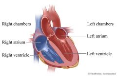
Right and Left Atria
Right and Left Ventricles |
|
|
The right atrium function |
Receives oxygen-poor blood from the body and pumps it to the right ventricle. |
|
|
The right ventricle function |
Pumps the oxygen-poor blood to the lungs. |
|
|
The left atrium function |
Receives oxygen-rich blood from the lungs and pumps it to the left ventricle. |
|
|
The left ventricle function |
Pumps the oxygen-rich blood to the body. |
|
|
Heart Valves and their Functions |
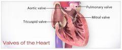
TRICUSPID VALVE PULMONARY VALVE MITRAL VALVE AORTIC VALVE |
|
|
ECG waves and what they represent. |
ELECTROCARDIOGRAM: •Recording of electricalchanges inheart P wave QRS Wave T wave |
|
|
Causesof Heart sounds |
The “ lub” is the first heart sound, commonly termed S1, and is caused by turbulence caused by the closure of mitral and tricuspid valves at the start of systole. The second sound,” dub” or S2, is caused by the closure of aortic and pulmonic valves, marking the end of systole. |
|
|
Functions of Leukocytes: NEUTROPHILS |
•60-70%of white cells •Firstat site of injury •Phagocytizeantibody-markedbacteria •Releaseinflammation-producing prostoglandins •Releasephagocyte-attracting leukotrines •Live 10hours |
|
|
Functions of Leukocytes: EOSINOPHILS |
•2-4% of white cells •Phagocytizeantibody-marked bacteria and protozoa anddebris •Releasetoxins to kill invaders •Killparasitic worms •Increaseduring allergicreactions •Reduce inflammation |
|
|
Functions of Leukocytes: BASOPHILS |
•1% of white cells •Releasehistamine-causinginflammation•Releaseheparinprevents clotting •Attractother cells to site of injury •Increaseduring allergicreactions, leukemias, diabetes, hypothyroidism |
|
|
Functions of Leukocytes: MONOCYTES |
•3-8% of white cells •Leaveblood to become Macrophages •Engulfmicrobes particularly viruses •Attractother white cells to site of attack •Clean debris, deadcells |
|
|
Functions of Leukocytes: LYMPHOCYTES |
•25-33% of whitecells •Movebetween tissues and blood •Threetypes •T cells •B cells •NK cells |
|
|
Plasma Proteins: Albumens |
- 55-60% –Viscosity, Osmotic pressure, Transport |
|
|
Plasma Proteins: Globulins |
– 35-38% –Immunoglobulins •Disease fighting antibodies –Transport globulins |
|
|
Plasma Proteins: Fibrinogen |
- 4-7% –Blood clotting |
|
|
*PLASMA versus SERUM |
Plasma: The fluid = water, proteins and other fluids Serum: The fluid without blood clotting factors (i.e. fibrinogen) or blood cells. |
|
|
TRICUSPID VALVE Function |
Closes off the upper right chamber (or atrium) that holds blood coming in from the body. Opens to allow blood to flow from the top right chamber to the lower right chamber (or from right atrium to right ventricle). Prevents the back flow of blood from the ventricle to the atrium when blood is pumped out of the ventricle. |
|
|
PULMONARY VALVE Function |
Closes off the lower right chamber (or right ventricle). Opens to allow blood to be pumped from the heart to the lungs (through the pulmonary artery) where it will receive oxygen. |
|
|
MITRAL VALVE Function |
Closes off the upper left chamber (or left atrium) collecting the oxygen-rich blood coming in from the lungs. Opens to allow blood to pass from the upper left side to the lower left side (or from the left atrium to the left ventricle). |
|
|
AORTIC VALVE Function |
Closes off the lower left chamber that holds the oxygen-rich blood before it is pumped out to the body. Opens to allow blood to leave the heart (from the left ventricle to the aorta and on to the body). |
|
|
What does this ECG wave mean? P wave |
-atrial depolarization |
|
|
What does this ECG wave mean? QRS wave |
- Ventricular depolarization |
|
|
What does this ECG wave mean? T wave |
- Ventricular repolarization |