![]()
![]()
![]()
Use LEFT and RIGHT arrow keys to navigate between flashcards;
Use UP and DOWN arrow keys to flip the card;
H to show hint;
A reads text to speech;
78 Cards in this Set
- Front
- Back
|
Pathway of olfactory nerve |
Becomes olfactory bulbs Through the cribiform foramina |
|
|
Pathway of the optic nerve |
Crosses at optic chiasm Through optic canal Ends at eye |
|
|
Pathway of oculomotor |
In cavernous sinus Through superior orbital fissure Motor to extraocular muscles Parasympathetic through ciliary ganglion and then to sphincter pupillae and ciliary muscle |
|
|
Pathway of trochlear nerve |
Through cavernous sinus Through superior orbital fissure Motor to superior oblique |
|
|
Parhway of trigeminal V1 (ophthalmic) |
Trigeminal ganglion Through cavernous sinus Through superior orbital fissure Somatic sensory: nasal, Frontal and lacrimal nerves Frontal gives Supraorbital (which goes through supraorbital notch) and supratrochlear nerve All nerves are somatic sensory |
|
|
Pathway of trigeminal V2 (maxillary) |
Trigeminal ganglion Cavernous sinus Foramen rotundum Gives off zygomatic Then gives lesser, greater and naso-palantine (all three go through the pterygopalantine ganglion) Then gives superior alveolar and lastly infraorbital (goes through infraorbital Foramen) All nerves are somatic sensory |
|
|
Pathway of trigeminal V3 (mandibular) |
Goes through Foramen ovale Gives motor branches to: tensor veil palatini, muscles of mastication, tensor tympani, mylohyoid and ant. belly of digastric. Somatic sensory branches: meningeal (which goes through Foramen spinosum), inferior alveolar (goes through mandibular and mental Foramen and then becomes the mental nerve) Then gives auriculotemporal, buccal, lingual branches |
|
|
Pathway of abducent nerve |
Cavernous sinus Superior orbital fissure Lateral rectus (extraocular) Motor nerve |
|
|
Pathway of facial nerve |
Through internal acoustic meatus Then facial canal Synapses at geniculate ganglion Motor: through styloid Foramen Gives posterior belly of digastric, stylohyoid, muscles of facial expression Special sensory: part of chorda tympani, hitchhikes w/ V3, innervates ant 2/3 of tongue Para: greater petrosal nerve feeds lacrimal gland and nasal mucosa and palate Part of Chorda tympani does submandibular and sublingual glands Also involved with the deep petrosal nerve (sympathetic) |
|
|
Pathway of vestibulocochlear |
Goes through internal acoustic meatus Special: cochlea and semicircular canals |
|
|
Pathway of glossopharyngeal |
Goes through jugular Foramen Motor: stylopharyngeus Special: post 1/3 tongue Somatic: carotid body and sinus, oropharynx, and post 1/3 Tympanic nerve has somatic and parasympathetic to tympanic plexus. Para continues and is lesser petrosal which goes through Foramen ovale and otic ganglion innervates parotid |
|
|
Pathway of Vagus nerve |
Jugular Foramen Pharyngeal branch gives special sensory to base of tongue and motor to pharyngeal constrictors and soft palate muscles Superior laryngeal: internal branch goes through thyrohyoid membrane and innervates mucosa of larynx (superior to vocal folds), external motor to cricothyroid Recurrent laryngeal turns into Inferior laryngeal: motor to intrinsic muscles of the larynx and somatic to mucosa of larynx inferior to vocal folds |
|
|
Pathway of spinal accessory nerve |
Foramen magnum Jugular Foramen Motor to: trapezius and sternocleidomastoid |
|
|
Pathway of hypoglossal nerve |
Through hypoglossal canal Motor to: intrinsic and extrinsic tongue (except palatoglossus) |
|
|
Features of the hyoid bone |
Body Greater horn Lesser horn C3 vertebral level |
|
|
What travels in the carotid sheath |
Common/internal carotid arteries Internal jugular vein Vagus nerve Deep cervical lymph nodes |
|
|
External jugular vein features |
Receives blood from scalp, face, neck and shoulder Superficial to SCM |
|
|
Where does the brachial plexus emerge from |
Emerges between anterior and middle scalene |
|
|
Features of the internal jugular vein |
Within carotid sheath receives blood from face, neck and head Join subclavian veins at venous angle to form brachiocephalic veins |
|
|
Subclavian veins |
Receives blood from UE, head and neck. Becomes the brachiocephalic veins |
|
|
Features of the common carotid arteries |
Within carotid sheath carotid body- chemoreceptors carotid sinus- baroreceptors Internal carotid- no branches in neck External carotid- occipital, facial, lingual, ascending pharyngeal, and superior thyroid arteries |
|
|
Subclavian arteroes |
Internal thoracic Vertebral Thyrocervical trunk- inferior thyroid, transverse cervical, suprascapular arteries |
|
|
Lymphatics of the neck |
Superficial cervical nodes- along external jugular veins, submandibular and submental nodes Deep cervical nodes- along internal jugular veins |
|
|
Features of the thyroid gland |
Deep to sternohyoid and sternohyoid C5-T1 vertebral levels Right lobe, left lobe, isthmus and pyramidal lobe ~50% Superior thyroid artery (external carotid) Inferior thyroid (thyrocervical trunk) Thyroid ima artery ~10% Superior and middle thyroid vein (to IJV) Inferior thyroid vein (to brachiocephalic vein) 2 superior and inferior parathyroid glands |
|
|
Sections of the pharynx |
Nasopharynx Oropharynx Laryngopharynx |
|
|
Features of the constrictor muscles |
Superior constrictor- pterygoid Hamilcar of sphenoid bone and mandible Middle constrictor- hyoid Inferior constrictor- thyroid and cricoid cartilage Constrictors fuse posteriorly at raphe |
|
|
What is the platysma innervated by |
Facial nerve |
|
|
Pharynx internal muscles |
Stylopharyngeus- inserts between superior and middle constrictor, elevates pharynx and larynx, innervated by glossopharyngeal. |
|
|
Innervation of the pharynx |
Glossopharyngeal nerve- motor to stylopharyngeus, sensory to mucosa of oropharynx, exits jugular Foramen Vagus- motor to constrictor muscles, sensory to mucosa of laryngopharynx (includes piriform recesses), exits jugular Foramen Gag reflex- motor is vagus sensory is glossopharyngeal |
|
|
Larynx features |
Function: phonation, respiration Inferior to hyoid (C3-C6) |
|
|
Laryngeal cartilages |
Thyroid cartilage- laryngeal prominence, superior horn, inferior horn, laminae. Motion: rotation/gliding of thyroid cartilage, affects length of vocal ligaments Cricoid cartilage Epiglottic cartilage Arytenoid cartilages- motion is adduction/abduction, tilting/rotation. Tenses vocal ligaments. |
|
|
Laryngeal ligaments and folds |
Thyrohyoid membrane Vocal folds- ( space makes up the rima glottidis) Quadrangular ligament- connects epiglottic and arytenoid cartilages. Cricotracheal ligament |
|
|
Three sections of the laryngeal cavity |
Vestibule- between inlet and vestibular folds Ventricle- between vestibule and vocal folds Infraglottic cavity- between vocal folds and inferior border of cricoid cartilage. |
|
|
Extrinsic muscles of the larynx |
Suprahyoid muscles (elevate larynx)- mylohyoid, digastric, stylohyoid, geniohyoid Infrahyoid muscles- thyrohyoid (elevates larynx and depresses hyoid) sternothyroid (depresses larynx and hyoid) |
|
|
Intrinsic muscles of the larynx |
Aryepiglottic: close laryngeal inlet Lateral cricoarytenoid, transverse arytenoid and oblique arytenoid- all adduct vocal folds Posterior cricoarytenoid- abduct vocal folds Cricothyroid- stretch/tense vocal folds Thyroarytenoid- relax vocal folds |
|
|
Innervation of the larynx |
Superior laryngeal nerve: internal branch- sensory to mucosa of larynx superior to vocal folds, pierces thyrohyoid membrane External branch- motor to cricothyroid Inferior laryngeal nerve: (continuation of recurrent laryngeal nerve) motor to most intrinsic muscles of larynx Sensory to mucosa inferior to vocal folds |
|
|
Vessels of the larynx |
Superior laryngeal artery: (branch of superior thyroid artery that travels with internal branch of superior laryngeal nerve through thyrohyoid membrane) Inferior laryngeal artery: (branch of inferior thyroid artery |
|
|
What invest the deep cervical fascia? |
Trapezius Sternocleidomastoid Scalenes Submandibular glands Parotid glands |
|
|
Sympathetic trunks |
Posterior to carotid sheath Only grey rami communicantes Three paravertebral ganglia: superior, middle and inferior/stellate |
|
|
What is horners syndrome |
Damage to sympathetic nervous system (cervical or thoracic) Patient presentation: ptosis, anhidrosis (denervation of sweat glands ipsilateral face and neck), miosis (denervation of dilator pupillae) and heterochromia (in infants) |
|
|
Infrahyoid muscles features |
Depresses hyoid Most are innervated by ansa cervicalis Sternohyoid Omohyoid sternothyroid (also depresses larynx) Thyrohyoid (innervated by C1) |
|
|
Suprahyoid muscles features |
Mylohyoid (mandibular nerve) Digastric (also depresses mandible, ant belly- mandibular nerve, post belly facial nerve) Stylohyoid (facial nerve) Geniohyoid (deep to mylohyoid, cervical plexus) |
|
|
Sternocleidomastoid features |
Action: unilateral- ipsilateral lateral flexion of neck, contralateral rotation of head Bilateral- flex neck Innervation: spinal accessory |
|
|
Muscles within the posterior triangle of the neck. |
Splenius capitis Levator scapulae Posterior scalene Middle scalene Anterior scalene Omohyoid (inferior belly) |
|
|
Scalenes features |
Action: laterally flex or elevate ribs Innervation: cervical plexus |
|
|
Features of the cervical plexus |
From ventral rami C1-C4 Emerges from posterior edge of SCM Lesser occipital Great auricular Transverse cervical Supraclavicular Ansa Cervicalis- (loop formed by C1-C3, superficial to carotid sheath, innervates infrahyoid. "Hitchhikes" but is not a branch of hypoglossal) |
|
|
Phrenic nerve features |
C3-C5 Superficial to anterior scalene, innervates diaphragm, parietal pleura, fibrous pericardium, and parietal layer of serous pericardium |
|
|
Where are the spinal cord enlargements |
Cervical: C4-T1 located at aame vertebral levels Lumbosacral: L1-S3 segments located at T11-L1 vertebral levels |
|
|
Where are the spinal cord enlargements |
Cervical: C4-T1 located at aame vertebral levels Lumbosacral: L1-S3 segments located at T11-L1 vertebral levels |
|
|
Spinal meninges and soaces |
Epidural space Spinal dura mater Spinal arachnoid mater Subarachnoid space (contains arachnoid trabeculae) Spinal pia mater (makes denticulate ligaments) |
|
|
What area is susceptible to an epidural hematoma |
Pterion |
|
|
What area is susceptible to an epidural hematoma |
Pterion (By the middle meningeal artery) |
|
|
Features of the middle cranial fossa (temporal and sphenoid bones) |
Temporal lobes Pituitary gland Optic canal: optic nerve and ophthalmic artery Superior orbital fissure: oculomotor, trochlear, ophthalmic, abducent nerves Ophthalmic veins Foramen rotundum: maxillary Foramen ovale: mandibular Foramen spinosum: middle meningeal artery and vein Foramen lacerum: covered by membrane, internal carotid passes horizontally |
|
|
Features of the posterior cranial fossa |
Brainstem Cerebellum Internal acoustic meatus: facial and vestibulocochlear nerves Jugular Foramen: vagus, spinal accessory, glossopharyngeal, internal jugular vein Hypoglossal canal: hypoglossal nerve Foramen magnum: medulla, meninges, vertebral arteries |
|
|
What two nerves are important for headache and migraine development? |
Trigeminal and Vagus (Innervates the dura mater) |
|
|
What two nerves are important for headache and migraine development? |
Trigeminal and Vagus (Innervates the dura mater) |
|
|
Features of the Dural reflections |
Falx cerebri Tentorium cerebelli Diaphragm sellae |
|
|
Dural venous sinuses |
Superior Sagittal sinus Inferior Sagittal sinus Straight vein Great cerebral vein (of Galen) Confluence of sinuses Occipital sinus Transverse sinuses Sigmoid sinuses Cavernous sinuses Superior petrosal sinus Inferior petrosal sinus |
|
|
Dural venous sinuses |
Superior Sagittal sinus Inferior Sagittal sinus Straight vein Great cerebral vein (of Galen) Confluence of sinuses Occipital sinus Transverse sinuses Sigmoid sinuses Cavernous sinuses Superior petrosal sinus Inferior petrosal sinus |
|
|
What all is in the cavernous sinus |
Oculomotor, trochlear, abducent, ophthalmic, maxillary nerves Optic chiasm posterior communicating artery Internal carotid hypophysis (pituitary gland) |
|
|
Blood supply of the brain |
Circle of Willis: anterior cerebral, anterior communicating, middle cerebral, posterior communicating, posterior cerebral, basilar, internal carotid Receives contributaries from posterior circulation (vertebral) and anterior circulation (internal carotid) Vertebral joins and becomes basilar artery |
|
|
5 layers of the scalp |
Skin Connective tissue Aponeurosis Loose connective tissue Periosteum |
|
|
Nerves of the scalp |
Trigeminal (V1, V2, V3) Cervical nerves (lesser and greater occipital) |
|
|
Blood supply to the scalp |
Branches from internal carotid (supraorbital) Branches from external carotid (Superficial temporal) Emissary veins can spread infection from fourth layer of scalp to the dural venous sinuses |
|
|
The muscles of facial expression |
Occipitofrontalis (Frontalis muscle belly, Epicranial Aponeurosis, occipitalis muscle belly) Orbicularis oculi (Palpebral and orbital part) Nasalis Orbicularis oris Buccinator Lip elevators (zygomaticus major) Lip depressors (depressor anguli oris) Platysma |
|
|
Nerves of the face |
Facial nerve: (motor stylomastoid Foramen behind parotid gland Posterior auricular nerve, temporal, zygomatic, buccal, mandibular, cervical) Bell's palsy is paralysis of the facial nerve Ophthlalmic (CN V1- supraorbital through supraorbital notch) Maxillary (CN V2- infraorbital) Mandibular (CN V3- buccal, mental, auriculotemporal) |
|
|
Arteries of the face |
Branches of external carotid: superficial temporal Facial artery- gives superior and inferior labial arteries and terminated as angular artery Internal carotid branch: Supraorbital arteries |
|
|
Veins of the face |
Angular vein becomes facial vein and receives blood from superior and inferior labial veins. Then joins anterior retromandibular vein and drains into internal jugular. Facial veins communicates with cavernous sinus and pterygoid plexus through superior and inferior ophthalmic veins and deep facial vein |
|
|
What passes through and emerges from the parotid gland |
Passes through: Facial nerve Retromandibular vein External carotid artery Emerges superior: auriculotemporal nerve Superficial temporal vessels Emerges anterior: 5 terminal branches of facial nerve, parotid duct Emerges posterior: Posterior auricular nerve |
|
|
What are the possible movements at he TMJ |
Depression Elevation Protrusion Retrusion Lateral side to side |
|
|
Muscles of mastication |
Motor innervation from mandibular nerve Temporalis- neurocranial bones to coronoid process Masseter- zygomatic arch to ramus of mandible Lateral pterygoid- sphenoid to TMJ joint capsule & mandibular condyle. (2 heads) Medial pterygoid- sphenoid to medial aspect of the ramus of mandible (2 heads) |
|
|
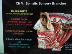
What are the somatic sensory branches of the mandibular nerve? |
|
|
|
What muscles get motor innervation from the inferior alveolar nerve? |
Mylohyoid Anterior digastric |
|
|
What innervation does the chorda tympani provide? |
Taste to anterior 2/3 of tongue Parasympathetics to submandibular and sublingual salivary glands via the submandibular ganglion |
|
|
What are the vessels of the infratemporal fossa |
Arteries: External carotid gives Maxillary which in turn gives Middle meningeal and Inferior alveolar Middle meningeal passes through Foramen spinosum and is the artery to worry about at the pterion Inferior alveolar supplies the mandibular teeth. Veins: pterygoid venous plexus to maxillary vein To retromandibular vein To external jugular vein (The pterygoid communicates with cavernous sinus and facial vein) |
|
|
Features of the pterygopalatine fossa |
Communicates with the nasal cavity, orbit and middle cranial fossa through the sphenopalatine Foramen, Inferior orbital fissure and Foramen rotundum respectively Contents: maxillary nerve (enters through Foramen rotundum and exits through the inferior orbital fissure as infraorbital nerve) Pterygopalatine ganglion (para) Vessels- maxillary artery gives: sphenopalatine, Descending palatine, Infraorbital, Superior alveolar |