![]()
![]()
![]()
Use LEFT and RIGHT arrow keys to navigate between flashcards;
Use UP and DOWN arrow keys to flip the card;
H to show hint;
A reads text to speech;
4 Cards in this Set
- Front
- Back
|
Capsules of kidney |
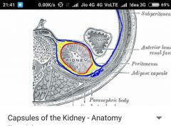
Mainly 3 coverings, If we take anatomical cross section, we can view FIBROUS CAPSULE adhering to kidney , outer PERINEPHRIC FAT (peri means around) and outer to it the RENAL FASCIA (Fuses with extra peritoneal fascia laterally and outer to it the PARANEPHRIC FAT (postero lateral) . |
|
|
Starting of internal structure of kidney |
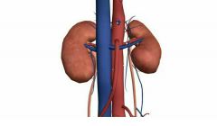
Abdominal aorta (red) and inferior venacava(blue) giving off renal arteries and renal veins through hilum( which is the point of entrance) From kidney ureter coming out. |
|
|
Hilum |

Vertical slit in medial aspect of kidney through where structures like renal vein, renal artery, blood vessels, lymphatics, nerves and ureter. |
|
|
How is kidney sliced for internal view? |
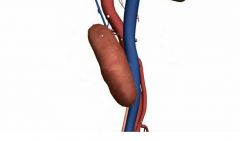
Through that arrow, downwards, Then take the cross section for internal study. |