Reading...
![]()
Play button
![]()
Play button
![]()
Use LEFT and RIGHT arrow keys to navigate between flashcards;
Use UP and DOWN arrow keys to flip the card;
H to show hint;
A reads text to speech;
149 Cards in this Set
- Front
- Back
|
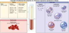
About ____ times more viscous (or thicker) than water.
|
About four times more viscous (or thicker) than water.
|
|
|
Considered a_______ because it contains cells, a liquid ground substance (called plasma), and dissolved protein fibers.
|
connective tissue
|
|
|
Erythrocytes (or red blood cells)
|
form the lower layer of the centrifuged blood
typically make up about 44% of a blood sample |
|
|
Buffy coat
|
makes up the middle layer
thin, slightly gray-white layer composed of cells called leukocytes (or white blood cells) and cell fragments called platelets forms less than 1% of a blood sample |
|
|
Plasma
|
straw-colored liquid that rises to the top
generally makes up about 55% of blood |
|
|
Considered a connective tissue because it contains cells, a liquid ground substance (called plasma), and dissolved protein fibers.
|
blood
|
|
|
Erythrocytes
|
(or red blood cells)
form the lower layer of the centrifuged blood typically make up about 44% of a blood sample |
|
|
Buffy coat
|
makes up the middle layer
thin, slightly gray-white layer composed of cells called leukocytes (or white blood cells) and cell fragments called platelets forms less than 1% of a blood sample |
|
|
Plasma
|
straw-colored liquid that rises to the top
generally makes up about 55% of blood |
|
|
formed elements
|
Erythrocytes and the components of the buffy coat
not “cells,” merely fragments broken off from a larger cell |
|
|
Functions of blood
|
regulation of body temp.
regulation of pH levels maintain fluid levels protection (immunity) |
|
|
Functions of Blood –Transportation
|
Transports numerous elements and compounds throughout the body.
|
|
|
erythrocytes carry
|
oxygen from the lungs to body cells and then transport carbon dioxide from the cells back to the lungs for expulsion from the body
|
|
|
blood plasma transports....
|
transports nutrients that have been absorbed from the GI tract
hormones secreted by the endocrine organs to their target cells |
|
|
plasma carries
|
waste products from the cells to organs such as the kidneys, where these waste products are removed
|
|
|
Regulates body temperature.
- plasma |
absorbs and distributes heat throughout the body
|
|
|
Regulates body temperature - the blood vessles
|
blood vessels in the dermis dilate and dissipate the excess heat through the integument
|
|
|
dermal blood vessels constrict
|
when the body needs to conserve and the warm blood is shunted to deeper blood vessels in the body
|
|
|
Functions of Blood – Regulation of pH Levels
|
Blood plasma contains compounds and ions that may be distributed to the fluid among tissues (interstitial fluid) to help maintain normal tissue pH.
|
|
|
Blood plasma pH is continuously regulated at
|
a value of 7.4 the pH level required for normal cellular functioning.
|
|
|
Functions of Blood – Maintenance of Fluid Levels
|
Constant exchange of fluid between the blood plasma and the interstitial fluid.
|
|
|
If too much fluid is absorbed in the blood
|
high blood pressure results.
|
|
|
If too much fluid escapes the bloodstream and enters the tissues
|
blood pressure drops to unhealthy low levels, and the tissues swell with excess fluid
|
|
|
blood pressure drops to unhealthy low levels, and the tissues swell with excess fluid
|
fluid(such as salts and some proteins) to prevent excess fluid loss in the plasma.
|
|
|
Leukocytes
|
(white blood cells) help guard against infection by mounting an immune response if a pathogen or an antigen is found.
|
|
|
antibodies
|
Plasma transports antibodies, which are molecules that can immobilize antigens until a leukocyte can completely kill or remove the antigen
|
|
|
Platelets and blood proteins
|
protect the body against blood loss by forming blood clots on damaged vessels.
|
|
|
Components of Plasma
|
Complex mixture of water, proteins, and other solutes
Water makes up about 92% of plasma’s total volume |
|
|
serum
|
When the proteins are moved from plasma, the remaining fluid is termed serum.
|
|
|
Plasma Proteins
|
Make up about 7% of the plasma.
6 and 8 grams of protein in a volume of 100 milliliters of blood (referred to as g/dl) albumins globulins fibrinogen regulatory proteins |
|
|
Smallest and most abundant of the plasma proteins
|
Albumins
|
|
|
which plasma protein Regulates water movement between the blood and interstitial fluid.
|
albumin
|
|
|
Albumins act as transport proteins
|
carry ions, hormones, and some lipids in the blood.
|
|
|
Globulins
|
Second largest group of plasma proteins, forming about 37% of all plasma proteins
|
|
|
3 types of globulins
|
alpha-globulins (smaller)
beta-globulins (larger) gamma-globulins |
|
|
gamma-globulins are also known as
|
immunoglobulins or antibodies
|
|
|
Globulins are Produced by some of our defense cells to
|
protect the body against pathogens that may cause disease.
|
|
|
Smaller alpha-globulins and the larger beta-globulins primarily act as a transporter protiens that
|
bind, support, and protect certain water-insoluble or hydrophobic molecules, hormones, and ions.
|
|
|
Fibrinogen
|
Makes up about 4% of all plasma proteins.
Responsible for blood clot formation |
|
|
Following trauma to the walls of blood vessels, _____ is converted into long, insoluble strands of fibrin, which is the essence of a blood clot
|
fibrinogen
|
|
|
Regulatory Proteins
|
Form a very minor class of plasma proteins.
<1% of total plasma proteins Include enzymes to accelerate chemical reactions in the blood and hormones being transported throughout the body to target cells. |
|
|
Solutes
|
last component of plasma that the other componenets are suspended in
|
|
|
Formed Elements in the Blood
|
Erythrocytes - make up more than 99% of formed elements
Leukocytes -make up less than .01% of formed elements Platelets make up less than 1% of formed elements and |
|
|
function of Erythrocytes
|
primary function is to transport respiratory gases in the blood
|
|
|
function of Leukocytes
|
contribute to defending the body against pathogens
|
|
|
function of Platelets
|
help with blood clotting
|
|
|
Hematocrit
|
Percentage of erythrocytes in the blood.
|
|
|
Altitude can affect the hematocrit
|
body compensates by making more erythrocytes
more erythrocytes in the blood can carry more oxygen to the tissues |
|
|
Percentage of erythrocytes in the blood. Adult males
|
range between 42% and 56%
|
|
|
Percentage of erythrocytes in the blood. Females
|
range from 38% to 46%.
|
|
|
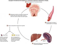
other Erythrocytes facts
|
Mature erythrocytes lack nuclei.
Transport oxygen and carbon dioxide to and from the tissues and the lungs. Lack of nuclei enables them to carry respiratory gases more efficiently. |
|
|
rouleaux
|
term that describes a pile of red blood cells
|
|
|
hemogloobin Transports oxygen and carbon dioxide, and is responsible for the characteristic bright red color of
|
arterial blood.
|
|
|
Every erythrocyte is filled with approximately 280 million molecules of a red-pigmented protein called
|
hemoglobin.
|
|
|
Hemoglobin that contains no oxygen
|
has a deep red color that is perceived as blue because the blood within these veins is observed through the layers of the skin and the subcutaneous tissue.
|
|
|
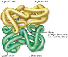
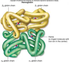
Each hemoglobin molecule consists of four protein building blocks called
|
globins - 2 alpha-chains and 2 beta-chains.
|
|
|
All globin chains contain a nonprotein) group that is in the shape of a ring, with an iron (Fe) ion in its center. this non proitein is called
|
heme
|
|
|
_____binds to these iron ions for transport in the blood.
|
Oxygen
|
|
|
Each hemoglobin molecule has four iron ions and is capable of binding
|
four molecules of oxygen
|
|
|
Oxygen binds to the hemoglobin when the erythrocytes pass through the .
|
blood vessels of the lungs
|
|
|
oxygen leaves the hemoglobin when the erythrocytes pass through the
|
blood vessels of body tissues.
|
|
identify this image
|
|
|
memorize
|
memorize
|
|
|
Each erethrocyte has how many hemoglobin molecules
|
280 million
|
|
|
Leukocytes
|
help initiate an immune response and defend the body against invading pathogens.
are true “cells” in that they contain a nucleus and cellular organelles |
|
|
Leukocytes also differ from erythrocytes
|
in that they are about 1.5 to 3 times larger, and they do not contain hemoglobin.
|
|
|
The five types of leukocytes are
|
Neutrophil
Eosinophils Basophils lymphocytes monocytes |
|
|
two distinguishable classes of leukocytes.
|
granulocytes and agranulocytes
|
|
|
agranulocytes
|
lymphocytes and monocytes.
no granules in the cytoplasm |
|
|
granulocytes
|
neutrophil
Eosinophils Basophils -has organelles called granules in the cytoplasm |
|
|
Neutrophil
|
60–70% of the total number of leukocytes
|
|
|
Eosinophils
|
have reddish, or pink-orange granules in their cytoplasm.
constitute about 2–4% of the total number of leukocytes nucleus usually has two lobes, which are connected by a thin strand |
|
|
Basophils
|
are
1.5 times larger than erythrocytes least numerous of the granulocytes constitute about 0.5–1% of the total number of leukocytes always exhibit a bilobed nucleus and abundant blue-violet granules in the cytoplasm |
|
|
T-lymphocytes (T-cells)
|
manage and direct an immune response
some directly attack foreign cells and virus-infected cells |
|
|
B-lymphocytes (B-cells)
|
stimulated to become plasma cells and produce antibodies
|
|
|
Natural killer cells (NK cells)
|
attack abnormal and infected tissue cells
|
|
|
Monocytes
|
Up to three times the diameter of an erythrocyte.
Constitute about 3–8% of all leukocytes. Nucleus is kidney-shaped or U-shaped. Macrophages phagocytize bacteria, cell fragments, dead cells, and debris. |
|
|
Macrophages
|
monocytes found outside the blood system
|
|
|
Platelets
|
Irregular, membrane-enclosed cellular fragments that are about 2 micrometers in diameter (less than one-fourth the size of an erythrocyte).
|
|
|
megakaryocytes
|
Platelets are Continually produced in the red bone marrow in these alls.
|
|
|
valves prevent
|
the unidirectional flow of blood in the heart
|
|
|
The right side of the heart send blood to the
|
lungs for oxygen
|
|
|
The left side of the heart sends blood to the
|
body
|
|
|
blood pressure is developed through
|
alternate cycles of the heart wall contraction and relaxation
|
|
|
minimum blood pressure is required to
|
push vessels to the body for nutrient and waste exchange.
|
|
|
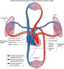
the pulmonary circuit consists of
|
the chambers on the right side of the heart (right atrium and ventricle) as well as the pulmonary arteries and veins.
|
|
|
The pulmonary circuit ...
|
conveys blood to the lungs via pulmonary arteries to reduce carbon dioxide and replenish oxygen levels in the blood before returning to the heart in pulmonary veins
|
|
|
the systemic circuit consists of
|
the chambers on the left side of the heart (left atrium and ventricle) along with all the other name blood vessels.
|
|
|
the systemic circuit...
|
carries blood to all the peripheral organs and tissues of the body.
|
|
|
The Aorta is
|
the largest systemic artery in the body.
|
|
|
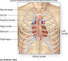
the heart is Rotated such that its right side or border (right atrium and ventricle) is located more _____, while its left side or border (left atrium and ventricle) is located more _______.
|
anteriorly, posteriorly
|
|
|
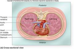
the heart is located
|
left of the body midline posterior to the sternum in the middle mediastinum.
|
|
|
the base of the heart is
|
The posterosuperior surface of the heart, formed primarily by the left atrium
|
|
|
The apex of the heart is
|
the inferior conical end
|
|
|
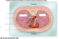
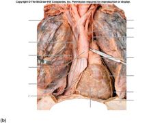
cross sectional view of the heart
|
|
|
|
|
|

|
know this
|
|
|
know this
|
|
|
|
|
|
know this
|
|
|
|
|

|
know this
|
|
|
|
|
|
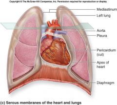
Pericardium
|
a fibrous, serous sac held in place within the mediastinum by connective tissue that supports the great vessels’ external walls superior to the heart and the diaphragm inferior to it
|
|
|
Restricts heart movements so that it doesn’t bounce and move about in the thoracic cavity, and prevents the heart from overfilling with blood
|
Pericardium
|
|
|
fibrous pericardium
|
Outer portion of the pericardium
a tough dense connective tissue layer |
|
|
serous pericardium
|
Inner portion of the pericardium
a thin, double-layered serous membrane parietal layer visceral layer |
|
|
Heart wall consists of three distinctive layers
|
external epicardium
middle myocardium, and internal endocardium |
|
|
outermost heart wall layer
|
Epicardium is the outermost heart layer and is also known as the visceral layer of serous pericardium
As we age, more fat is deposited in the epicardium, and so this layer becomes thicker and more fatty. |
|
|
the middle layer of the heart wall
|
Myocardium and is composed chiefly of cardiac muscle tissue
it lies deep to the epicardium and superficial to the endocardium |
|
|
the thickest of the three heart wall layers
|
Myocardium
|
|
|
endocardium
|
Internal surface of the heart and the external surfaces of the heart valves are covered by a thin endothelium
|
|
|
subendocardial layer
|
between the endocardium and myocardium, which is composed of areolar connective tissue
|
|
|
Fibers are relatively short, branched fibers that usually house one or two central nuclei and numerous mitochondria for ATP supply.
|
Cardiac Muscle Tissue
|
|
|
Fibers are arranged in spiral bundles and wrapped around and between the heart chambers.
|
Cardiac Muscle Tissue
|
|
|
Resembles skeletal muscle in that fibers in both muscles are striated, with extensive capillary networks that supply needed nutrients and oxygen.
|
Cardiac Muscle Tissue
|
|
|
Fibers contract as a single unit because muscle impulses are distributed immediately and simultaneously throughout all fibers either of the atria or of the ventricles.
|
Cardiac Muscle Tissue
|
|
|
Specialized cell–cell contacts called intercalated discs electrically and mechanically link the fibers together and permit the immediate passage of nerve impulses.
|
Cardiac Muscle Tissue
|
|
|
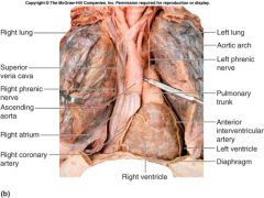
External Anatomy of the Heart
|
Composed of four hollow chambers: two smaller atria and two larger ventricles
|
|
|
Atria
|
thin-walled chambers that are located superiorly
|
|
|
auricle
|
anterior part of each atrium is a wrinkled, flaplike extension
|
|
|
Atria receive blood
|
returning to the heart through both circulatory circuits
|
|
|
right atrium receives blood from the
|
systemic circuit
|
|
|
left atrium receives blood from the
|
pulmonary circuit
|
|
|
Blood that enters an atrium is passed to the
|
ventricle on the same side of the heart.
|
|
|
Ventricles are the
|
inferior chambers
|
|
|
Two large arteries that exit the heart at the basal surface.
|
, the pulmonary trunk and the aorta
|
|
|
pulmonary trunk carries blood from the right ventricle
|
into the pulmonary circuit.
|
|
|
aorta conducts blood from the left ventricle
|
into the systemic circuit.
|
|
|
coronary sulcus (or atrioventricular sulcus)
|
Atria are separated from the ventricles externally by a relatively deep coronary sulcus (or atrioventricular sulcus) that extends around the circumference of the heart.
|
|
|
anterior interventricular sulcus and the posterior interventricular sulcus are located
|
between the left and right ventricles.
|
|
|
anterior interventricular sulcus and the posterior interventricular sulcus extend inferiorly from the coronary sulcus
|
toward the heart apex.
|
|
|
fibrous heart skeleton
|
is formed from dense irregular connective tissue.
|
|
|
fibrous heart skeleton is located
|
located between the atria and the ventricles
|
|
|
fibrous heart skeleton also
|
separates the atria and ventricles
anchors heart valves by forming supportive rings at their attachment points provides electrical insulation between atria and ventricles insulation ensures that muscle impulses are not spread randomly throughout the heart, and thus prevents all of the heart chambers from beating at the same time |
|
|
fibrous heart skeleton Provides
|
a rigid framework for the attachment of cardiac muscle tissue.
|
|
|
There are four heart chambers:
|
right atrium
right ventricle left atrium left ventricle |
|
|
Valves
|
permit the passage of blood in one direction and prevent its backflow
|
|
|
Three major vessels empty into the right atrium.
|
superior vena cava drains blood from the head, upper limbs, and superior regions of the trunk
inferior vena cava drains blood from the lower limbs and trunk coronary sinus drains blood from the heart wall |
|
|
interatrial septum
|
forms a wall between the right and left atria.
|
|
|
Right Atrioventricular (AV) Valve
|
Also called the tricuspid valve
Separates the right atrium from the right ventricle |
|
|
Receives deoxygenated venous blood from the right atrium.
|
Right Ventricle
|
|
|
interventricular septum
|
forms a wall between the right and left ventricles.
|
|
|
papillary muscles
|
internal wall surface of each ventricle has three cone-shaped, muscular projections
|
|
|
anchor chordae tendineae
|
attach to the cusp of the right AV valve and prevent everting and flipping into the atrium when contracting
|
|
|
pulmonary semilunar valve marks the
|
end of the right ventricle and the entrance into the pulmonary trunk.
|
|
|
Pulmonary trunk divides shortly into
|
right and left pulmonary arteries. carries deoxygenated blood to the lungs
|
|
|
Semilunar Valves
|
Located within the walls of both ventricles immediately before the connection of the ventricle to the pulmonary trunk and aorta.
|
|
|
Once gas exchange occurs in the lungs, the oxygenated blood travels through the pulmonary veins to the _____
|
left atrium
|