![]()
![]()
![]()
Use LEFT and RIGHT arrow keys to navigate between flashcards;
Use UP and DOWN arrow keys to flip the card;
H to show hint;
A reads text to speech;
116 Cards in this Set
- Front
- Back
|
What are the characteristics of anatomical position? |
Hands, toes, penis, and face facing forward. Pads of thumbs are perpendicular to pads of fingers, which are together and in a single plane. Mouth is closed, and eyes are open and focussed in the distance. Inferior margin of the orbit is level with the top of the external acoustic meatus. |
|
|
What is a transverse section? |
A section that crosses the body axis, or the long axis of a limb segment. May be transverse (horizontal) or oblique. |
|
|
What is protraction/retraction? |
Rotation of a limb segment in a parasagittal plane |
|
|
What is protrusion/retrusion? |
Anterior or posterior movement of a body part without rotation. Usually only used to refer to the position of the mandible. |
|
|
What is flexion/extension of the thumb? |
Movement of the thumb in the plane of the fingers. |
|
|
What is adduction/abduction of the thumb? |
Movement of the thumb perpendicular to the plane of the fingers. |
|
|
What is movement of the thumb in the plane of the fingers? |
Flexion/extension |
|
|
What is movement of the thumb perpendicular to the plane of the fingers? |
Adduction/abduction |
|
|
How does fertilisation occur? |
A single sperm dissolves penetrates the zona pellucida of the secondary oocyte, causing a chemical change that cross-links the proteins of the zona pellucida and keeps other sperm out. Meiosis II then completes, resulting in two polar bodies and one mature oocyte. Unfertilised follicles will collapse into the corpus luteum, which will produce progesterone to cause the endometrium to thicken and vascularise until progesterone production is taken over by the placenta. The sperm and the egg are now known as the male and female pronuclei, and as the male pronucleus enlarges, the two haploid pronuclei fuse to produce a zygote, which immediately divides. |
|
|
How does embryonic cleavage occur? |
The first cell division occurs shortly after fertilisation. The individual cells are now known as blastomeres. Before the wall between these cells is even complete, the second division occurs. At this stage, all cells are still totipotent, but at the 8-cell stage, compaction occurs, causing cells to communicate via their cytoplasm and become fated. After compaction, the embryo is referred to as a morula. As divisions continue, the morula draws in fluid, producing a blastocyst consisting of an inner embryoblast at the embryonic pole, and an outer trophoblast. Before implantation in the uterus, this blastocyst "hatches" as the zona pellucida degenerates. |
|
|
How does implantation occur? |
At day 6, the trophoblast attaches to the endometrial epithelium at the embryonic pole, and differentiates into the syncytiotrophoblast and the cytotrophoblast. The syncytiotrophoblast penetrates the endometrial epithelium, forming structures that begin to receive maternal nourishment by the end of the first week (until this point, the embryo has fed off of the yolk sac??). The hypoblast, a layer of cuboidal cells, appears on the surface of the embryoblast, on the inner surface of the blastocyst. |
|
|
Where does fertilisation usually occur? |
In the ampullae of the fallopian tubes |
|
|
At what stage does the embryo enter the uterus? |
At the blastula stage |
|
|
What is the best location for embryonic implantation in the uterus? |
On the posterosuperior uterine wall |
|
|
How is an embryo nourished in an ectopic pregnancy? |
Local blood supply can sustain an embryo almost to the third trimester |
|
|
How do spontaneous abortions occur? |
The corpus luteum (produced from a collapsed unfertilised follicle) produces progesterone, causing the endometrium to thicken and vascularise. If progesterone secretion is not taken over by the developing placenta, or if levels dip too low before the placenta takes over, the embryo may spontaneously abort after implantation. |
|
|
How does the bilaminar disk form? |
After implantation of the blastocyst, the syncytiotrophoblast releases hCG, signalling successful implantation, and serving to maintain the corpus luteum a little longer before the placenta takes over progesterone secretion. The hypoblast (primary endoderm) and epiblast (primary ectoderm) form from the cytotrophoblast. An amniotic cavity forms as fluid collects within the epiblast, and amnioblasts separate from the epiblast to line the amniotic cavity and produce amniotic fluid. The embryoblast becomes a flattened, circular disk, called "bilaminar" because it consists of the epiblast, associated with the amniotic cavity, and hypoblast, associated with the exocoelomic cavity. |
|
|
How does the primary yolk sac develop?? |
At day 9, once the embryo is completely embedded in the endometrium, the syncytiotrophoblast surrounds the embryo completely (though a small pore remains to communicate with the lumen of the uterus ??). The amniotic cavity expands, and cells migrate from the hypoblast to form the exocoelomic membrane, to match the amniotic membrane. The exocoelomic membrane becomes the primary yolk sac, which produces cells that form the extraembryonic mesoderm surrounding both the amnion and yolk sac. |
|
|
How do originate?? |
After the embryo is completely embedded, isolated cavities begin to form in the syncytiotrophoblast, that diffuse blood and cellular debris to the embryonic disk. At day 13, the lacunae fuse to form networks. Extraembryonic coelomic spaces also form in the extraembryonic mesoderm, which fuse to form the extraembryonic coelom, surrounding the amnion and yolk sac except for a connecting stalk at the embryonic pole. This splits the extraembryonic mesoderm into somatic (outer) and splanchnic (inner) mesoderm. The primary yolk sac pinches and splits into a primary and secondary yolk sac, and the primary shrinks and degenerates. |
|
|
What is the chorion? |
Part of an embryo that consists of the somatic extraembryonic mesoderm, cytotrophoblast, and syncytiotrophoblast, after the extraembryonic coelom forms. |
|
|
What is the prechordal plate? |
A thickened layer of endodermal cells at one end of the hypoblast, that forms at day 14 of embryonic development. This indicates the future site of the mouth. |
|
|
What is gastrulation? |
The process by which the bilaminar disk is converted to a trilaminar embryonic disk. First, the primitive streak forms from the epiblast as a rapid proliferation of cells that involute and build up against hypoblast cells. The first few cells push the hypoblast out of the way and become the endoderm, and the next few cells contact the endoderm and become the mesoderm. The primitive streak migrates up the midline of the bilaminar disk, forming a primitive groove, and a primitive node and pit that travels up the bilaminar disk. The primitive node extends into a canal that will develop within the notochord. About 2/3 of the way up, the primitive streak stops, and the notochordal process forms. As the notochord develops, the primitive streak regresses. In one crucial step, the notochord briefly contacts the yolk sac and closes again. |
|
|
What is neurulation? |
The process involved in the formation of the neural plate and neural tube, which is completed by the fourth week. After the notochord briefly opens up to the yolk sac and pinches off again, it starts to secrete chemical messengers into the surrounding tissues. The concentration of these chemicals causes the closest mesodermal tissue to condense into somites, while farther away mesoderm develops into the intermediate and lateral plate mesoderm. Ectoderm closest to the notochord becomes the neural groove, while further away ectoderm differentiates into the neural crest. Eventually, the edges of the neural crest meet in the sagittal plane and slough off, forming the neural tube. The intraembryonic coelom also forms in the lateral plate mesoderm, splitting it into somatic and splanchnic mesoderm. |
|
|
How many somites are there? |
44 pairs of somites form in total: 7 somitomeres, four occipital somites, 8 cervical somites, 12 thoracic somites, 5 lumbar somites, 5 sacral somites, and 3 coccygeal somites. |
|
|
How does embryonic folding in the median and horizontal planes occur? |
At the head end of the embryo, the oropharyngeal membrane is anterior to the neural fold, and the tissue that will become the heart is anterior to both, with the septum transversum at the anteriormost end. At the tail end, the cloacal membrane is posterior to the neural tube. As both the head and tail ends begin to curl, the oropharyngeal membrane (mouth) ends up below the neural tube (brain), and the heart ends up below the oropharyngeal membrane, on top of the septum transversum, which later becomes the diaphragm. Similarly, the cloacal membrane ends up superior to the end of the neural tube, leaving the neural tube to form a post-anal tail. |
|
|
What are the main layers of the body? |
Skin (epidermis and dermis), superficial fascia (subcutaneous), deep fascia (epimysium), muscles and bones. |
|

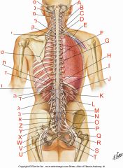
What is feature A? |
A retroperitoneal organ |
|

What is feature B? |
Bone |
|

What is feature C? |
Epidermis |
|

What is feature D? |
Dermis |
|

What is feature E? |
Skin |
|

What is feature F? |
Superficial fascia |
|

What is feature G? |
External layer of deep fascia |
|

What is feature H? |
Muscle |
|

What is feature I? |
Internal layer of deep fascia |
|

What is feature J? |
Deep fibrous lamina of the superficial fascia |
|

What is feature K? |
An internal organ |
|

What is feature L? |
Coelomic cavity |
|
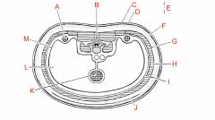
What is feature M? |
Coelomic sac |
|
|
What are the first three subdivisions of somites? |
Sclerotome (closest to neural tube), epaxial dermomyotome, hypaxial dermomyotome |
|
|
How does the epaxial dermomyotome develop? |
Migrates posteriorly to form the epaxial muscles and associated skin, becoming innervated by the dorsal rami of the spinal nerves |
|
|
How does the hypaxial dermomyotome develop? |
Migrates anteriorly to form all skeletal muscles except for some in the back, head, and neck. Becomes innervated by ventral rami of spinal nerves |
|
|
How many spinal nerves are there? |
31: 8 cervical, 12 thoracic, 5 lumbar, 5 sacral, 1 coccygeal |
|
|
What are somatic motor neurons? |
Efferent neurons that carry impulses to skeletal muscles, causing them to contract |
|
|
What are somatic sensory neurons? |
Afferent neurons that carry information about sensations and stimuli in the body wall |
|
|
What are visceral motor neurons? |
Efferent neurons that carry impulses to glandular tissues, causing them to secrete |
|
|
What are visceral sensory neurons? |
Afferent neurons that carry information about sensations and stimuli from visceral structures (in the body cavities AND body wall) |
|
|
What is the pathway of a somatic motor neuron? |
Cell body located in the ventral horn, axon leaves spinal cord through ventral root. Innervates epaxial muscles via dorsal rami, or hypaxial muscles via ventral rami |
|
|
What is the pathway of a somatic sensory neuron? |
Cell body is located in the dorsal root ganglion. Neuron receives an axon from the periphery, and synapses with the CNS in the dorsal horn. |
|
|
What structures are found in the ventral root of the spinal cord? |
Somatic motor axons |
|
|
What structures are found in the dorsal root of the spinal cord? |
Somatic sensory axons |
|
|
What structures are found in the ventral ramus of a spinal nerve? |
Somatic motor and somatic sensory axons |
|
|
What structures are found in the dorsal ramus of a spinal nerve? |
Somatic motor and sensory axons |
|
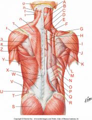
Where is the trapezius muscle? |
ה |
|

Where is the latissimus dorsi muscle? |
Y |
|

Where are the rhomboid muscles? |
I+F |
|

Where is the levator scapulae muscle? |
E |
|

Where is the iliocostalis muscle? |
Z (J, K, O) |
|
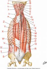
Where is the longissimus muscle? |
Y (E, I, N, ד) |
|

Where is the spinalis muscle? |
X (G, M) |
|

Where is the dura mater? |
G |
|

Where is the arachnoid mater? |
H |
|

Where is the pia mater? |
I |
|

Where is the dorsal root? |
B, O |
|

Where is the ventral root? |
A, T |
|

Where is the dorsal root ganglion? |
C, Q |
|
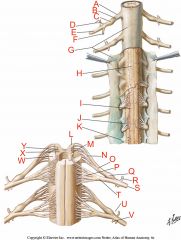
Where are the denticulate ligaments? |
K |
|
Where is the conus medullaris? |
ד |
|

Where is the cauda equina? |
ג |
|

Where is the filum terminale? |
Y, W |
|
|
What is the origin of the trapezius? |
Superior nuchal line (occipital), external occipital protuberance, ligamentum nuchae, spinous processes of CVII to TXII |
|
|
What is the insertion of the trapezius? |
Lateral third of the clavicle, acromion process, and scapular spine |
|
|
What is the innervation of the trapezius? |
Motor innervation from the spinal accessory nerve (CN XI), proprioception from C3 and C4 |
|
|
What is the function of the trapezius? |
Assists in rotating the scapula during abduction of the humerus, elevates scapula (upper fibers), adducts scapula (middle fibers), depresses scapula (lower fibres). |
|
|
What is the origin of the latissimus dorsi? |
Spinous processes of TVII to LV and sacrum, iliac crest, ribs X-XII. |
|
|
What is the insertion of the latissimus dorsi? |
The floor of the intertubercular sulcus. |
|
|
What is the innervation of the latissimus dorsi? |
The thoracodorsal nerve (C6-C8). |
|
|
What is the function of the latissimus dorsi? |
Extends, adducts, and medially rotates the humerus. |
|
|
What is the origin of the levator scapulae? |
Transverse processes of CI to CIV |
|
|
What is the insertion of the levator scapulae? |
The upper portion of the medial border of the scapula. |
|
|
What is the innervation of the levator scapulae? |
C3 to C4 and dorsal scapular nerve (C4, C5). |
|
|
What is the function of the levator scapulae? |
Elevates the scapula |
|
|
What is the origin of the rhomboid major? |
Spinous processes of TII to TV |
|
|
What is the insertion of the rhomboid major? |
Media border of the scapula, between spine and inferior angle. |
|
|
What is the innervation of the rhomboid major? |
The dorsal scapular nerve (C4, C5). |
|
|
What is the function of the rhomboid major? |
Retracts (adducts) and elevates scapula |
|
|
What is the origin of the rhomboid minor? |
The lower portion of the ligamentum nuchae, spinous processes of CVII and TI |
|
|
What is the insertion of the rhomboid minor? |
Medial border of scapula at scapular spine |
|
|
What is the innervation of the rhomboid minor? |
The dorsal scapular nerve (C4, C5). |
|
|
What is the function of the rhomboid minor? |
Retracts (adducts) and elevates scapula |
|
|
What is the origin of the serratus posterior superior? |
Lower portion of ligamentum nuchae, spinous processes of CVII to TIII, and supraspinous ligaments. |
|
|
What is the insertion of the serratus posterior superior? |
Upper border of ribs II to V just lateral to their angles |
|
|
What is the innervation of the serratus posterior superior? |
Anterior rami of upper thoracic nerves (T2 to T5). |
|
|
What is the function of the serratus posterior superior? |
Elevates ribs II to V |
|
|
What is the origin of the serratus posterior inferior? |
Spinous processes of TXI to LIII and supraspinous ligaments. |
|
|
What is the insertion of the serratus posterior inferior? |
Lower border of ribs IX to XII just lateral to their angles. |
|
|
What is the innervation of the serratus posterior inferior? |
Anterior rami of lower thoracic nerves (T9 to T12). |
|
|
What is the function of the serratus posterior inferior? |
Depresses ribs IX to XII, and may prevent lower ribs from being elevated when the diaphragm contracts. |
|
|
What is the origin of the iliocostalis lumborum? |
Sacrum, spinous processes of lumbar and lower two thoracic vertebrae and their supraspinous ligaments, and the iliac crest |
|
|
What is the insertion of the iliocostalis lumborum? |
Angles of the lower six or seven ribs |
|
|
What is the origin of the iliocostalis thoracis? |
Angles of the lower six ribs |
|
|
What is the insertion of the iliocostalis thoracis? |
Angles of the upper six ribs and the transverse process of CVII |
|
|
What is the origin of the iliocostalis cervicis? |
Angles of ribs III to VI |
|
|
What is the insertion of the iliocostalis cervicis? |
Transverse processes of CIV to CVI |
|
|
What is the origin of the longissimus thoracis? |
Blends with the iliocostalis in the lumbar region, and is attached to transverse processes of lumbar vertebrae. |
|
|
What is the insertion of the longissimus thoracis? |
Transverse processes of all thoracic vertebrae, and just lateral to the tubercles of the lower nine or ten ribs |
|
|
What is the origin of the longissimus cervicis? |
Transverse processes of upper four or five thoracic vertebrae. |
|
|
What is the insertion of the longissimus cervicis? |
Transverse processes of CII to CVI |
|
|
What is the origin of the longissimus capitis? |
Transverse processes of upper four or five thoracic vertebrae, and articular processes of lower three or four cervical vertebrae. |
|
|
What is the insertion of the longissimus capitis? |
Posterior margin of the mastoid process |
|
|
What is the origin of the spinalis thoracis? |
Spinous processes of TX or TXI to LII |
|
|
What is the insertion of the spinalis thoracis? |
Spinous processes of TI to TXIII |
|
|
What is the origin of the spinalis cervicis? |
Lower part of ligamentum nuchae and spinous process of CVIII (sometimes TI to TII) |
|
|
What is the insertion of the spinalis cervicis? |
Spinous process of CII (axis) |
|
|
What is the origin of the spinalis capitis? |
Usually blends with the semispinalis capitis |
|
|
What is the insertion of the spinalis capitis? |
With semispinalis capitis |