![]()
![]()
![]()
Use LEFT and RIGHT arrow keys to navigate between flashcards;
Use UP and DOWN arrow keys to flip the card;
H to show hint;
A reads text to speech;
24 Cards in this Set
- Front
- Back
Front (Term) |
Endolimax nana (Cystic Stage) |
|
|
Endolimax Nana (Cystic stage) Shape: |
Oval |
|
|
Entamoeba Histolytica (Trophozoite stage) Size: |
10-60 u |
|
|
Entamoeba Coli (Trophozoite stage) Shape: |
Condensed, Thickened, Rounded mass |
|
|
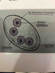
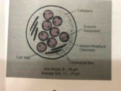
Entamoeba Coli (Cystic stage) Structures: |
1. Definite and Thick cystic wall 2. Nucleus:1-8 with eccentric karyosome 3. Chromatoidal Bars with splintered ends in younger cysts 4. Glycogen mass may be present in younger cyst |
|
|
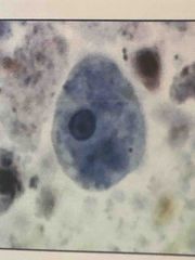
Entamoeba Histolytica (Trophozoite stage) Shape: |
Irregular |
|
Front (Term) |
Entamoeba coli (Cystic stage) |
|
|
Entamoeba histolytica (Trophozoite stage) Size: |
10-60u |
|
|
Endolimax Nana (Cystic stage) Structures: |
1. Smooth, definite cystic wall 2. Nuclei are darkly stained dots 3. Chromatoidal bodies seen as deeply stained, small, slightly curved rods |
|
|
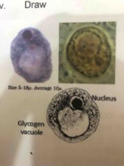
Iodamoeba buetschlii ( Cystic stage) |
|
|
Iodamoeba Buetschlii (Cystic stage) Shape: |
Irregular |
|
|
Entamoeba Histolytica (Cystic stage) Size: |
5-20u |
|
|
Entamoeba coli (Trophozoite stage) Size: |
15-20u |
|
|
Endolimax Nana (Cystic Stage) Size: |
6-10u |
|
|
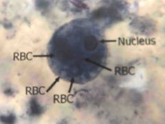
Entamoeba Histolytica (Trophozoite Stage) Structures: |
1. Clear demarcation between the ectoplasm and endoplasm 2. Endoplasm is briefly granular with ingested rbc 3. Single sphercial nucleus 4. Nuclear membrane lined with fine 5. Karyosome is relatively small |
|
|
Iodamoeba Buetschlii (Cystic Stage) Structures: |
1. Smooth, definite cystic wall 2. Single Nucleus, Rarely with Two Nuclei 3. Large glycogen 4. Usually absent chromatoidal bodies |
|
|
Entamoeba Histolytica (Cystic stage) Shape: |
Spherical |
|
|
Entamoeba Coli (Cystic stage) Shape: |
Spherical |
|
Front (Term) |
Entamoeba Coli (Trophozoite Stage) |
|
Front (Term) |
Entamoeba Histolytica (Trophozoite Stage) |
|
|
Iodamoeba Buetschlii (Cystic stage) Size: |
6-15u |
|
|
Entamoeba Coli (Trophozoite Stage) Structures: |
1. No clear demarcation between the ectoplasm and endoplasm 2. Endoplasm is highly vacuolated 3. Nucleus is spherical 4. Karyosome is moderately large |
|
|
Entamoeba Coli (Cystic stage) Size: |
10-33u |
|
|
Entamoeba Histolytica (Cystic stage) Structures |
1. Definite, smooth, relatively thin cystic wall 2. Nucleus 1-4 with centrally located karyosome 3. Chromatoidal bars with rounded ends in younger cysts 4. Glycogen mass may be present in younger cyst |