Reading...
![]()
Play button
![]()
Play button
![]()
Use LEFT and RIGHT arrow keys to navigate between flashcards;
Use UP and DOWN arrow keys to flip the card;
H to show hint;
A reads text to speech;
145 Cards in this Set
- Front
- Back
|
nervous system
|
* transmits information very rapidly from one area of the body to another by nerve impulses conducted from one body area to another.
divided into: 1. central nervous system (CNS) 2. peripheral nervous system (PNS) |
|
|
central nervous system (CNS)
|
* contains the brain and spinal cord
* occupies the center or midline of the body , located in the dorsal body cavity * Intemeurons are located only in the Central Nervous System * brain and spinal cord are protected in this cavity by bone, fluid, and protective membranes * receives impulses, interprets them, and sends them out to the appropriate organ systems of the body. |
|
|
peripheral nervous system (PNS)
|
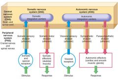
made up of the cranial nerves (12 pairs) and spinal nerves (31 pairs) that branch from the central nervous system (the brain and spinal cord
* contains all the long nerves that extend throughout the body. * These nerves are the collecting link between the body and the central nervous system (CNS). two subdivisions: 1. Somatic nervous system - voluntary 2. Autonomic nervous system - involuntary |
|
|
function of the peripheral nervous system
|
the (PNS) peripheral nervous system functions either voluntarily or involuntarily.
1. somatic nervous system - voluntary division 2. autonomic nervous system or ANS - involuntary division |
|
|
somatic nervous system
|
* (PNS) peripheral nervous system- course says its CNS)
* voluntary division of the central nervous system * composed of somatic or voluntary motor neurons * motor neurons that affect skeletal muscle and cause voluntary actions * contains both sensory and motor neurons * which conducts information to and from nerves that are involved in voluntary activities such as skeletal muscle action * uses only one neuron to conduct impulses all the way from the spinal cord or brainstem to effectors with no intervening synapse |
|
|
autonomic nervous system or ANS
|
* (PNS) peripheral nervous system
* involuntary division of the central nervous system * composed (made up) of the autonomic neurons located in the gray matter of the spinal cord or brainstem, * contains both sensory and motor neurons * division of the nervous system that regulates involuntary actions or automatic actions. * stimulates organs and glands and is responsible for involuntary activities * functions chiefly in the innervation of smooth muscle in the viscera * motor neurons that conduct impulses from the spinal cord or brainstem to th cardiac and smooth muscle tissue and glandular epithelial tissue ie. the heart is an ANS organ, stomach contractions, secretions from glands. two subdivisions: 1. Sympathetic 2. Parasympathetic also includes: norepinephrine and adrenergic effectors: blood vessels, sweat glands, the iris of the eye. |
|
|
cranial nerves
|
* PNS
* 12 pairs attached to the brain, the majority extending from the brainstem. * can send information directly to the brain without going through the spinal cord * transmit sensory information and voluntary and involuntary messages throughout the body * Each cranial nerve is assigned a Roman numeral that indicates the order in which the nerves exit the brain from front to back |
|
|
spinal nerves
|
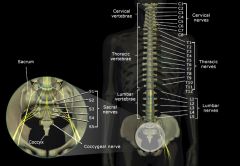
* 31 pairs in the PNS
* extend from the spinal cord to all the different parts of the body. * exit from the vertebral column through openings in the vertebrae called foramina * send impulses to the spinal cord and up to the brain and parts of the body not supplied by cranial nerves. * function to make sensations and movements possible. * contain both motor and sensory fibers * Each nerve is named for the level of the spinal cord from which it emerges: Cervical (C1 to C8): 8 pairs Thoracic (T1 to T12): 12 pairs Lumbar (L1 to L5): 5 pairs Sacral (S1 to S5): 5 pairs Coccygeal (CX): 1 pair |
|
|
autonomic nerves
|
* In the PNS
* carry information about involuntary activities such as blood pressure and digestion |
|
|
somatic motor nerves
|
* carry information about voluntary muscle movements to the brain
|
|
|
sensory nerves
|
* carry sensory information such as touch, pressure, and hot/cold temperatures to the brain
* can run directly to the brain or through the spinal cord first. * CNS and PNS work together to regulate normal body functions. ie. the eyes are a sense organ |
|
|
two cell types in the nervous system:
|
1. Neurons - nerve cells that conduct impulses
2. Glia - support cells, support neurons |
|
|
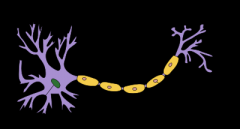
Neurons
|
* one of the two cell types found in the nervous system
* nerve cells that conduct impulses Composed of three main parts: 1. Cell Body -The cell body is the core of the neuron. 2. Dendrites - Extending out from the cell body are one or more projections called dendrites that transmit impulses to the cell body. 3. Axon - a single elongated projection that carries nerve impulses away from the body. Depending on its location, the structure of the axon varies: * Neuron pathway for impulse conduction - dendrite, cell body, then axon. |
|
|
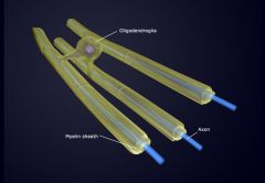
axon
|
* a single elongated projection that carries nerve impulses away from the body.
* axon transmits impulses away from the body of the neuron (or dendrite). Depending on its location, the structure of the axon varies: 1. Outside the CNS - On many neurons outside the CNS, the axon is surrounded by a white, fatty substance known as myelin that is formed by Schwann cells. Nodes of Ranvier are indentations between adjacent Schwann cells. The outer cell membrane of a Schwann cell (the nuclei and cytoplasm) is called the neurilemma and is necessary for the regeneration of nerve cells. 2. Within the CNS - Neurons within the CNS have axons that are missing the outer cell membrane of a Schwann cell—the neurilemma—which is necessary for the regeneration of nerve cells. This is a primary reason for the lack of regeneration of nerves when there is damage or trauma to the CNS. |
|
|
Neuron classification:
|
Neurons are classified according to the direction in which they transmit impulses:
1. Sensory neurons (afferent neurons) transmit impulses to the spinal cord and brain from all parts of the body and are also known as afferent neurons. 2. Motor neurons (efferent neurons) transmit impulses away from the brain and spinal cord to only two kinds of tissue- muscle and glandular epithelial tissue. 3. Interneurons (central neurons or connecting neurons) are connecting neurons and conduct impulses from sensory neurons to motor neurons. They are located only in the central nervous system. |
|
|
cell body
|
* main part of the neuron
* the nucleus of the neuron lies within the cell body |
|
|
Dendrites
|
* Extending out from the cell body are one or more projections called dendrites that transmit impulses TO OR TOWARD the neurons cell body or axons.
* can be one or many dendrites that project from the cell body of the neuron * highly branched part of the neuron that carries impulses toward the cell body. |
|
|
Nodes of Ranvier
|
* indentations between adjacent Schwann cells
* areas of the axon that are not covered by myelin |
|
|
Glia (neuroglia)
|
* support cells, support neurons
* regulate neuron function * cell type found in the nervous system * do not specialize in transmitting impulses but instead, serve as supporting cells to hold the functioning neurons of the CNS together (glue) and protects them. 3 types of Glia: 1. Astrocytes 2. Microglia 3. Oligodendrocytes |
|
|
Astrocytes
|
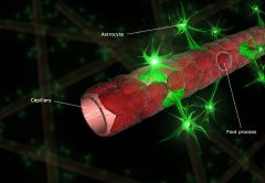
* hold neurons and small blood vessels close to each other
* star shaped, with many threadlike extensions or branches out of their surface, which accounts for their name astrocyte. * help build the blood-brain barrier |
|
|
Microglia
|
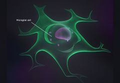
* provide phagocytosis for the system
* consume microbes by phagocytosis in inflammed or degenerating brain tissue * smaller that Astrocytes |
|
|
Oligodendrocytes
|
* produces the myelin sheath for nerve fibers in the brain and spinal cord, or and for axons inside the CNS central nervous system
* hold nerve fibers together |
|
|
Motor Neurons
|
* transmit impulses away from the brain and spinal cord.
|
|
|
myelin
|
* a fatty, white substance formed by Schwann cells
|
|
|
Nerves
|
a group of peripheral nerve fibers (axons) bundled together like the strands of a cable
* usually have a myelin sheath and look white because of this. |
|
|
CNS Nerve Fibers
|
The nerve fibers of the CNS are white and gray
1. The white matter appears white due to the fatty substance myelin, which forms the myelin sheath appear on the brain and cord 2. The gray matter is primarily cell bodies, interneurons, and unmyelinated axons or fibers and dendrites, which have a gray appearance |
|
|
PNS nerves
|
usually have myelin sheath and are therefore predominantly white matter.
|
|
|
Nerve impulse (action potential)
|
* are initiated by a stimulus
* can travel trillions of routes * an electrical signal that conveys information along a neuron's plasma membrane * the series of events that causes the electrical charge inside a nerve cell to convey information and go from a resting state to a depolarized state and back to a resting state. * Normally, a resting neuron has a slight positive charge on the outside and a negative charge on the inside * Sodium (an ion) is rushed into the neuron Once the stimulus has passed along the neuron, repolarization occurs. |
|
|
polarization phase (resting phase)
|
* Normally, a resting neuron has a slight positive charge on the outside and a negative charge on the inside
* chief intracellular ions include the positively charged potassium ions (K+) and several negatively charged ions or anions. * potassium ions tend to leak out of the cell * when they leak they take positive charges with them, resulting in a neg. environment within a cell. |
|
|
salatory conduction
|
* if the traveling impulse encounters a section of membrane covered with insulating myelin, it jumps around the myelin.
* this type of impulse travel is much faster than is possible in nonmyelinated sections. |
|
|
depolarization
|
* During a stimulus, this reverses temporarily; neuron has a slight negative charge on the outside and a positive charge on the inside
* When an impulse is initiated (neuron is stimulated), the cell membrane then becomes permeable to sodium ions (Na+), which rapidly enter the cell and depolarize the membrane, rendering it an optimum environment for a stimulus. |
|
|
repolarization
|
* after the impulse has passed through, the cell membrane again changes. It no longer allows the diffusion of sodium in to the cell
* sodium begins to be removed by pumps locate within the membrane of the neuron * allows potassium to once again diffuse out of the cell. this outward movement of the positively charged potassium ions removes the positive charge from the inside of the cell and leaves behind the neg. charged ions which causes repolarization |
|
|
Synapse
|
* Transmission of stimuli (information) from one neuron to another.
* requires that it pass over a space or junction where the two neurons come into close proximity with each other. Three synapse structures: 1. A synaptic knob 2. A synaptic cleft 3. The plasma membrane |
|
|
synaptic knob
|
* located on an axon
* contains many small vesicles that release chemical compounds known as neurotransmitters, which allow neurons to communicate. * tiny bulge at the end of the terminal branch of a presynaptic neuron's axon * contains many small sacs or vesicles which contain a very small quantity of a chemical compound called a neurotransmitter |
|
|
synaptic cleft
|
* the space between the two neurons
* narrow extracellular cleft between the presynaptic and postsynaptic membranes * when a nerve impulse arrives at the synaptic knob, neurotransmitter molecules are released from the vesicles into the synaptic cleft. |
|
|
Reflex
|
a rapid way of responding to a situation
|
|
|
postsynaptic membrane
|
the plasma membrane of the receiving dendrite.
|
|
|
Neurotransmitters function
|
by assisting, stimulating, or inhibiting impulses to cross over to postsynaptic neurons
|
|
|
reflex arc
|
* a type of specialized involuntary neuron conduction pathway for a reflex action
* simplest reflex arc consists of only two neurons (sensory and motor neuron) * allow impulse conduction in only one direction; like a one way street * many reflex arcs have at least three or more (sensory, interneurons, and motor neurons) * allow us to make swift adjustments to our environment or our body to maintain homeostasis ie. heart rate adjustments, blood pressure adjustments, coughing, reactions to painful stimuli such as withdrawing a hand from a hot stove. |
|
|
reflex arc of a patella
|
"knee-jerk," impulse
* simplest reflex arc (2 neuron) Involves the following components: 1. receptor (sensory muscle) it stimulates the receptors in the muscles of the thigh 2. afferent or sensory neuron receives the impulse. the nerve impulse is carried by the sensory neuron to the spinal cord. 3. integration center. consists of one or more synapses in the central nervous 4. An efferent or motor neuron receives the impulse. the nerve impulse is carried by the motor nerve to the muscles of the thigh 5. the effector organ receives the stimulus. The muscles of the thigh are the effector organs, and in response to the nerve impulse, the muscles contract and move the leg in an upward movement. |
|
|
neuron pathway
|
the route that nerve impulses travel
|
|
|
monosynaptic
|
when the reflex has two neurons involved
|
|
|
polysynaptic
|
when a reflex has more than 2 neurons involved
|
|
|
Reflex arc sequence
|
1. Stimulus
2. Receptors 3. Sensory neuron 4. Intemeuron 5. Motor neuron 6. Withdrawal reflex. |
|
|
Disorders of Nervous Tissue (myelin disorders)
|
Disorders that injure or destroy myelin, abnormal growths, and deficiencies in neurotransmitters are among the most common diseases affecting the nervous system.
examples of Nervous tissue disorders: 1. Multiple sclerosis 2. Tumors 3. Parkinson disease |
|
|
Parkinson Disease (PD)
|
* Parkinson disease is a chronic nervous disorder resulting from a deficiency of the neurotransmitter dopamine in certain parts of the brain.
* Dopamine belongs to a group of compounds called catecholamines that may play a role in sleep, motor function, mood, and pleasure recognition Signs: forward tilt of trunk, rigidity and trembling of head and extremities, reduced arm swing, and shuffling galt with short steps. * medications - levodopa or L-dopa (Sinemet) increases dopamine leves in afflicted patients. |
|
|
Multiple Sclerosis (MS)
|
* most common myelin disorder
* characterized by myelin loss and destruction accompanied by varying degrees of oligodendrocyte injury and death. * most common in women ages 20 - 40. * thought to be caused by autoimmunity and to viral infections * Multiple sclerosis (MS) is characterized by destruction of oligodendrocytes, resulting in demyelination throughout the white matter of the CNS. Hard, plaquelike lesions replace destroyed myelin, inflammation occurs, and nerve conduction is impaired as a result. |
|
|
Tumors
|
* Tumors generally develop from glia.
* They may be usually benign can be malignant. * Most malignant tumors are secondary and develop as a result of metastasis from other areas. Multiple neurofibromatosis is an example of noncancerous tumors. They appear as small nodules in the Schwann cells of cutaneous nerves, and while benign, the tumors can be large and disfiguring. A famous case of this disease was the "Elephant Man," whose crippling and tragic life was depicted in books and plays. |
|
|
endocrine system
|
transmits information from one area of the body to another more slowly than the nervous system by chemicals secreted by ductless glands into the bloodstream and then circulated from the glands to other parts of the body.
|
|
|
Schwann cells
|
* form the substance Myelin
* forms the myelin sheath surrounding the axon of myelinated nerve fibers only in the peripheral nervous system. (outside the CNS) two types: 1. Myelinating Schwann cells wrap around axons of motor and sensory neurons to form the myelin sheath 2. Nonmyelinating Schwann cells are involved in maintenance of axons and are crucial for neuronal survival. Some group around smaller axons and form Remak bundles |
|
|
neurilemma
|
* the outer cell membrane of a Schwann cell
* clinically significant because it plays an essential part in the regeneration of cut and injured axons. * Missing in axons of the brain and spinal cord. |
|
|
glioma
|
* most common type of brain tumor develops from glia
|
|
|
neuroma
|
tumors arising in nervous system structures
|
|
|
multiple neurofibromatosis
|
* an inherited disease characterized by numerous fibrous neuromas throughout the body
* benign tumors * appear first as small nodules in the Schwann cells of cutaneous nerves * most malignant tumors of glia and other nervous tissues do not originate there but instead are secondary tumors resulting from metastasis of cancer cells from the breast, lung, or other organs. |
|
|
tracts
|
bundles of axons in the CNS
|
|
|
endoneurium
|
a thin wrapping of fibrous connective tissue that each axon in a nerve is surrounded by
|
|
|
fascicles
|
groups of wrapped endoneurium
|
|
|
perineurium
|
* surrounds each fascicle
* a thin, fibrous perineurium |
|
|
epineurium
|
tough, fibrous sheath that covers the whole nerve
|
|
|
receptors
|
* the beginnings of dendrites of sensory neurons
* often located some distance from the spinal cord (in skin, tendons, and mucous membranes) |
|
|
ganglion
|
a group of nerve-cell bodies located in the PNS
* located near the spinal cord * each ganglion contains hundreds of sensory neuron cell bodies |
|
|
depression
|
* can be caused by a deficiency of serotonin
* drugs for severe depression by blocking the uptake of serotonin back into presynaptic neurons to increase the amount of serotonin in the synapse. |
|
|
presynaptic neuron
|
the neuron where impulses are transmitted from
|
|
|
postsynaptic neuron
|
the neuron where impulses are transmitted to.
|
|
|
neurotransmitters
|
* chemicals by which neurons communicate
1. noreplinephrine dopamine and serotonin - belong to the catecholamines group 2. morphine-like neurotransmitters - endorphins and enkephalins (inhibit conduction of pain impulses) 3. nitric oxide (NO) - very small molecules - diffuses directly across a plasma membrane rather than released from vesicles. (ie. Viagra) |
|
|
acetylcholine
|
* a neurotransmitter
* substance that is released at some of the synapses in the spinal cord and at neuromuscular junctions. |
|
|
catecholamines
|
compounds that play a role in sleep, motor function, mood, and pleasure recognition.
|
|
|
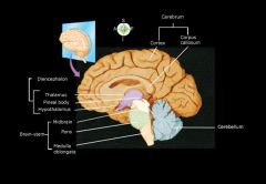
brain consists of several major divisions:
|
1. brainstem
2. cerebellum 3. diencephalon |
|
|
diencephalon
|
composed of the thalamus and hypothalamus and is located between the midbrain and the cerebrum.
|
|
|
brainstem
|
* The brainstem connects the spinal cord with higher brain structures
* lies just inside the cranial cavity above the large hole in the occipital bone called the foramen magnum. * serves as a conduction pathway; midportion of two-way conduction pathway * responsible for breathing, heart rate, vital centers, and vasomotor control. * relay for visual and auditory impulses consists of: (from lowest to highest location) 1. medulla oblongata 2. the pons 3. the midbrain |
|
|
Brain picture
|
.
|
|
|
medulla oblongata
|
* connects the spinal cord with the pons
* lowest part of the brain stem * consists of gray and white matter * acts as a two-way conduction pathway and relay for sensory and motor information * The cardiac, respiratory, and vasomotor centers (collectively called the vital centers) are also located in the medulla. |
|
|
pons
|
* extends from the midbrain to the medulla oblongata
* Pons means "bridge," * it functions as a bridge or two-way conduction pathway for information traveling to and from several brain structures * It also influences respiration rate and rhythm. |
|
|
midbrain
|
* extends from the lower diencephalon to the pons
* It is a two-way conduction pathway and relay for visual and auditory impulses. |
|
|
Diencephalon
|
* responsible for body temperature, appetitie, and sleep-cycle control.
* located between the midbrain below and the cerebrum above * alerting or arousal mechanisms * located between the midbrain and the cerebrum consists of: (from lowest to highest location) 1. thalamus - It associates sensations with emotions, and it plays a part in the arousal or alerting mechanism. 2. hypothalamus - regulate many processes such as body temperature, water balance, sleep-cycle control, appetite, and sexual arousal. |
|
|
thalamus
|
* located above the hypothalamus - dumbell-shaped section of gray matter
* serves as a relay station for sensory fibers traveling from the lower brain and spinal cord to the sensory areas of the cerebrum * It associates sensations with emotions, produces sensations, and it plays a part in the arousal or alerting mechanism. |
|
|
hypothalamus
|
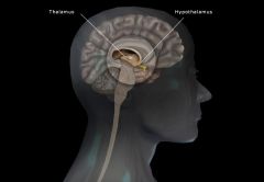
* located below the thalamus
* It helps to regulate many processes such as body temperature, water balance, sleep-cycle control, appetite, sexual arousal and emotions like pleasure, fear, anger and pain. * influence the release of hormones of the endocrine system * vital functions: controls heartbeat, constriction and dilation of blood vessels, and contractions of the stomach and intestines. |
|
|
Cerebellum
|
* the second largest part of the brain and is located under the occipital lobe of the cerebrum
* The primary function of the cerebellum is the coordination of voluntary muscle activity so that movements are smooth and coordinated * It also assists with the maintenance of equilibrium and posture. * gray matter composes the outer layer * white matter composes the bulk of the interior functions of the cerebllum: balance, coordination, and skilled motor activity. |
|
|
Cerebrum
|
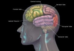
functions: Sensory perception, emotions, willed movements, consciousness, and memory
* the largest part of the brain, uppermost part of the brain * has superficial ridges called convolutions or gyri and grooves known as sulci * The deepest sulci are called fissures * the longitudinal fissure divides the cerebrum into right and left haves or hemispheres * The hemispheres are connected by a structure called the corpus callosum. Four lobes make up the structure of the cerebrum: 1. frontal lobe 2. parietal 3. occipital 4. temporal |
|
|
frontal lobe
|
* the motor area and affects the personality, conscious thought, emotions, problem solving, speaking, expressive language and memory as well as other functions.
|
|
|
parietal lobe
|
* the sensory area and affects body sense perception including taste, speech, reading, and other functions.
* interprets our sense of touch, identifies size, shapes and colors, and provides spatial perception |
|
|
occipital lobe
|
* affects vision and vision-related reflexes and functions.
* where vision is interpreted. |
|
|
temporal lobe
|
* affects hearing, smell, taste, and memory storage as well as other functions.
* assists us in sequencing, hearing, and memory |
|
|
Physiology of the Brain
|
* The brain serves as a conduction pathway and a regulator of body temperature, water balance, and sexual arousal.
* coordinates muscles, maintains equilibrium and posture, and directs sensory perception, emotions, consciousness, and memory. |
|
|
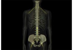
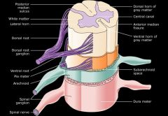
Spinal Cord
|
* a continuation of the brainstem
* fragile and vulnerable and must be protected to avoid injury that may be permanent or life-threatening. The spinal cord: * Is approximately 17 to 18 inches long. * Lies inside the spinal column in the spinal cavity. * Extends from the occipital bone to the bottom of the first lumbar vertebra. * Serves as a sensory and motor pathway and reflex center. * Is protected by the vertebral column, cerebrospinal fluid, and the meninges. |
|
|
Meninges
|
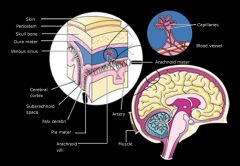
* fluid-containing membranes that surround the brain and spinal cord and offer protection to them.
Three types: 1. Dura mater 2. Arachnoid 3. Pia mater |
|
|
subarachnoid space
|
Cerebrospinal fluid fills the spaces between the pia mater and the arachnoid
|
|
|
Cerebrospinal fluid
|
is formed continuously from fluid filtering out of the blood in a network of brain capillaries known as the choroid plexus and into the ventricles and eventually entering the subarachnoid space.
|
|
|
choroid plexus
|
a network of brain capillaries
|
|
|
Dura mater
|
outermost layer of the meninges that lines the vertebral canal
|
|
|
Arachnoid
|
* middle meninges that covers the brain
* means "cobweb-like" |
|
|
Pia mater
|
innermost layer of the meninges that covers the brain and spinal cord
|
|
|
descending tracts
|
conduct impulses from the brain to the spinal cord
|
|
|
ascending tracts
|
conduct impulses from the spinal cord to the brain.
|
|
|
Brain and Spinal Cord Disorders
|
* can be caused by neuron injury or destruction
common brain and spinal cord disorders: * Destruction of brain tissue * Seizure disorders * Infection of the meninges * Accumulation of spinal fluid in the ventricles |
|
|
Destruction of brain tissue
|
* Injury or disease can destroy neurons. A common example is a cerebrovascular accident (CVA), or stroke. When the oxygen supply to portions of the brain is disrupted—such as in the case of a CVA—neurons cease to function or are destroyed.
* Motor control can be impacted and paralysis can result. Hemiplegia—paralysis of one whole side of the body—is a common result of a stroke. Other brain disorders that can destroy brain tissue are: * Alzheimer disease, with its accompanying dementia * Huntington disease, an inherited disease characterized by chorea (involuntary, jerky movements) that progresses to severe dementia and death by 55. symptoms appear ages 30 - 40 * Cerebral palsy, a common crippling disease that appears during childhood. CP is caused by events before, during, or after birth. |
|
|
Seizure disorders
|
* Seizure disorders cause sudden bursts of abnormal neuron activity that result in temporary changes in brain function. They can be mild or quite severe as in the case of convulsions or unconsciousness.
* Chronic seizure episodes are referred to as epilepsy. Tumors, trauma, or chemical imbalances can be the cause of epilepsy, but often the reason for the seizures is unknown. * Diagnosis and evaluation of seizure disorders are often assisted by an electroencephalogram (EEG). |
|
|
Infection of the meninges
|
Inflammation or infection of the meninges is meningitis. This disorder is caused by viruses as well as bacteria. Depending on the primary cause, meningitis may be mild or progress to a severe, even fatal, condition.
|
|
|
Accumulation of spinal fluid in the ventricles
|
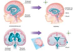
If the pathway for the cerebrospinal fluid is narrowed or blocked—by a tumor, trauma, or at birth—retention of CSF in the ventricles occurs. This condition is hydrocephalus. It is usually surgically treated by placing a shunt or tube to drain the excess fluid to another location in the body.
|
|
|
blood brain barrier (BBB)
|
*formed by an astrocyte wall along with the vessel wall
* allows water, oxygen, carbon dioxide, and a few other substances - such as alcohol to move between the blood and the tissue of the brain. |
|
|
cerebral cortex
|
* thin layer of gray matter made up of neuron dendrites and cell bodies
* makes up the surface of the cerebrum |
|
|
cerebral nuclei (basal ganglia)
|
* essential for producing automatic movements and postures
* islands of gray matter * Parkinson is a disease of the cerebral nuclei |
|
|
hemiplegia
|
paralysis of one whole side of the body
|
|
|
spastic paralysis
|
charcterized by involuntary contractions of affected muscles
may affect many areas: paraplegia - both legs triplegia - both legs and one arm quadriplegia - all four extremities |
|
|
demensia
|
adversely affects memory, attention span, intellectual capacity, personality and motor control
ie. Alzheimer disease - caused by lesions developing in the cortex during middle to late adult years. |
|
|
seizure
|
* sudden bursts of abnormal neuron activity that result in temporary changes in brain function
* EEG or electroencephalogram is used to diagnose and evaluate seizure disorders |
|
|
epilepsy
|
recurring or chronic seizure episodes
|
|
|
convulsions
|
severe seizures, resulting in jerky, involuntary muscle contractions
|
|
|
spiral tracts
|
bundles of myelinated nerve fibers
|
|
|
spinal cord reflexes
|
reflexes that result from conduction over arcs whose centers lie in the spinal cord
two common kinds: 1. withdrawal - pulling hand away from hot stove 2. jerk reflex -knee jerk |
|
|
anesthesia
|
loss of sensation
|
|
|
paralysis
|
the loss of ability to make voluntary movements
|
|
|
meningitis
|
* infection or inflammation of the meninges
* often caused by the bacteria Neisseria meningitidis (meningococcus), streptococcus pneumoniae, or Haemophilus influenzae * can also be caused by viral infections |
|
|
(cerebral) ventricles
|
* spaces in the brain that are located between portions of the brain
* filled with cerebrospinal fluid (CSF) |
|
|
hydrocephalus
|
"water on the brain"
* a build up of fluid in the ventricles * headaches are common because of pressure and swelling on the brain caused by a narrowing or blockage of the pathways for cerebrospinal fluid, causing retentionof the CSF in the ventricle. * treatment involves surgical placement of a shunt in the blocked channel so that CSF can drain into another location in the body. must stay in place for life since CSF is produced throughout life. |
|
|
Cranial nerve list
|
mnemonic- on old olympus' tiny tops, a friendly viking grew vines and hops.
I. olfactory nerve - carries information form the nose to the brain and is concerned with the sense of smell II. optic nerve - carries visual information from the eye to the brain for the sense of sight III. oculomotor nerve - causes movement of the eyeball and contraction of the extrinsic eye muscles to raise the eyelid. it also constricts the pupil of the eye IV. trochlear nerve - also causes contraction of the extrinsic eye muscles to move the eyeball V. trigeminal nerve - carries information regarding touch, pressure and pain form the face, scalp, and teeth to the brain. It also innervates the muscles of mastication VI, abducens nerve - controls eye movements by innervating an extrinsic eye muscle that causes eye movement and by carrying information regarding the sensation of eye movement to the brain. VII. facial nerve - carries sensory informatio regarding taste and performs motor function by contracting the muscles of facial expression, secretion of saliva, and tears. VIII. vestibulocochlear nerve - carries information for hearing and sense of balance from the ear to the brain IX. glossopharyngeal nerve - carries sensory information regarding taste from the posterior tongue and aids in reflex control of blood pressure and respiration. Motor functions of the glossopharyngeal include stimulating the secretion of salivary glands and also stimulating sensations of the throat and the swallowing movements. X. vagus nerve - carries sensations to and produces movements of the organs supplied by it. examples include slowing the heart, increasing peristalsis, contracting muscles for voice production, and aiding in the swallowing process. XI. accessory nerve - stimulates shoulder movements, turning movements of the head, movements of viscera, and voice production. XII. hypoglossal nerve - stimulates tongue movements |
|
|
plexus
|
As spinal nerves exit from the vertebral column, sometimes nerve fibers from several spinal nerves are reorganized to form a single peripheral nerve. This reorganization can be seen as a network of intersecting branches called a plexus
The three major nerve plexuses are: 1. Cervical 2. Brachial 3. Lumbosacral |
|
|
Cervical nerve plexus
|
Innervates skin, muscles of the neck and shoulder, and the diaphragm
|
|
|
Brachial nerve plexus
|
Innervates skin and muscles of the upper extremities
|
|
|
lumbosacral nerve plexus
|
Innervates skin and muscles of the lower torso and lower extremities
|
|
|
dermatome
|
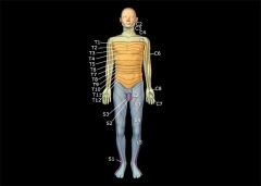
* the area of skin surface supplied with afferent nerve fibers (sensory fibers) by a single posterior spinal root nerve.
* Each spinal nerve innervates a particular area on the surface of the body * each dermatome is named for the nerve that serves it. |
|
|
Peripheral Nerve Disorders
|
* diseases and disorders to the peripheral nervous system are often far more treatable than those dealing with the CNS
* medication and/or surgical procedures offer successful recovery in many disorders of the PNS. common peripheral nerve disorders include: 1. Neuritis 2. shingles 3. Bell's palsy 4. Tic douloureux |
|
|
Neuritis
|
Simply stated, neuritis is inflammation of a nerve. Symptoms may include neuralgia, paralysis, muscular atrophy, and hyperesthesia. An example is sciatica, or inflammation of the sciatic nerve.
|
|
|
Shingles
|
Also known as Herpes zoster, shingles is caused by a Varicella zoster virus of chickenpox and is usually very painful. It affects the skin of a single dermatome and lies dormant after an episode of chickenpox. It reactivates under stress or when the immune system is compromised.
|
|
|
Bell's palsy
|
* Bell's palsy presents as unilateral paralysis of the facial nerve that may be due to trauma, compression, or infection of the nerve.
* Patients are often unable to close the eyelid or make facial movements (mouth) on the affected side. * most often temporary but, in some cases, may be irreversible. * compression, degeneration, or infection of the seventh cranial nerve, the facial nerve * plastic surgery is sometimes used to correct a permanent disfigurement. |
|
|
Tic douloureux
|
Also known as trigeminal neuralgia, this disorder causes pain that is due to compression or degeneration of the fifth cranial nerve, the trigeminal. It is characterized by recurring episodes of stabbing pain along the angle of the jaw, branching sometimes over the forehead, around the eyes, in the tongue, and lower lip. The pain can be so severe that it has sometimes been referred to as the "suicide disease."
|
|
|
sciatica
|
painful inflammation of the spinal nerve branch in the thigh called the sciatic nerve - the largest nerve in the body.
|
|
|
neuralgia
|
nerve pain, may lead to atrophy of the leg muscles.
|
|
|
somatic response
|
contractions of skeletal muscles.
|
|
|
Sympathetic
|
* autonomic nervous system - PNS
* adapts the body to periods of stress or threats and is often referred to as the "fight or flight system." * Sympathetic responses generally have widespread effects on the body because preganglionic fibers synapse with several postganglionic fibers |
|
|
Parasympathetic
|
* autonomic nervous system - PNS
* more regulatory * It is most active during ordinary, relaxed conditions and is sometimes called the "rest and repose system." * It conserves energy and assists in returning functions to a normal state after being stimulated by the sympathetic nervous system. |
|
|
ganglia
|
peripheral "junction boxes" -where axons extend from the gray matter and terminate in
|
|
|
preganglionic neuron
|
a neuron whose axon terminates in contact with another nerve cell located in a peripheral ganglion
|
|
|
postganglionic neuron
|
a neuron that is distal to or beyond a ganglion
|
|
|
autonomic or visceral effectors
|
the tissues to which autonomic neurons conduct impulses.
ie. visceral effectors are cardiac muscle that makes up the wall of the heart, |
|
|
sympathetic preganglionic neuron
|
* have dendrites and cell bodies in the gray matter of the thoracic and upper lumbar segments of the spinal cord.
* the final site for integration of information that arises from central sympathetic premotor neurons * aka. thoracolumbar system |
|
|
sympathetic postganglionic neuron
|
*have dendrites and cell bodies in sympathetic ganglia that is located in front of and at each side of the spinal column
* look like two chains of beads often referred to as sympathetic chain ganglia. |
|
|
sympathetic nervous system
|
* functions: as an emergency system
* impulses over sympathetic fibers take control of many internal organs when we exercise strenuously and when strong emotions like anger and hate are elicited. * ie. heart beats faster, blood vessels constrict increasing blood pressure, sweat and adrenal glands secrete * prepare for strenuous muscular work "fight or flight" * autonomic, adrenergic, norepinephrine * sympathetic preganglionic axons release acetylcholine * sympathetic postganglionic axons release acetylcholine * sympathetic preganglionic neuron * sympathetic postganglionic neuron |
|
|
fight-or-flight response
|
group of changes induced by sympathetic control.
* ie. heart beats faster, blood vessels constrict increasing blood pressure, sweat and adrenal glands secrete |
|
|
Parasympathetic Nervous System (craniosacral system)
|
* dendrites and cell bodies of Parasympathetic preganglionic neurons are located in the gray matter of the brainstem and the sacral segments of the spinal cord.
* functions- dominates control of many visceral effectors under normal, everyday conditions. * preganglionic axons release the neurotransmitter acetylcholine and postanglionic axons release norepinephrine (noradrenaline) neurotransmitters ie. impulses over parasympathetic fibers, tend to slow heartbeat, increase peristalsis, and increase secretion of digestive juices and insulin, cause contractions of the urinary bladder, relaxation of the sphincters of the digestive tract, and increase salivation. |
|
|
cholinergic fibers
|
* an autonomic neurotransmitter (axon)
* fibers that employ (release) acetylcholine as their neurotransmitter three types: 1. parasympathetic preganglionic axon 2. parasympathetic postganglionic axon 3. sympathetic preganglionic axon |
|
|
adrenergic fibers
|
* an autonomic neurotransmitter (axon)
* fibers that employ (release) the neurotransmitter norepinephrine (noradrenaline) * norepinephrine is liberated at most sympathetic postganglionic nerve endings |
|
|
autonomic Nervous System (ANS) disorders
|
* stress has been cited as an indirect cause or an important risk factor in conditions
Conditions: 1. Heart disease - high blood pressure or hypertension, that can weaken the heart and blood vessels 2. Digestive problems - Colitis (colon inflammation) and gastric ulcers from changes in digestive secretion and movement along with increased susceptibility to infection 3. Reduced resistance to disease - hormones call glucocorticoids that are released by adrenal glands during prolonged or repeated stress episode depress the activity of the immune system. leads to increased risk of infection and cancer. |
|
|
neuroblastoma
|
* malignant tumor of the sympathetic nervous system.
* occurs most often in you children * metastasizes rapidly to other parts of the body * symptoms include: exaggerated or inappropriate * sympathetic effects. ie. increased heart rate, sweating, and high blood pressure |
|
|
limbic system (emotional brain)
|
* influenced directly or indirectly by impulses from neurons located above them, notably by some in the hypothalamus and in the parts of the cerebral cortex
* It appears to be primarily responsible for our emotional life, and has a great deal to do with the formation of memories. * sent through conduction paths |