![]()
![]()
![]()
Use LEFT and RIGHT arrow keys to navigate between flashcards;
Use UP and DOWN arrow keys to flip the card;
H to show hint;
A reads text to speech;
130 Cards in this Set
- Front
- Back
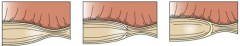
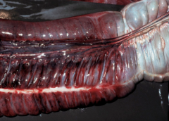
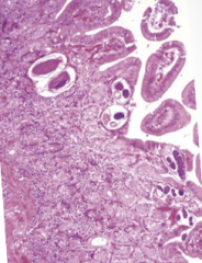
|
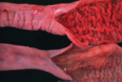
duodenum |
|
|
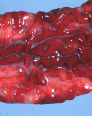
jejunum |
|
|
ileum |
|
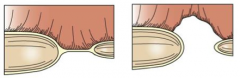
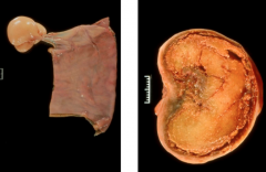
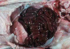
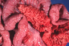
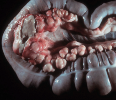
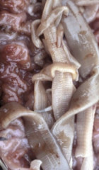
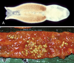
Identify each of the problems |
L to R: stenosis, stenosis with partial membrane, atresia with membrane |
|
|
L to R: atresia with cord, blind ending atresia |
|
|
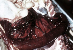
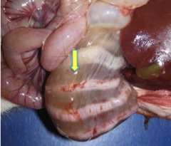
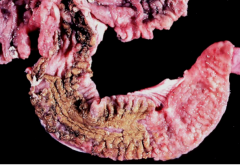
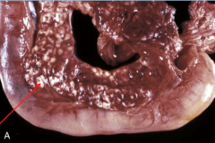
atresia coli with recto-vaginal fistula. Prominent megacolon |
|
What is this? What are the two broad causes? |
Megacolon - can be congenital or acquired |
|
|
With congenital megacolon, what does the term aganglionosis mean? |
Myenteric or submucosal plexus fails to develop in the colon and rectum. The colon is non-peristaltic. |
|
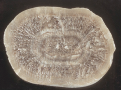
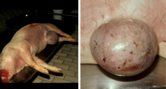
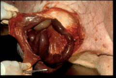
What is this? What causes it? What is it made of? |
Equine enterolith. Caused by diets made up of bran or alfalfa Made of ammonium magnesium phosphate (struvite) deposited in concentric lamellae. |
|
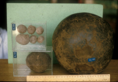
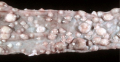
What are these? Who gets them? Where are they found? Why are they bad? |
- Trichobezoars (hairballs) - Found in cattle - In the abomasum - Can result in pyloric obstruction |
|
|
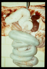
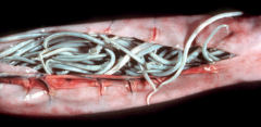
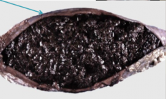
Ascarids impaction in a foal (Parascaris equorum) |
|
|
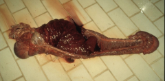
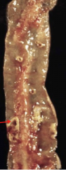
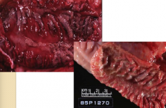
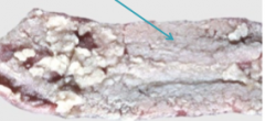
intestinal stricture in a horse following perforation. |
|
|
Strictures occur due to... |
mucosal injury - trauma, ischemia after infarction, or healing by fibrosis can all create mucosal injury |
|
|
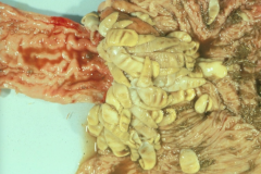
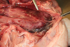
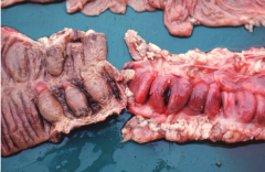
Colonic stricture due to salmonellosis in pigs |
|
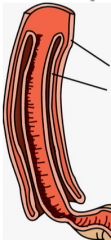
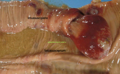
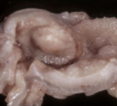
What is this process? What's the outside called? What's the inside called? |
Itussusception Outside = Intussuscipiens Inside = Intussusceptum |
|
|
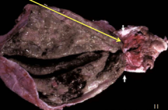
Explain the problem with intussusceptions |
Pressure pinches off venous return. High pressure of the arteries can still provide blood to the intussusceptum, but the intussuscipiens doesn't get blood and undergoes infarction, then necrosis. |
|
|
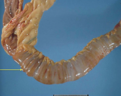
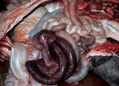
intestinal intussusception, foal |
|
|
intestinal intussusception, foal |
|
|
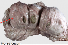
Cecocolic intussusception (cecum is inside the right ventral colon) |
|
|
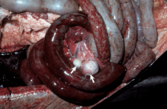
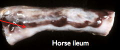
Anoplocephala perfoliata. Can be associated with intussusception |
|
|
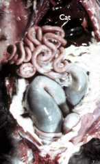
scrotal (inguinal) hernia in a pig |
|
|
What are the 5 broad stages of intestinal displacements |
Displacement --> Incarceration --> Strangulation --> Ischemia --> Necrosis |
|
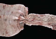
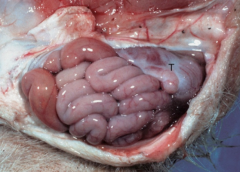
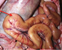
What anatomic feature is the star above? What is this area susceptible to? |
Epiploic foramen (horse) - susceptible to herniation (internal) of small intestines |
|
|
Epiploic herniation of small intestine (horse) |
|
|
Name 2 of the 4 possible locations where external hernias can occur |
- umbilical - inguinal - scrotal - hiatal |
|
|
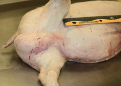
Scrotal hernia, pig |
|
Describe what happened here |
Incarceration of a piece of small intestine --> venous infarction --> necrosis --> sepsis --> death (Scrotal hernia in pig) |
|
|
umbilical hernia, pig |
|
|
Strangulated loop of small intestine - umbilical hernia, pig |
|
|
What does eventration mean? |
Displacement of viscera without an outpouching of peritoneum (type of hernia) |
|
|
What 3 places can eventrations occur? |
Diaphragmatic Perineal Post-op sites |
|
|
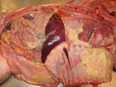
Diaphragmatic hernia due to trauma. Cat. - liver and intestines are now in the thoracic cavity. Lungs have been compressed |
|
|
What is volvulus? |
Rotation of guts around the mesenteric attachment. The small intestine is most vulnerable. |
|
|
What is torsion? |
Rotation along the axis of the gut. Cecum, colon, and abomasum most vulnerable. |
|
|
Is the horse's large intestine, which side is most vulnerable to torsion? Why? |
The left side. There is basically no attachments/anchor points on this side so it can easily displace |
|
|
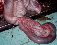
Torsion of the left colon |
|
|
colonic torsion |
|
|
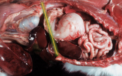
Intestinal volvulus in a foal - small intestine is twisted around the root of the mesentery |
|
|
Volvulus in a calf |
|
|
Intestinal strangulation by pedunculated lipomas |
|
|
Pedunculated lipomas |
|
|
Diverticula. Weak spots in the muculara allow for outpouching of mucosa, which can then rupture |
|
|
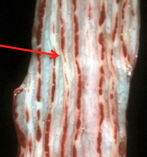
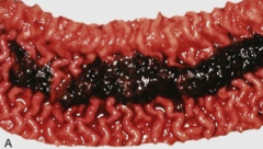
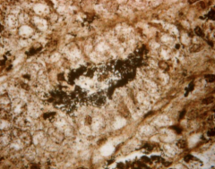
Hemomelasma ilei. Insignificant finding. Depigmentation causes discoloration. We don't know for sure why they happen, but it's theorized that it's from Stongyle larval migration |
|
|
Intestinal ceroidosis - Brown dog gut due to high PUFA and low vit E. - looks like jaundice but the rest of the animal will have no discoloration |
|
|
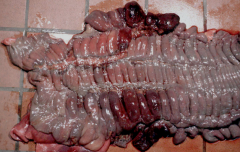
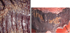
Tiger striping (congestion) - from tenesmus |
|
What is this? What does it cause? 2 types? |
Lymphangiectasia - cause of protein-losing enteropathy in dogs - congenital (breed disposed) or acquired (blockage of lacteals due to granulomas, neoplasia, or idiopathic) |
|
|
Pathogenesis of non-inflammatory diarrhea (ETEC) |
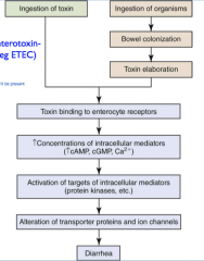
Pathogens disrupt the normal absorptive and secretory properties of the enterocytes, but don't kill them. Often occurs in the proximal small intestine. Creates enterotoxin-induced diarrhea (ETEC) |
|
|
Pathogenesis of inflammatory diarrhea (Invasive or cytotoxin-induced diarrhea)
|
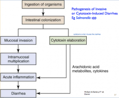
due to Salmonella, Lawsonia, and/or Brachyspira |
|
|
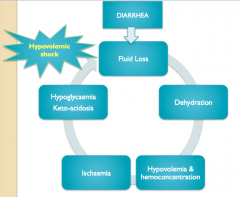
How can diarrhea lead to hypovolemic shock? |
|
|
|
Name 3 pathogens that target the villi tip (6 possibilities) |
Rotavirus Coronavirus Adenovirus E. coli Intracellular bacteria Intracellular protozoa |
|
|
Name 3 bacterial toxins that affect junctional complexes of villous and crypt enterocytes |
Salmonella Clostridia Anthrax EHEC Osteragia (not bacterial though) |
|
|
Name 3 pathogens that affect the enterocytes of the intestinal crypts |
Parvovirus FeLV BVD Rinderpest Mycotoxins ETEC |
|
|
Name 3 pathogens that target Peyer's patches |
BVD Salmonella Yersinia Rhodococcus |
|
|
Name 2 pathogens that target the tissue around the Peyer's patches |
MAP (Johne's) BVD Rhodococcus equi |
|
|
Which cytotoxin has non specific target cells or tissues? |
Clostridium cytotoxin |
|
|
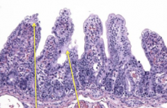
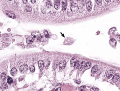
Describe enteric rotaviral infections |
Subclinical infections are common in piglets (first 7 weeks) and calves (up to one week). Can cause diarrhea in young animals of any species. Targets enterocytes at villi tips in upper SI. Causes villous blunting and fusion. Loss of fluid and Cl occurs. Increased peristalsis. Co-infections are common (E. coli, coccidia, crypto, etc). |
|
|
Blunted and fused villi due to rotaviral enteritis |
|
|
Coronaviral infections in calves |
Similar epidemiology and pathology to rotavirus, but more pathogenic. Also affects colon. |
|
|
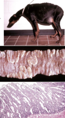
What does coronavirus cause in piglets? Why is this bad? |
Causes Transmissible Gastroenteritis (TGE). This is highly pathogenic (up to 100% mortality). |
|
|
TGE enteritis in piglet |
|
What causes these lesions? What are the cellular targets? |
Parvovirus enteritis. Target is rapidly dividing cells in the crypt enterocytes and bone marrow. |
|
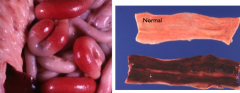
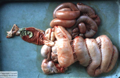
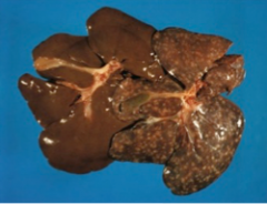
bovine colon |
BVD causing ulcerative colitis |
|
|
Peyer's patch necrosis due to BVD |
|
|
What is the primary cellular targets of BVD? What does the cytolytic virus target? |
BVD targets peyer's patches and the tissue around them. Cytolytic virus targets epithelial cells and lymphocytes |
|
What is this important cause of enteritis in neonatal animals? Why are there different clinical syndromes? |
Escherichia coli Bacterial strains have different virulence factors, which results in different clinical syndromes. These virulence factors promote colonization or adhesion, metabolic dysfucntion or death of enterocytes, and affect vasculature which can promote septicemia. |
|
|
Edema disease (enterotoxemic colibacillosis) |
|
|
Edema disease (enterotoxemic colibacillosis) |
|
|
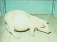
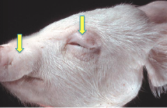
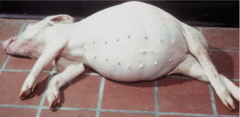
Why do these pigs get edema? What is the disease associated with? |
The bacterial enterotoxin (verotoxin) is angiotoxic which causes endothelial cell injury in arterioles and results in fluid loss/edema. Associated with change in diet at weaning (6-14 weeks old). Morbidity - 35%. Mortality - 100% |
|
|
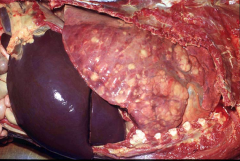
What is the most common Salmonella species that affect cattle, pigs, and horses? |
S. Typhimuirum |
|
|
What does S. Typhimuirum do in the body? |
Causes secretory diarrhea with enterocyte necrosis and cytokin mediated inflammation. Endotoxin-induced thrombosis occurs. Paratyphoid nodules (granulomas) in the liver arise + mesenteric lymphadenopathy. |
|
|
What is a classic lesion of S. Typhimuirum in calves? |
Fibrinous cholecystitis |
|
|
Salmonellosis in horse |
|
|
Salmonella causing fibrino-necrotizing entero-colitis |
|
|
Embolic mycotic pneumonia (sequel of Salmonellosis) in horse |
|
|
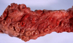
"button ulcers" in a pig due to chronic salmonellosis |
|
|
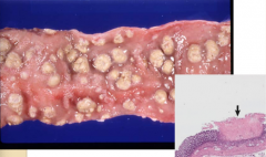
Rectal stricture after salmonellosis |
|
|
Megacolon and rectal stricture. Sequel of chronic Salmonella Typhimurium infection |
|
|
Rectal stricture + megacolon after Salmonellosis |
|
|
What pathogen causes clostridial enterotoxemia? |
Clostridium perfringens |
|
|
What type of Clostridium perfringens is the most common in mammals and birds? |
Type A (grouped A to E based on exotoxin produced) |
|
|
What is odd about which animals are affected by clostridial enterotoxemia? |
It affects the best nourished animals in the group (probably due to over eating, high carbs or protein) |
|
|
Which does C. perfringens type D produce? What kind of lesions does this make? What disease does is cause in sheep? |
type D produces an angiotoxin (epsilon toxin) - Causes intestinal lesions (fibrinonecrotic enterocolitis) and focal symmetrical encephalomalacia (FSE) in sheep. Called "blind staggers". Also causes pulpy kidney disease in sheep |
|
|
Enterotoxemia due to Clostridium perfringens type C |
|
|
Quail disease (Clostridium colinum) - affects farmed game birds |
|
|
What two Clostridiums cause Colitis X. Although it causes hemorrhagic necrotizing typhlocolitis in horses, which species do the two Clostridiums come from? |
C. difficile (humans, primates, cats, dogs) C. spiriforme (lagomorphs and rodents) |
|
|
Proliferative ileitis due to Lawsonia intracellularis infection in a pig |
|
|
Lawsoniasis causes which disease in pigs? |
Porcine Proliferative Enteropathy (PPE) |
|
|
Necrotic enteritis due to Lawsonia |
|
|
Proliferative hemorrhagic enteropathy from Lawsonia |
|
|
Proliferative ileitis from Lawsonia |
|
|
Glasser's disease (Haemophilus parasuis) |
|
|
Swine dysentery (spirochetal colitis) Due to Brachyspira hyodysenteriae. Characterized by large bowel diarrhea with mucos and blood in the feces. |
|
|
Swine dysentery (Brachyspira hyodisenteriae) |
|
|
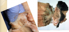
Pyogranulomatous colitis from Rhodococcus equi in a foal |
|
|
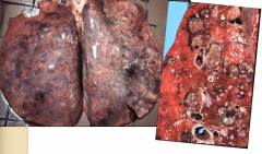
What type of pneumonia does R. equi cause in foals? What are the enteric lesions? |
Causes suppurative pyogranulomatous pneumonia. Enteric lesions are ulcerative and pyogranulomatous lesions centred on Peyer's patches and are associated with prominent mesenteric lymphadenitis |
|
|
Pyogranulomatous mesenteric lymphadenitis (R. equi) |
|
|
R. equi pneumonia |
|
|
What pathogen causes Johne's? |
Mycobacterium avium Paratuberculosis (MAP) |
|
|
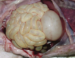
top jejunum is one with Johne's. Diffuse granulomatous enteritis bottom one is normal |
|
|
Johne's jejunum |
|
|
Where else can Johne's lesions be found besides the jejunum? |
ileum, cecum, proximal colon, and ileocecal valve |
|
|
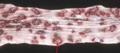
3 presentations of coccidiosis |
Proliferative enteritis - small ruminants Hemorrhagic - calves, dogs, cats Fibronecrotic enteritis - pigs and poultry |
|
|
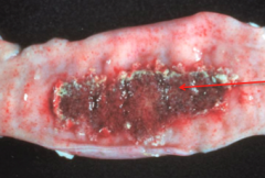
Hemorrhagic coccidiosis |
|
|
Fibronecrotic coccidiosis |
|
|
Proliferative coccidiosis |
|
|
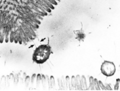
What is the hallmark lesion of cryptosporidium on histopath? |
Villous atrophy and fusion |
|
|
Which species of crypto is the cause of cryptosporidium? |
C. parvum |
|
|
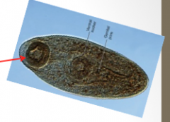
Giardia |
|
|
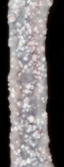
Milk spots due to Ascaris suis. Fibrotic tracks left by migrating larvae. |
|
|
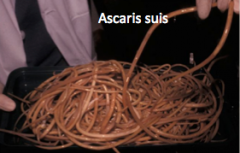
Name 2 of the 4 parasites that can cause Ascariasis |
- Ascaris suis - Parascaris equorum - Toxocara - Toxocaris leonina |
|
I am a hookworm! What kind of pathology do I cause? |
Hemorrhagic enteritis and anemia |
|
|
Which hookworm is most pathogenic in dogs?
|
Ancylostoma caninum. Uncinaria stenocephala also affects dogs, but it's much more mild. |
|
|
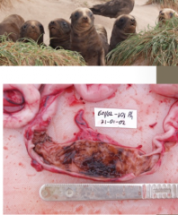
Uncinaria in NZ sea lion pups - life cycle and pathology similar to A. caninum in dogs |
|
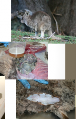
I am a little kangaroo with bottle jaw and blood in my duodenum. What do I have? What would my clin path look like? |
Globocephaloides, due to adult hookworms in the duodenum. Clin path will show anemia and hypoproteinemia. |
|
|
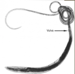
Whipworm (Trichuris sp.) |
|
|
Strongyloides westeri in the SI of a foal |
|
|
Where are trichostrongyles found in the body of ruminants? What other animals do they affect |
Ruminant abomasum and SI - also found in horses, rabbits, and hares |
|
|
Compare Large Strongyles and Small strongyles |
Large - most pathology is associated with larval migration (esp. in the cranial mesenteric artery). S. vulgaris most common species here. Small - (aka cyathostomins). Not pathogenic. Little red guys |
|
|
cestodes. Diphyllobothrium found in fur seals |
|
|
Nanophyteus salmincola - carries a rickettsia that causes salmon poisoning (pacific NW) |
|
|
Alaria sp. in dog SI |
|
|
cat colon - lymphoma |
|
|
mast cell tumour in cat SI |
|
|
Which animal is more susceptible to acute pancreatitis and pancreatic necrosis, cats or dogs? |
Dogs |
|
|
acute pancreatic necrosis/pancreatitis - will see coagulative necrosis |
|

|
chronic pancreatitis. - will see fat necrosis, saponification of fat, and intestinal fibrosis |
|
|
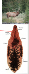
Dicrocelium dendriticum - affects cattle, sheep, and elk - found in biliary and pancreatic ducts |
|
|
Nodular hyperplasia - not significant - would need to do histo to distinguish from neoplasia |
|
|
carcinoma - epithelium of the ducts or acinus - firm white to grey nodular masses - locally invasive and widely metastatic |
|
|
Explain the pathogenesis of acute pancreatitis |
Obstruction --> direct insult to acinar cells (ischemia, toxins, trauma, migrating strongyle larvae, ascending bacteria from SI) --> disturbances of enzyme trafficking within acinar cells (trypsin activated in the cytoplasm causes autodigestion) --> vasculitis and DIC, hepatic necrosis, and chronic pancreatitis |