![]()
![]()
![]()
Use LEFT and RIGHT arrow keys to navigate between flashcards;
Use UP and DOWN arrow keys to flip the card;
H to show hint;
A reads text to speech;
49 Cards in this Set
- Front
- Back
|
Enteritis |
inflammation of large and small intestine |
|
|
typhlitis |
inflammation of caecum |
|
|
colitis |
inflammation of colon |
|
|
proctitis |
inflammation of rectum |
|
|
Diarrhea |
Passage of the feces with increased bulk/fluid content. *enteritis can occur without diarrhoea and vice versa |
|
|
SI epithelial renewal (how?) |
Epithelial progenitor cells in crypts continually divide. New epithelial cells move up to surface/villus tips. |
|
|
Villus atrophy |
Malabsorption of nutrients/water. |
|
|
Villus atrophy with intact crypt glands *example of virus?? |
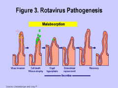
Rotavirus- necrosis of villous enterocytes. has villus atrophy and fusion. coronavirus coccidia some nematodes |
|
|
Villus atrophy with damage to crypts *example of virus? |
Primary insult to crypt cells. Production impaired. Insufficient cells on villi. Enterocytes lost to lumen. Replaced by increasingly immature cells. Malabsorption. i.e. Canine parvovirus Feline parvovirus |
|
|
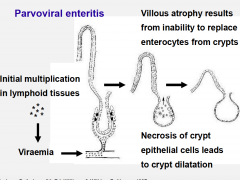
Parvovirus |
Viremia to the crypt cells and lymphoid areas. Epithelial cells are not replaced. Crypts dilated and damaged. Macroscopically: thickened SI, oedema Histologically: crypts dilated with debris, irregular flat epithelial cells. |
|
|
5 types of Enteritis |
1. Secretory and osmotic diarrhea 2. Haemorrhagic 3. Granulomatous 4. Necrotising 5. Ulcerative "SHUNG" Enteritis |
|
|
1. Secretory and osmotic diarrhea |
Colibacillosis (E. Coli) - bacteria adhere to enterocytes - toxins stimulate loss of NaCl and water in intestinal secretions |
|
|
2. Haemorrhagic Enteritis |
1. Clostridial Enterotoxemia Types A-E exotoxins 2. Canine Parvovirus -virus targets crypt cells and lymphoid areas. -small intestine -can be in bone marrow and lymphoid destruction--> pancytopenia |
|
|
3. Granulomatous Enteritis |
1. Johne's Disease/paratuberculosis Macrophages, giant cells in musosa, submucosa, and lymph nodes chronic granulomatous enteritis acid fast organisms 2. FIP (feline coronavirus) a. WET: white, miliary granuloma, fibrin, high protein exudate b. DRY: granulomatous masses on intestine, multifocal pyogranulomata, lymphocytes, plasma cells, macrophages, neutrophils, necrotising vasculitis. |
|
|
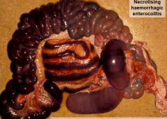
4. Necrotising Enteritis |
1. Salmonellosis - many serovars -Necrosis and exudation of fibrin. -infiltration of lamina propria with neutrophils and macrophages 2. Coccidiosis -Oocyts |
|
|
5. Ulcerative Enteritis |
Cyathostomosis (horses) Small strongyles. Larval development in nodules in mucosa/submucosa Diarrhoea Infiltration by eosinophils, neutrophils, macrophages Oedema Mucosal ulceratoin |
|
|
Inflammatory bowel disease |
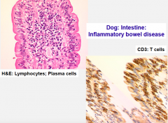
Results in: malabsorption and chronic diarrhoea 1. Lymphocytic/plasmacytic enteritis (dog, cat, horse) 2. Eosinophilic gastroenteritis: dog, cat, horse, idiopathic. 3. Lymphangiectasia: Dogs.****** Lymphatic obstruction DILATATION OF LYMPHATIC VESSELS AND LACTEALS. ACCUMULATION OF MACROPHAGES AND GRANULOMAS IN LYMPHATIC VESSELS. Congenital/acquired Dilation of lymphatic vessels including lacteals Oedema Accumulation of lipid laden macrophages in granulomas |
|
|
Intestinal adenoma |

SI, LI Grow into lumen Benign Polyp-like i.e. rectal adenoma/polyp |
|
|
Intestinal adenocarcinoma |
Dog, cat, sheep. Malignancy of intestinal epithelial cells. Aggressive. Spread via lymphatic vessels to lymph nodes, lung, liver. Trancoelemic spread. |
|
|
Adenocarcinoma of apocrine glands of anal sac |

May be associated with hypercalcemia of malignancy |
|
|
Hyperplasia/adenoma of perianal glands |
Dog Common Benign Old, entire males |
|
|
Lymphoma |

Cat, dog, horse Diffuse infiltration by neoplastic lymphocytes |
|
|
Gastrointestinal stromal tumour |
Increasingly being recognised. Derived from interstitial cells of Cajal. |
|
|
Peritoneum, normally is... |
Smooth, clear Small volume of clear fluid for lubrication. Lined by single layer of mesothelial cells. |
|
|
Post mortem change and autolysis of peritoneum lead to... |
Increased volumes of red/brown fluid in abdomen. No roughening of surfaces of organs or peritoneum. |
|
|
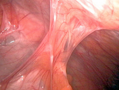
Peritonitis |
Usually secondary to other abdominal pathology. Local/general. |
|
|
Acute peritonitis |
Increased fluid in abdominal cavity and roughening of serosal surface of abdominal organs and parietal peritoneum. |
|
|
Acute peritonitis Types (3) |
1. Fibrinous 2. Purulent 3. Haemorrhagic i.e. splenic rupture |
|
|
Chronic peritonitis |
Fibrous adhesions in: serosal surfaces omentum mesentary peritoneum i.e. Granulomatous |
|
|
Causes of peritonitis (4) |
1. Chemical (bile, barium, urine) 2. Bacterial (abcess, bacteremia) 3. Viral (FIP) 4. Parasitic (Strongyle) |
|
|
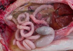
Parasitic cysts (3) |
1. Taenia hydatigena: ruminants, liver, mesentary, peritoneum 2. Taenia ovis: sheep, heart, muscle, including diaphragm 3. Hydatid cyst: Any mammal, humans, usually ruminants and horses |
|
|
Neoplasia of peritoneum |
1. Mesothelomia - malignant, serosa 2. Lipoma- from mesentary, large, pedunculated, instetinal strangulation 3. Secondary tumours - trancoelomic spread. Metastasis on peritoneum. |
|
|
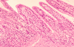
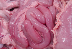
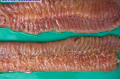
Pig rotavirus enteritis Example of villous atrophy with intact, hyperplastic crypt glands |
|
|
Canine parvovirus Virus targets crypt cells and lymphoid areas. Production is impaired. Insufficient cells for villi. Small intestine. Exampls of Haemorrhagic enteritis. |
|
|
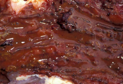
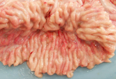
Haemorrhagic enteritis by Clostridium perfringes Type A. Necrotising and haemorrhagic lesions. |
|
|
Canine parvovirus Virus targets crypt cells and lymphoid areas. Production is impaired. Insufficient cells for villi. Small intestine. |
|
|
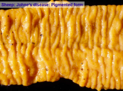
Granulomatous enteritis Johne's Disease/ ParaTB. Mycobacterium avium. Infects macrophages. Chronic ganulomatous enteritis. Epitheliod macrophages, and multinucleate giant cells in mucosa, submucosa, and lymph nodes. |
|
|
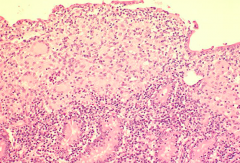
Bovine granulomatous enteritis. Histo: sheets of macrophages, multinucleate cells, some lymphocytes and plasma cells. |
|
|
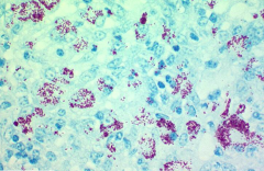
Johne's Disease Acid Fast staining Appears red on Zhiel Neelsen Stain Bacteria found in macrophages. |
|
|
FIP (feline coronavirus) WET: white, miliary granuloma, fibrin, high protein exudateb. DRY: granulomatous masses on intestine, multifocal pyogranulomata, lymphocytes, plasma cells, macrophages, neutrophils, necrotising vasculitis. |
|

|
Necrotising enteritis Salmonellosis- many serovars Necrosis and exudation of fibrin Infiltration of lamina propria with neutrophils and macrophages |
|
|
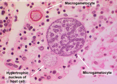
Necrotising Enteritis Coccidiosis Eimeria, isospora species |
|
|
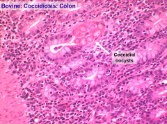
Necrotising Enteritis Coccidiosis |
|

|
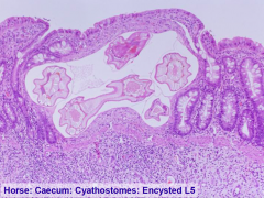
Ulcerative Enteritis Cyathostomosis Small strongyly ins horse. Larva develop in nodules in mucosa/submucosa of cecum an colon. |
|
|
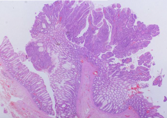
Rectal Polyp Intestinal adenoma. Usually grow into the lumen, benign, polyp like. |
|

|
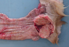
Intestinal Adenocarcinoma Dog, cat, sheep. Malignancy of intestinal epithelial cells. Aggressive. Spread via lymphatic vessels to lymph nodes, lung, liver. Trancoelemic spread. |
|
|
Adenocarcinoma of apocrine glands of anal sac |
|

|
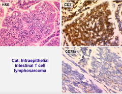
Lymphoma Diffuse infiltration by neoplastic lymphocytes |
|
|
Lymphosarcoma. |