![]()
![]()
![]()
Use LEFT and RIGHT arrow keys to navigate between flashcards;
Use UP and DOWN arrow keys to flip the card;
H to show hint;
A reads text to speech;
143 Cards in this Set
- Front
- Back
|
CNS
|
brain & spinal cord
|
|
|
PNS
|
all other neurons outside brain and spinal cord
|
|
|
Primary somatosensory system
|
tactile, deep pressure, pain, proprioception, kinesthesia
|
|
|
Cortical somatosensory system
|
two point discrimination, stereognosis
|
|
|
Reflex arc
|
governs automatic reflex actions, receptor, sensory neuron, interneuron, motor neuron, effector
|
|
|
Meisner’s corpuscles
|
fingers, soles, palms, best to detect textured objects moving aross skin
|
|
|
Free nerve ending
|
all over skin, feel pain, heat, cold, chemicals
|
|
|
Sensory nerve fiber
|
x |
|
|
Pacinian corpuscles
|
deep pressure, found in skin, mesenteries around gut, joint capsules, egg shaped structure with many concentric layers of tissue respond to pressure quickly – action potential initiated when corpuscle is deformed by pressure **Provide CNS with joint position information—PROPRIOCEPTION!
|
|
|
Hair follicle receptor
|
hair root plexus? Sens message to skin through touch to hair
|
|
|
Merkel’s discs
|
prominent in fingertips, light touch, diminish quickly, MOSQUITO
|
|
|
Affected side of the body from CNS lesions
|
contralateral (opposite)
|
|
|
Indicators of CNS damage
|
diminished or absent sensation, clumsy or uncoordinated movement, poor fine motor control, diminished appetite, social isolation
|
|
|
Affected area from PNS damage
|
specific nerves such as drooping eyelid from damage to oculomotor nerve
|
|
|
Indicators of PNS damage
|
muscle weakness, atrophy, lack of sensation, pain, loss of ability to perspire, changes in quality of skin and nails, hyper/hypoesthesia
|
|
|
Hypoesthesia/hypesthesia
|
decreased or dulled sensation
|
|
|
Hyperesthesia
|
increased or hypersensitivity
|
|
|
Anesthesia
|
complete loss of sensation
|
|
|
Parasthesia
|
abnormal sensation such as tinging
|
|
|
Anelgesia
|
complete loss of pain sensation
|
|
|
Hypoalgesia/hypalgesia
|
diminished pain sensation
|
|
|
Sensory recovery in peripheral nerves
|
rule of thumb is 1 inch per month, no guarantees
|
|
|
Sensory recovery in central nerves
|
CNS does not regenerate cells, but reeducation is sometimes possible
|
|
|
Sensory reeducation
|
rehabilitative treatment, provide appropriate sensory input to promote recovery of dulled or absent sensation
|
|
|
Graded desensitization
|
may reduce sensory hypersensitivity
|
|
|
Most important precaution with sensory problems
|
SAFETY – self awareness, protect affected body part during tasks
|
|
|
Sensory reeducation for impairments in CNS
|
electrical stimulation to promote feeing and awareness of movement, progressive object discrimination is when client uses all senses to understand an object then grades up until they are only using one sense, functional activities to incorporate the involved body part into occupations
|
|
|
Sensory reeducation for impairments of the PNS
|
graded stimulation to expose client to feeling and identify it as in running a pencil eraser across hand, localization of stimuli is when client is asked to identify where they are being touched, tactile discrimination/visual tactile integration is when client used sense of touch to locate object in rice or sand then uses touch and sight to locate object
|
|
|
How are sensory reeducation techniques for PNS and CNS the same or different?
|
P. 436-437 Early book
|
|
|
Difference between vision and visual perception
|
vision is reception of sensory information through visual receptors, visual perception is integrating that information and some from other senses into the brain
|
|
|
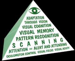
Hierarchy of visual perceptual skill development
|
|
|
|
Visual acuity
|
sharp, clear, accurate vision
|
|
|
Oculomotor contol |
coordination of eye movements with eye muscle |
|
|
visual field |
reception of the complete visual environment |
|
|
visual attention |
fixating gaze on object as long as rquired, shifting to other objects as needed |
|
|
visual scanning |
shifting attention from one target to another smoothly, in focus, regardless of eye movemet |
|
|
pattern recognition |
identify important features of objects, distinguish objects from its surroundings and other objects |
|
|
visual memory |
brain creates and holds an image short term and uses it to produce a response, later store in long term memory for later recognition or recall, like a photograph |
|
|
visual cogniton |
ability to manipulate visual information mentally and understand and integrate other sensory information, **Foundation for reading and writing, SEE IT AND USE IT |
|
|
myopia |
nearsightedness, can see near |
|
|
hyperopia |
farsightedness, can see far |
|
|
presbyopia |
farsightedness associated with aging, as in reading glasses |
|
|
diplopia |
double vision, movement of one eye does not match other so see two images visual stress - headache, eye pain or strain **eyepatch must be prescribed by dr, but therapist can make one just for a session |
|
|
astigmatism |
variations in the curvature of the eye, like a funhouse mirror |
|
|
cataracts |
clouding of lens, hard to tolerate glare |
|
|
macular degeneration |
loss of center vision, visual detail loss, hard to see faces or read |
|
|
glaucoma |
increased pressure in eye, loss of peripheral vision |
|
|
hypertension |
hardening of arteries in retinal blood vessels=damage to sight |
|
|
diabetic retinopathy |
dilation and breakdown, leakage of blood from retinal vessels floater, blurred vision, then loss of vision |
|
|
color blindness |
more common in males, can be dulled colors not always total loss... |
|
|
environmental treatment for visual acuity |
high contrast-light food, dark plate illumination - non glare or shadow, task lighting, fluorescent...? decrease clutter, plain rather than heavily patterned background |
|
|
homonymous hemianopsia |
loss of visual field in corresponding right or left half or each eye, most common after CVA, poor visual scanning (report to OTR) bump into objects, difficulty reading, misplace objects in one field |
|
|
treatment for visual field loss |
increase awareness of deficit, retrain scanning pattern, (thick red line on rading material), increase attention to field cut area |
|
|
visual inattention |
contralateral affect lost the attentional CNS mechanism that drive the search for visual information, no attempt to search for information, no head turning or eye movements, asymmetrical scanning patterns (not an eye problem - brain is not searching for info) |
|
|
CNS Left side injury visual deficit |
visual agnosia - aware of object but have difficulty identifying them |
|
|
CNS Right side injury |
fail to recognize an object or pattern because they do not perceive or "see" it |
|
|
visual memory deficit treatment |
provde a variety of other sensory information to functon wel in ADL, like when you block out eyes in a photo for anonymity - brain needs eyes to recognize pattern of face |
|
|
visual perceptual deficit treatment |
learn to take in visual info in consistent organized manner, teach scanning patterns in combination with physical manipulation of objects, "touch it" |
|
|
food viscosity categories |
nectar, honey, pudding |
|
|
NPO |
nothing per orifice |
|
|
aesthesiometer |
adjustable, tests 2 point discrimination, disc-riminator |
|
|
mono filament nerve tester |
filament bends, can you feel this sensation |
|
|
CVA |
stroke, loss of blood supply to the brain resulting brain cell damage and death, deficits are in area of brain where stroke occurred **Single largest diagnostic group sen by OT working in physical dysfunction |
|
|
CVA F.A.S.T |
face - facial muscles droop arm - one arm drooping speech - slurred time - is of the essence, call 911 now! |
|
|
CVA symptoms |
symptoms are sudden weakness of face, arm, leg, esp. one one side of body, confusion, trouble understanding and speaking, trouble seeing in one or both eyes, trouble walking, dizziness, loss of balance, coordination, severe headache no known cause |
|
|
frontal lobe of the brain |
responsible for cognitive thought process - knowing, thinking, learning, judging regulates voluntary movements prefrontal areas responsible for intelligence and personality |
|
|
parietal lobes of the brain |
associated with sense of touch and balance, important in interpreting sensory info from all parts of the body and in the manipulation of objects |
|
|
temporal lobes of the brain |
near ears, deal with hearing |
|
|
left brain |
controls right side of the body speech, academic, analysis |
|
|
right brain |
controls left side of body artistic, imaginative, facial perception, music |
|
|
ischemic cva -2 types |
BLOOD CLOT (most common) thrombolic- clot forms in artery in brain, diseased artery embolic- clot forms elsewhere and travels to brain |
|
|
hemorrhagic cva-2 types |
BLOOD VESSEL BREAKS (poorer outcome) intracerebral - burst blood vessel bleeds into brain, more common subarachnoid - burst blood vessel bleeds outside brain, usually caused by aneuryism |
|
|
risk factors hemorrhagic stroke |
high blood pressure, alcohol, drugs, anti blood clot drugs, blood clotting disorder such as hemophilia or sickle cell |
|
|
modifiable risk factors cva |
obesity, cholesterol, smoking, alcohol, hypertension, diabetes |
|
|
non modifiable risk factors cva |
age, gender (male), race (af am and hispanic), genetic predisposition
|
|
|
medical treatment for ischemic |
tPA- clot buster medication, must be given fast MERCI- retrieval system, corkscrew, can use after 3 hours penumbra system - suction, must be used w/in 8 hrs |
|
|
TIA |
transient ischemic attack, temporary blockage of artery in brain, stroke-like symptoms, few min to 24 hrs, warning that stroke might occur |
|
|
residual effects |
symptoms left after episode balance, fall risk |
|
|
hemiplegia |
cva effect one sided paralysis, contralateral |
|
|
spasticity |
cva effect constant contraction or contraction and release, involuntary, |
|
|
danger zone |
shoulder in adduction and internal rotation, elbow in flexion, forearm in pronation/supination, wrist and fingers in flexion |
|
|
flexor synergy |
limbs move as one unit as in the danger zone |
|
|
medical complications of stroke |
DVT - use SCD sequential compression device, inflates n legs and allows circulation subluxation - GHJ/rotator cuff paralysis or weakness, spasticity of scapular muscles, shoulder muscles are not holding joint together, positioning very important, (neutral, ext rot) measured by # fingers |
|
|
treatment for motor pathway damage |
neuroplasticity high repetition makes new pathways, uncovers paths not usually used use dependent, use it or lose it |
|
|
DVT |
MOST LIKELY TO OCCUR IN AFFECTED LEG red/warm/swollen/painful --elevate area |
|
|
cva ot goals |
pg 471 Early |
|
|
cva symptoms and management |
edema - use retrograde massage, let gravity help pain visual deficits - like HH, spatial neglect (left most common) tactile changes fatigue sublux - positioning, knesiotaping joint ROM to prevent deformity flaccid paralysis, hypotonic, low tone spasticity, hypertonic, synergies balance imairment |
|
|
motor function return |
proximal to distal |
|
|
Brunstrom's stages of motor recovery |
p. 103-106 |
|
|
dysarthria |
difficulty with speech, pronouncing words, slurred speech |
|
|
anarthria |
loss of motor ability for speech |
|
|
dysphasia |
trouble swallowing drooling, pocketing, coughing, gurgly voice high risk for aspiration |
|
|
anomia |
difficulty with word finding |
|
|
alexia |
difficulty with reading comprehension |
|
|
agraphia |
difficulty with written expression |
|
|
Broca's aphasia |
difficulty with expressive area |
|
|
Wernicke's aphasia |
difficulty with receptive area |
|
|
global aphasia |
global language deficit with expressive and receptive |
|
|
cva behavior manifestations |
impulsivty, persevoration, mood, pseudobulbar effect, combative, poor coping, poor adjustment |
|
|
cva cognitive deficits |
initiation, motivation, memory, confusion, attention, lck of insight, lack of judgement and safety awareness, thought inflexibility, trouble solving problems, sequencing, planning, organization |
|
|
cva perceptual/perceptual motor deficits |
problems interpreting information fro the environment, hemi-inattention, apraxia, difficulty with spatial relations |
|
|
differences and similarities between L and R CVA |
pg 482 |
|
|
# 1 cause of TBI and most damaged areas |
falls, frontal and temporal lobes from falls on side and front mva, struck, assault |
|
|
criteria for TBI |
external force impinging upon head and brain documented loss of consciousness amnesia for the event glasgow coma scale of less than 15 for first 24 hours |
|
|
stint vs. shunt |
stint-short term shunt - longer term |
|
|
most likely for TBI |
kids 0-4, 15-24 yr old elderly - falls af am, males substance abuse |
|
|
brain characteristics |
floating, supported by fluid within the skull brain can be compressed, pulled, stretched sharp bones which pose threat to brain, esp. frontal lobe |
|
|
types of TBI |
open- penetrating, gunshot wound closed - nonpenetrating, fall, mva, assault |
|
|
diffuse axonal injury |
when brain cells are torn from one anther, localized damage - brain bounce against skull |
|
|
effects of TBI |
based on area of brain that is injured and severity of injury |
|
|
contusion vs. concussion |
contusion - bruising concussion - bruising and swelling of brain, ice, stint to drain fluid |
|
|
hematoma, definitions and types |
blot clot formed subdural-below protective layer of brain epidural - between layer and brain intercerebral - within the brain tissue causing increased pressure in the brain |
|
|
hypoxia anoxia |
decreased oxygen to brain no oxygen to brain |
|
|
craniotomy |
hole in skull, brings swelling/intracranial pressure down |
|
|
intracranial hematoma |
secondary brain injury bleeding in the brain, often requires surgery |
|
|
cerebral edema |
secondary brain injury swelling of the brain tissue, needs medical intervention |
|
|
post traumatic amnesia |
amount of time it takes for continuous memory to return after an injury best guide to the severity of the diffuse damage longer ptas are associated with worse damage |
|
|
retrograde amnesia |
decreased ability to recall info occurring before the brain injury (eposodic) |
|
|
anterograde amnesia |
decreased ability to recall new information (episodic) |
|
|
functional assessments for tbi |
glasgow outcome scale disability rating scale (DRS) Functional independence measure (FIM) Rancho Los Amigos Scale of Cognitive Functioning |
|
|
glasgow coma scale |
objective tool to measure the conscious state does not indicate recovery limits rated on eyes, verbal, motor severe 8 or below moderate 9-12 mild 13-15 |
|
|
deceberate posturing |
|
|
|
decorticate posturing |
|
|
|
Rancho Los Amigos levels of cognitive functioning scale |
|
|
|
post traumatic vision syndrome (PTVS) |
affects visual acuity, oculomotor control, binocular vision double vision, reduced gaze stability, poor attention, poor visual memory, impaired balance, visual-spatial misperceptions |
|
|
visual midline shift syndrome (VMSS) |
distortion of the perception of the midline causes person to lean to one side, forward, backward |
|
|
self-care in tbi |
broken down into small segments, structured, backward chaining, sequence cards |
|
|
x |
x |
|
|
x |
x |
|
|
x |
x |
|
|
x |
x |
|
|
babinski reflex |
positive for motor neuron damage if big toe flexes and little toes flay, normally seen only in children |
|
|
ankle clonus |
forcibly flex foot, foot will move up and down indicates upper motor neuron damage |
|
|
stages of pressure ulcers |
x |
|
|
x |
x |
|
|
MS |
demylenated lesions scattered through CNS white matter, plaques of scar tissue after myelin loss |
|
|
ALS |
degeneration of motor neurons affects voluntary muscles |
|
|
x |
x |
|
|
bradykenesia |
slowness, difficulty with movements, reduced speed, range, amplitude |
|
|
akinesia |
absence of movement, freezing |
|
|
hypokinesia |
reduced amplitude of movement, mask-like face, dec trunk rotation, arm swing |
|
|
dyskinesia |
purposeless wriggling |
|
|
x |
x |