Reading...
![]()
Play button
![]()
Play button
![]()
Use LEFT and RIGHT arrow keys to navigate between flashcards;
Use UP and DOWN arrow keys to flip the card;
H to show hint;
A reads text to speech;
19 Cards in this Set
- Front
- Back
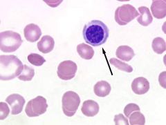
Identify the cell indicated by the arrow.
|
Nucleated Red Blood Cell
|
|
|
Give a brief description of Howell Jolly Bodies.
|
Basophilic nuclear remnants seen in young erithrocytes during the response to anemia.
|
|
|
Give a brief description of nucleated red blood cells.
|
Immature rbc, represent early release immature cells during anemia.
|
|
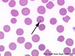
Identify the abnormal RBC indicated by the arrow.
|
Howell Jolly Bodies
|
|
|
How would you differentiate between a NRBC and lymphocyte?
|
NRBC: cytoplasm will be similar color to the rbc around it and the nucleus will be darker with looser chromatin structure. Lymphoctyes: sky blue cytoplasm less than in amount than the nrbc.
|
|
|
What are some general situations that result in anemia’s?
|
Parasitic infestation and iron-deficiency.
|
|
|
What are the 2 classifications of bone marrow response?
|
Nonregenerative and regenerative anemias.
|
|
|
How does the retic count relate to the anemia classification?
|
If there are a large amount of retics then more than likely the animal has a regenerative anemia.
|
|
|
Give a brief description of Heinz Bodies.
|
Round structures representing denatured hemoglobin caused by certain oxidant drugs or chemicals.
|
|
|
Give a brief description of Basophilic stippling.
|
The presence of small, dark blue bodies of residual RNA within the erythrocyte.
|
|
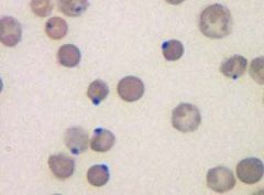
Identify the abnormal RBC in this photo.
|
Heinz Bodies
|
|
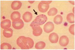
Identify the abnormal RBC indicated by the arrow.
|
Basophilic stippling
|
|
|
Define anemia
|
A decrease in the number of red blood cells (RBCs) or less than the normal quantity of hemoglobin in the blood.
|
|
|
Name at least four abnormal red blood cells that would fall under the category, poikilocytes.
|
Acanthocyte, Schistocyte, Keratocyte, Spherocyte, Target Cell, Heinz Bodies, Eccentrocyte, Stomatocyte, Dacrocyte.
|
|
|
RBC's with a smaller than normal diameter and decreased MCV; seen in iron deficiency.
|
Microcytes
|
|
|
A grouping of erythrocytes in stacks; seen with increased fibrinogen or globulin.
|
Rouleaux
|
|
|
Darkly staining red cells with reduced or no central pallor; not easily detected except in dogs; suggest IMHA.
|
Spherocyte
|
|
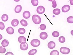
Identify the abnormal RBC indicated by the arrows.
|
Target Cell
|
|
|
Give a brief description of Agglutination.
|
An antibody coats the erythrocyte causing bridging and clumping.
|