![]()
![]()
![]()
Use LEFT and RIGHT arrow keys to navigate between flashcards;
Use UP and DOWN arrow keys to flip the card;
H to show hint;
A reads text to speech;
125 Cards in this Set
- Front
- Back
|
What are the organs in the GI tract? |
- mouth - pharynx - esophagus - stomach - s.i. - l.i. - rectum - anus |
|
|
What are the accessory organs of the GI tract? |
- salivary glands - liver - gallbladder - pancreas |
|
|
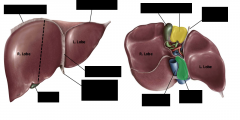
What are the anatomical lobes of the liver? |
right lobe - right of falciform lig. left lobe - left of falciform lig. quadrate lobe - between gall bladder and falciform lig. caudate lobe - between IVC and falciform lig |
|
|
What are the functional lobes of the liver? |
right lobe - right anatomical left lobe - left anatomical, quadrate and caudate |
|
|
|
|
|
What quadrants is the liver in? |
right hypochondriac epigastric left hypochondriac right lateral |
|
|
What is the porta hepatis? |
entrance/exit on inferior side of liver for hepatic portal v, proper hepatic a., and hepatic duct |
|
|
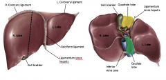
What are coronary ligaments? |
lines of reflection of visceral peritoneum onto diaphragm define the bare area |
|
|
What is the falciform ligament? |
midline fusion of right and left coronary ligaments on anterior surface of liver |
|
|
What is the ligamentum teres hepatis? |
contained in inferior free edge of falciform ligament |
|
|
What is the falciform ligament embryological remains of? |
ventral mesentery |
|
|
What is the ligamentum teres hepatis embryological remains of? |
umbilical vein of embryo |
|
|
What is the bare area? |
area of liver not encased in visceral peritoneum |
|
|
How does the bare area form? |
it was fused with the diaphragm during development |
|
|
What is the glisson's capsule? |
collagenous capsule covering external surface of the liver exposed only in bare area |
|
|
What is the triangular ligament? |
lateral fusion of anterior and posterior coronary ligaments on right and left side |
|
|
|
|
|
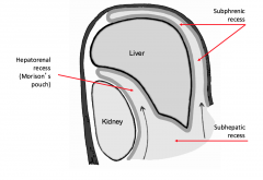
What recess does most fluid tend to go to in a supine position? |
heptorenal recess (Morrison's pouch) |
|
|
What is the function of the spleen? |
lymphatic organ lymphocyte proliferation immune surveillance blood reservoir |
|
|
When is the spleen suspect to injury? |
most delicate in abdomen susceptible to damage when blow to left upper quadrant |
|
|
What is the spleen protected by? |
9th and 10th ribs |
|
|
What happens in a spleen rupture? |
bleeding |
|
|
What region is the spleen in? |
left hypochondriac (left upper quadrant) |
|
|
What is the order food passes through in the digestive tract? |
esophagus stomach duodenum jejunm ileum ascending colon transverse colon descending colon sigmoid colon rectum |
|
|
Where does the esophagus pass into the abdomen at? |
esophageal hiatus level of T9-10 |
|
|
What accompanies the esophagus through the esophageal hiatus? |
anterior and posterior vagal trunks |
|
|
What is the gastroesophageal junction? |
where the esophagus joins the cardiac region of the stomach |
|
|
What is a hiatal hernia? |
movement of cardiac portion of stomach up into thorax |
|
|
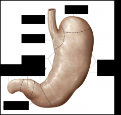
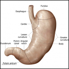
What is the stomach covered by? |
peritoneum |
|
|
What is the sphincter between the stomach and duodenum? |
pyloric sphincter |
|
|
What are rugae? |
folds of mucosa in stomach |
|
|
What are the purpose of the rugae? |
increase surface area of stomach |
|
|
|
|
|
Where is the lesser sac located? |
small pouch posterior to stomach |
|
|
How is the lesser sac formed? |
rotation of GI tract |
|
|
What is the lesser sac remnant of? |
right half of coelomic cavity |
|
|
What is the greater sac? |
remainder of peritoneal cavity |
|
|
How do the greater sac and lesser sac communicate? |
epiploic foramen |
|
|
How does a peptic ulcer form? |
breach in mucosal lining of GI structure due to high content of stomach acid |
|
|
What is the mucosal lining susceptible to? |
infections by H. phylori bacteria |
|
|
What is gastric carcinoma? |
malignant cells in lining of the stomach |
|
|
What are some symptoms of a gastric carcinoma? |
heartburn upper abdominal pain loss of appetite weight loss blood in stool |
|
|
What regions is the stomach in? |
epigastric umbilical left hypochondriac |
|
|
Where is bile formed? |
liver |
|
|
What conducts bile? |
right hepatic duct and left hepatic duct |
|
|
What is the common hepatic duct? |
external to liver formed by right and left hepatic |
|
|
Where is the gall bladder? |
embedded in inferior side of right lobe of liver |
|
|
What is the function of the gall bladder? |
stores and concentrates bile |
|
|
What is the cystic duct? |
duct from gall bladder |
|
|
What is the common bile duct formed from? |
formed from cystic duct and common hepatic duct |
|
|
Where does the common bile duct empty? |
duodenum |
|
|
What closes the common bile duct? |
sphincter of Oddi |
|
|
What is the biliary tree? |
system of ducts that drain bile produced by liver to gallbladder for storage and duodenum to enter digestive tract |
|
|
What is the hepatopancreatic ampulla of Vater? |
swelling where bile duct and pancreatic duct meet |
|
|
Where does the bile duct come into the intestine? |
duodenal papilla |
|
|
What are gallstones? |
concretions of bile components in the gallbladder |
|
|
When do gallstones become symptomatic? |
when blocking cystic duct - cholecytisis causing inflammation |
|
|
Where will you feel pain from gallstones? |
epigastric region right hypochondriac region |
|
|
What does it mean to be secondarily retroperitoneal? |
started out suspended by peritoneum but during development rotation of the gut forced them against the dorsal body wall where they lost peritoneum anteriorly |
|
|
What foregut organs are secondarily retroperitoneal? |
pancreas 2nd and 3rd part of duodenum |
|
|
What foregut parts are suspended by peritoneum? |
1st and 4th parts of duodenum |
|
|
What is function of the main pancreatic duct? |
conducts pancreatic digestive enzymes into the GI tract at duodenal papilla in descending part of duodenum |
|
|
What is the function of the accessory pancreatic duct? |
if present empties into duodenum proximal to main pancreatic duct |
|
|
What regions is the pancreas in? |
epigastric umbilical left hypochondriac |
|
|
What regions is the gallbladder in? |
right hypochondriac and right lateral |
|
|
What are the three parts of the small intestine? |
duodenum jejunum ileum |
|
|
How are the jejunum and ileum suspended? |
suspended from posterior body wall by mesentery - mobile |
|
|
Where is the duodenojejunal junction? |
as duodenum emerges from being retroperitoneal |
|
|
How can you tell the difference between the jejunum and ileum? |
jejunum has mesenteric windows and ileum does not |
|
|
What are mesenteric windows? |
where fat is kept away from intestinal wall |
|
|
Where does the ileum end? |
ileocecal junction |
|
|
What are the vesa recta? |
straight vessels in organ that come from loops |
|
|
What regions is the small intestine in? |
all of them |
|
|
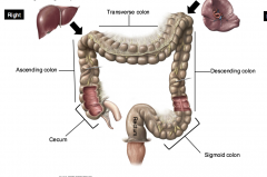
What are the regions of the large intestine? |
vermiform appendix ascending,transverse, descending & sigmoid colon rectum anal canal |
|

|
|
|
|
What side of the large intestine is the liver on? |
right |
|
|
What side of the large intestine is the spleen on? |
left |
|
|
What are taeniae coli? |
3 evenly spaced longitudinal bands of external longitudinal muscle on the colon |
|
|
What is the function of the taeniae? |
gather colon into haustra because it is shorter in length than the colon |
|
|
What is the function of the haustra? |
facilitates water resorption from feces |
|
|
What happens to the taeniae at the appendix and rectum? |
fuse into a complete smooth muscle layer |
|
|
What does the inner layer of muscle of the colon do? |
ring shaped, act like wringing a towel |
|
|
What are appendages epiploicae? |
tags of fat that creep onto the wall of the colon and attach |
|
|
What is apendicitis? |
infection of appendix |
|
|
Where is pain from apendicitis felt? |
McBurney's point |
|
|
What regions is the colon in? |
starts in right inguinal and goes all the way around hitting everything except umbilical |
|
|
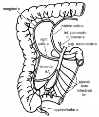
What are the three unpaired arteries of the descending aorta that supply the gut? |
celiac a. - foregut superior mesenteric a. - midgut inferior mesenteric a. - hindgut |
|
|
What are the break offs for foregut, midgut and hindgut? |
foregut ends at 2nd part of duodenum midgut ends at distal 3rd of transverse colon |
|
|
What structures does the celiac artery supply? |
liver stomach pancreas duodenum spleen |
|
|
At what level does the celiac artery branch off? |
T12 |
|
|
What is the celiac artery surrounded by? |
celiac plexus - autonomic plexus of nerve fibers and postganglionic neurons |
|
|
What does the superior mesenteric artery supply? |
duodenum jejunum ileum large intestine |
|
|
What level does the superior mesenteric artery branch off at? |
L1 |
|
|
What surrounds the superior mesenteric artery? |
superior mesenteric autonomic plexus |
|
|
What does the inferior mesenteric artery supply? |
descending colon sigmoid colon superior part of rectum |
|
|
Where does the inferior mesenteric artery split from? |
3-4 cm above bifurcation of aorta |
|
|
What do the inferior phrenic arteries supply? |
diaphragm |
|
|
What do the renal arteries supply? |
kidneys suprarenal branch to suprarenal gland |
|
|
What do the testicular/ovarian arteries supply? |
gonads |
|
|
What are the common iliac arteries? |
terminal branches of abdominal aorta |
|
|
What branches from the common iliac arteries? |
external iliac artery internal iliac artery |
|
|
What are the lumbar arteries? |
5 segmental arteries supplying posterior body wall |
|

|
1) abdominal aorta 2) celiac artery 3) common hepatic 4) splenic 5) left gastric 6) gastrodudenal 7) proper hepatic 8) cystic 9) right gastric 10) superior pancreaticoduodenal 11) right gastroepiploic 12) left gastroepiploic |
|
|
What are the major branches of the superior mesenteric artery? |
intestinal arteries ileocolic right colic middle colic |
|
|
Where does the marginal artery run? |
anastomoses along border of large intestine |
|
|
What branches is the marginal artery from? |
ileocolic middle colic right colic inferiormesenteric branches of superior and inferior mesenteric a. |
|
|
How is blood supply of the jejunum different from the ileum? |
fewer arcades and longer vesa recti |
|
|
|
|
|
What are the major branches of the inferior mesenteric artery? |
middle colic sigmoidal superior rectal |
|
|
What are watershed areas? |
regions supplied by most distal branches of two arteries |
|
|
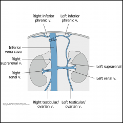
What does the inferior vena cava drain? |
lower limbs trunk kidneys gonads |
|
|
What are the tributaries of the IVC? |
common iliac renal lumbar r. gonadal |
|
|
Where does the left gonadal v drain? |
left renal vein |
|
|
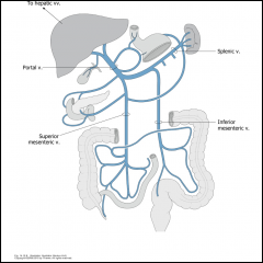
What drains into the hepatic portal vein? |
superior mesenteric splenic inferior mesenteric |
|

|
|
|
|
What is blood carried to the liver for? |
storage metabolism detoxification |
|
|
|
|
|
What happens when return of blood through the liver is obstructed? |
increase in hydrostatic pressure in venous portal system alternative pathway found through portacaval anastomoses |
|
|
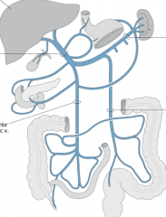
Where are portacaval anastomoses found? |
superior rectal v. (inf. mesenteric) w inf. rectal v. (internal iliac v) branches of colic veins (sup. or inf. mesenteric) w renal veins remnant of l. umbilical v. with paraumbilical and sup./inf. epigastric veins esophageal veins (gastric veins) w esophageal veins (azygos veins) |
|
|
What can cause blockage of venous drainage to liver? |
pregnancy or cirrohosis |
|
|
What happens to flow of blood in a liver blockage? |
reverses and flows into caval/systematic system causing enlargement of caval vessels |
|
|
What does the enlargement of rectal vessels lead to? |
hemorrhoids |
|
|
What does the enlargement of esophageal veins lead to? |
esophageal varices |
|
|
What does the enlargement of paraumbilical/epigastric veins result in? |
caput medusa |
|
|
What is caput medusa? |
enlarged radiated veins on abdomen - need to be drained |