![]()
![]()
![]()
Use LEFT and RIGHT arrow keys to navigate between flashcards;
Use UP and DOWN arrow keys to flip the card;
H to show hint;
A reads text to speech;
161 Cards in this Set
- Front
- Back
|
What does Cardiopulmonary Circulation refer to? |
blood flow between the heart & lungs |
|
|
What does Systemic / Blood Flow Cycle refer to? |
blood flow throughout entire body. |
|
|
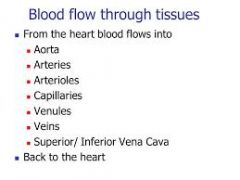
State the blood flow cycle starting at Aorta, ending at IVC/SVC |
Aorta Arteries Arterioles Capillaries Venules Small veins Large veins IVC/SVC |
|
|
What does Portal Circulation refer to? |
blood flow to and from the LIVER |
|
|
Path of Venous Blood in Portal Circulation |
- digestive tract & spleen - liver to be FILTERED - IVC - then the heart |
|
|
Does Venous Blood go directly to the IVC? |
No. It has to go to the LIVER to be filtered from toxic digested products |
|
|
What is the FIRST system to develop and function in utero? |
Cardiovascular -by end of 3rd gestational week -from mesodermal cells |
|
|
When does the IVC develop in utero? |
weeks 6-8 |
|
|
When does the portal system develop in utero? |
8th week |
|
|
3 vessel wall layers (arteries and veins) |
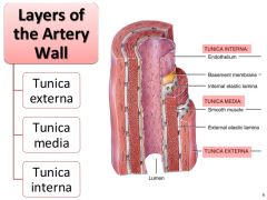
- tunica intima (inner) - tunica media (middle) - tunica adventitia (outer) |
|
|
Tunica Intima |
innermost THINNEST layer - single layer of endothelial cells |
|
|
In what fashion do the cells run in the Tunica Intima? Why? |
longitudinal (up & down) - allowing smooth surface that offers no obstruction to flow |
|
|
Tunica Media |
middle layer - composed of collagen & smooth muscle |
|
|
In what fashion do the fibers run in the Tunica Media? Why? |
circular - allowing for control of diameter to regulate blood flow |
|
|
In Arteries, the Tunica Media is the ________ layer |
thickest |
|
|
In Veins, the Tunica Media is the _________ layer |
thinnest |
|
|
Tunica Adventitia AKA Externa |
outermost layer - elastic tissue with thin fibrous layer |
|
|
In what fashion do the fibers run in the Tunica Adventitia? Why? |
longitudinal - gives elasticity |
|
|
The Tunica Adventitia contains tiny internal vessels that supply nutrients and remove waste, what is this called? |
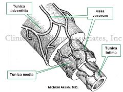
the Vaso Vasorum "vessels of vessels" |
|
|
Arteries |
carry blood AWAY from the heart to the capillaries |
|
|
Arterioles |
Smallest type of arterial vessel - considered resistance vessels because they assist with regulating BF |
|
|
2 layers of Arterioles |
- tunica media - tunica intima |
|
|
Capillaries |
microscopic network of the smallest arterioles (metarterioles) |
|
|
Capillaries only consist of 1 layer, what is it? |
tunica intima |
|
|
Capillary beds AKA Vascular beds |
complex network that connects arterioles with venules |
|
|
What is the function of the Capillary beds? |
- supply oxygen and nutrients to the tissues - remove carbon dioxide and waste |
|
|
Which vessel wall layer contains valves? |
tunica intima (VEINS) |
|
|
Veins |
transport blood from capillary beds to the HEART |
|
|
What is the reason for collapsibility of veins? |
the tunica media is much thinner |
|
|
Venules |
smallest venous vessel |
|
|
Venules consist of which 2 layers? |
- tunica media - tunica intima |
|
|
What is the general rule regarding abdominal arteries and veins? |
Arteries lay posterior to veins |
|
|
Aorta |
main artery - arising from LEFT ventricle of heart |
|
|
4 Sections of the Aorta |
1. ascending aorta 2. aortic arch 3. descending / thoracic aorta 4. abdominal aorta |
|
|
Major branches of the Thoracic Aorta feed.... |
pericardium lungs esophagus intercostal spaces |
|
|
Where does the Abdominal Aorta enter? |
@ aortic hiatus of diaphragm @ level of T12 |
|
|
The Abdominal Aorta decends _________ slightly ____ of midline |
caudally; left |
|
|
The Abdominal Aorta runs adjacent to.... |
IVC |
|
|
The Abdominal Aorta runs ________ to the curvature of the spine |
Anterior |
|
|
Where does the Abdominal Aorta terminate? |
L-4 (umbilicus) |
|
|
What does the Abdominal Aorta bifurcate into? |
right and left common iliac arteries |
|
|
The diameter of the Abdominal Aorta tapers as it courses ___________ |
inferiorly |
|
|
2 aspects of Aorta |
- anterior aspect - lateral aspect |
|
|
What is found along the Anterior aspect of the Abdominal Aorta? |

- celiac trunk - SMA - IMA |
|
|
What is found along the Lateral aspect of the Abdominal Aorta? |
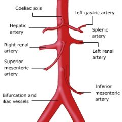
- renal artery - gonadal artery |
|
|
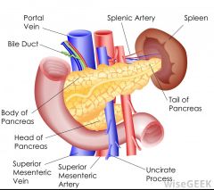
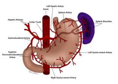
Celiac Artery |
1st anterior branch of Abdominal Aorta |
|
|
The Celiac Artery divides into what 3 branches? |
1. Splenic 2. Left Gastric 3. Common Hepatic |
|
|
Which of the 3 Celiac branches is the largest? |
Splenic |
|
|
Which of the 3 Celiac branches is the smallest? |
Left Gastric |
|
|
In which direction does the Splenic Artery course? |

- Left (very tortuous) - horizontally from Celiac to Splenic Hilum |
|
|
Describe the course of the Splenic Artery in relation to surrounding structures |
- posterior to stomach - along superior border of pancreas - anterior to upper part of L kidney |
|
|
The Splenic Artery gives rise to smaller arterial branches which feed the.... |
pancreas stomach omentum |
|
|
Describe the course of the Left Gastric Artery |
anterior
superior & to the left |
|
|
What structures does the Left Gastric Artery supply blood to? |
esophagus stomach |
|
|
Is the Left Gastric Artery normally visualized by Ultrasound? |
NO |
|
|
Describe the course of the Common Hepatic Artery |
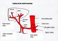
anterior & right from CA passes superior duodenum & portal vein enters liver @ porta hepatis |
|
|
The Common Hepatic Artery branches into what? |
Gastroduodenal Artery (GDA) |
|
|
Describe the course of the Gastroduodenal Artery |
- inferior along postero-medial duodenum - toward antero-lateral surface of pancreas head |
|
|
After branching of the Gastroduodenal, the Common Hepatic then becomes the.... |
proper hepatic artery |
|
|
The Proper Hepatic gives rise to the.... |
Right Gastric Artery |
|
|
The Proper Hepatic continues into the liver to branch into the..... |
Left Hepatic & Right Hepatic |
|
|
Where is the Right Hepatic located? |
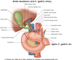
between the Common Bile Duct and Portal Vein |
|
|
What does the Right Hepatic give rise to? |
Cystic Artery |
|
|
Where does the Cystic Artery supply blood to? |
Gallbladder |
|
|
What is the Portal Triad? |
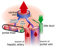
- hepatic artery - portal vein - bile duct |
|
|
Superior Mesenteric Artery |
2nd anterior branch of Abdominal Aorta |
|
|
Describe the course of the SMA |
- caudally - parallel to aorta - posterior to pancreatic neck - anterior to uncinate process of pancreas |
|
|
What does the SMA feed? |
portions of small intestines portions of large intestines pancreatic head |
|
|
Inferior Mesenteric Artery |
last major anterior branch of Abdominal aorta |
|
|
Describe the course of the IMA |

arises from slightly left aorta distal to renal arteries 3-4 cm superior to AO bifur |
|
|
Where does the IMA supply blood to? |
majority of large intestines rectum |
|
|
Which arteries arise from the terminal end of the Aorta? |
Left & Right Common Iliacs |
|
|
Each Common Iliac artery bifurcates into.... |
Internal and External Iliacs |
|
|
What do the Internal Iliacs supply blood to? |
pelvic viscera |
|
|
What do the External Iliacs supply blood to? |
lower limbs |
|
|
Where do the Renal Arteries arise from? |
the Lateral aspect of the Aorta |
|
|
Which arteries are located 1-1.5cm DISTAL to the Superior Mesenteric Artery? |
Renal Arteries |
|
|
What do the Renal arteries supply blood to? |
kidneys adrenal glands ureters |
|
|
Which Renal Artery is longer? |
Right |
|
|
How does the Right Renal Artery course by the IVC? |
posterior |
|
|
How does the Left Renal Artery course? |
Directly from Aorta to the renal hilum |
|
|
Describe the Intrarenal Vasculature |
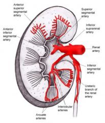
Renal Arteries -> Segmental Arteries AKA Lobar Arteries -> Interlobar Arteries -> Arcuate Arteries -> Interlobular Arteries -> Microscopic branches |
|
|
What is the function of the Microscopic branches in the kidney? |
nutrients/wastes |
|
|
Name the 3 common Arterial Variants |
1. duplicating renal arteries 2. CHA arises somewhere other than celiac trunk 3. SMA and celiac share the same trunk |
|
|
What is the Central / Great Vein? |
Inferior Vena Cava (IVC) |
|
|
How much longer is the IVC compared to the Aorta? |
7-8cm |
|
|
Where is the IVC located in the Abdomen? |
- right of midline - parallel & anterior to Aorta |
|
|
Where does the IVC originate? |
inferior abdomen by union of Right and Left iliac veins |
|
|
Are the Right and Left Iliac veins anterior or posterior to their Arteries? |
posterior |
|
|
General rule for positioning of Veins and Arteries in the lower body |
Veins will be posterior to their arteries (opposite from upper abdomen) |
|
|
What does the IVC do with the blood from the body regions inferior to the diaphragm? |
Returns the blood to the heart via the right atrium |
|
|
What happens to the IVC with respirations? |
diameter changes |
|
|
What is the diameter of the IVC? |
2.5-3.5cm |
|
|
Where does the Right Gonadal vein drain? |
Directly to IVC |
|
|
Where does the Left Gonadal vein drain? |
Left Renal Vein |
|
|
Which veins are the Largest branches of the IVC? |
Hepatic |
|
|
Where do the Hepatic Veins drain blood? |
from the liver INTO the IVC |
|
|
The Hepatic Vein consists of which 3 vessels? |
1. Right hepatic 2. Middle hepatic 3. Left hepatic |
|
|
Where do the Hepatic vessels enter the IVC? |
anterior aspect |
|
|
Where do the Renal Veins enter the IVC? |
lateral sides |
|
|
Which Renal Vein is longer? |
Left |
|
|
Why is the Left Renal Vein longer than the Right? |
it courses posterior to IVC and anterior to the aorta |
|
|
Right Renal Vein flow |
directly to IVC |
|
|
The Right kidney is less likely to get infections, why? |
it flows directly to the IVC (unlike the Left) |
|
|
Which Venous system is completely separate from the Systemic Circulation? |
Portal Venous System |
|
|
Do the venous branches that form the Portal vein directly join the IVC? |
NO |
|
|
Hepatopetal Flow |
into the liver |
|
|
Hepatofugal Flow |
away from the liver |
|
|
What are the 4 major vessels in the Portal Venous System? |
1. portal vein 2. splenic vein 3. superior mesenteric vein 4. inferior mesenteric vein |
|
|
Which vein forms posterior to pancreas from the junction of the SMV and SV? |
Portal Vein AKA Main Portal Vein |
|
|
Portal Confluence |
Superior Mesenteric Vein + Splenic Vein |
|
|
How long is the Portal Vein? |
5-7cm |
|
|
Does the Portal Vein pass anterior or posterior to the IVC? |
anterior |
|
|
The Portal Vein bifurcates into.... |
Right and Left portal veins |
|
|
The Right Portal Vein branches further into which sections? |
anterior and posterior |
|
|
The Left Portal Vein branches further into which sections? |
medial and lateral |
|
|
What 3 veins does the Portal Vein collect from? |
1. SMV 2. IMV 3. SV |
|
|
Where is the start of the Portal Vein? |
Portal Confluence |
|
|
Which vein joins with the SMV to form the Portal Vein? |
Splenic |
|
|
Which vein courses transversely to the right, superior to pancreatic tail and continues posterior to pancreatic body? |
Splenic |
|
|
Which vein is an important collateral route in patients with Hypertension? |
Coronary Vein AKA Left Gastric Vein |
|
|
Which vein courses cephalad from the small intestines, coursing anteromedial to join with the SV to form the Portal Vein? |
Superior Mesenteric |
|
|
How does the SMV run compared to the SMA? |
parallel and to the right of SMA |
|
|
Where does the SMV drain blood from? |
small intestines cecum colon |
|
|
Which vein passes posterior to the pancreatic neck and anterior to the uncinate process of pancreas? |
Superior Mesenteric |
|
|
Where does the Inferior Mesenteric vein drain blood from? |
Colon |
|
|
Which vein travels cephalad along the posterior left abdominal wall and joins the SV posterior to the pancreas? |
Inferior Mesenteric |
|
|
Where does the Portal System lay in the abdomen? |
Anterior to the Aorta and IVC |
|
|
What are the Venous Variants? |
- accessory hepatic veins - transposition of the IVC - azygous continuation of IVC - agenesis of IVC - retro-aortic left renal vein |
|
|
In which Venous Variant will you see the "medusas" sign sonographically and chronic DVTs? |
Agenesis of IVC |
|
|
Which Venous Variant is associated with polysplenia syndrome? |
Azygous continuation of IVC |
|
|
Name some of the indications for Abdominal Vascular exams |
- pulsatile abdomen mass - abdominal bruit - compromised flow in lower limbs - hypertension - swelling of lower limbs |
|
|
Fasting times for patients having an Abdominal Vascular Exam? (adults, kids, infants) |
adults - 8-12 hrs kids - 6 hrs infants - 4 hrs |
|
|
Transducer selection for patients getting an Abdominal Vascular exam? (average, thin/children, obese) |
average - 3-5 MHz thin/children - 4-6 MHz obese - 2 MHz |
|
|
Doppler Assessments in arteries - angle correction? |
less than or equal to 60 degrees |
|
|
Doppler Assessments in veins - angle correction? |
not required unless a velocity is required |
|
|
Normal Sonographic appearance of Sagittal Abdominal Aorta? |
- anechoic - tubular - echogenic walls |
|
|
Normal Sonographic appearance of Transverse Abdominal Aorta? |
- round - anechoic lumen - echogenic walls |
|
|
What are the normal AP diameter measurements of Aorta? (prox, distal, iliacs) |
prox - 2-2.5cm distal - 1.5-2cm iliacs - 1cm or less |
|
|
Which measurement is considered aneurysmal in the Aorta? |
greater than 3cm |
|
|
The Doppler signals in the Aorta are.... |
high resistance/impedance |
|
|
Any vessel that feeds an organ usually has what type of resistance? |
LOW |
|
|
Any vessel that feeds limbs usually has what type of resistance? |
HIGH |
|
|
Where does the Celiac Artery course from? |
inferior from anterior surface of Aorta |
|
|
The Celiac Artery looks like a "seagull/dove" in Transverse. What arteries branch off on each side? |
right - hepatic left - splenic |
|
|
What are the Doppler Signals at the trifurcation of the Celiac? |
Low resistant |
|
|
Prandial |
relating to eating |
|
|
Should the Doppler signals at the Celiac change in a post prandial patient? |
No |
|
|
3 parts of the Portal Triad |
1. hepatic artery 2. Portal vein 3. splenic vein |
|
|
Doppler signals in Hepatic Artery |
low resistant |
|
|
The Hepatic Artery may be visibly enlarged in a patient with... |
Portal Hypertension |
|
|
Which Artery is seen in the "seagull" and is tortuous in nature? |
Splenic Artery |
|
|
Doppler Signals in Splenic Artery and what do we see due to the tortuosity? |
low resistant - increased spectral broadening due to turbulence |
|
|
What does the SMA look like in Sagittal? How does it course? |
tubular - coursing inferior |
|
|
What does the SMA look like in Transverse? How does it course? |
round, anechoic with highly echogenic walls - Anterior to Aorta - Inferior to Splenic Vein |
|
|
Doppler Signals in SMA (pre and post prandial) |
can be high or low resistant - pre-prandial: High - post-prandial: Low |
|
|
Doppler Signals in IMA |
high resistant |
|
|
In what image are the Renal Arteries best seen? |
Transverse Aorta |
|
|
How does the RRA pass the IVC? |
posteriorly |
|
|
LRA takes direct course from Aorta to.... |
renal hilum |
|
|
Doppler Signal in Renal Arteries |
low resistant |