Reading...
![]()
Play button
![]()
Play button
![]()
Use LEFT and RIGHT arrow keys to navigate between flashcards;
Use UP and DOWN arrow keys to flip the card;
H to show hint;
A reads text to speech;
30 Cards in this Set
- Front
- Back
- 3rd side (hint)
|
abdomen
|
large oval cavity extending from diaphragm down to brim of the pelvis.
|
|
|
|
line a alba
|
tendinous seam of the flat muscles of the ventral wall.
|
|
|
|
Rectus abdominis
|
forms a strip extending the length of the midline and it's edge is soft and palpable. The muscles protect the organs.
|
|
|
|
viscera
|
All the internal organs are called this.
|
|
|
|
solid viscera
|
organs that maintain shape I.e. liver, pancreas, spleen, adrenal glands, kidneys, ovaries, uterus.
|
|
|
|
Hollow viscera
|
Not palpable. examples are stomach, gallbladder, sm. intestine, colon and bladder.
|
|
|
|
spleen
|
mass of lymphatic tissue on the posterolatetal wall of the abdominal cavity immediately under the diaphragm.
|
|
|
|
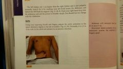
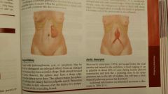
Aorta
|
left of midline in the upper part of the abdomin. Defends behind the peritoneum and 2 cm. below the umbilicus.
|
|
|
|
pancreas
|
soft lobulated gland behind the stomach. stretches obliquely across the posterior abdominal wall in the upper quadrant.
|
|
|
|
costovertebral angle
|
The 12th rib forms an angle with the vertebral column.
|
|
|
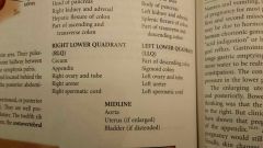
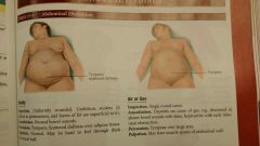
quadrants & midline.
|
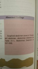
abnormal findings...
|
|
|
abnormal findings continued....
|
abnormal findings....
|
|
|
abnormal findings continued...
|
Auscultation bowel sounds. Movement of air and fluid through the small intestines. stomach growling is termed "borborgmus."
|
|
|
|
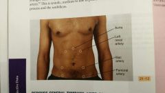
vascular sounds
|
Pretense of bruits. using firm pressure check over the aorta, arteries, iliac and femoral arteries. especially with people with hypertension
|
|
|
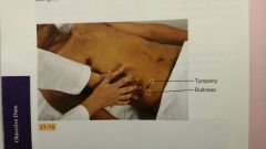
Percuss general tympany - liver span and splenic dullness.
|
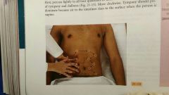
First lightly percuss four quadrants. move clockwise. tympany should predominant because air in the intestines rises to the surface when a person is supine.
|
|
|
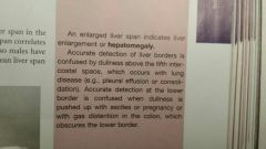
Liver Span
|
Percuss to Map out boundaries of certain organs. measure height of liver in the right midclavicular line. begin in area of lung resonance and percuss down the interspaces until sound changes to dull quality. usually 5th intercostal space. Then find abdominal tympany and purcuss up the midclavicular line. mark where sound changes from tympany to dull sound - normally right costal margin. Normal liver span is 6-12 cm. height of liver span correlates with height of the person. Taller people have larger livers. Men have larger livers than woman of same height. Average span (Men 10.5 cm - woman 7 cm.)
|
|
|
|
Normal Findings
|
People with chronic emphysema the liver is displaced downward by the hyperinflation of the lungs. Overall span is still within normal limits.
|
|
|
Abnormal Findings...
|
Splenic dullness observed over 9th to 11th intercostal space. normally not wider than 7 cm. in an adult and should not encroach on the normal tympanic over the gastric air bubble. Ask pt. to take deep breath - normally Tynan remains through full inspiration.
|
|
|
Abnormal findings....
|
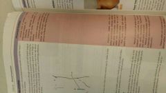
Fluid Wave - Stand on pt. right side place Ulmer edge of examiner hand or pt. own hand on firmly on abdomin midline. Place hand on pt. left flank, with right hand reach across abdomin and give flank a firm strike.
|
|
|
|
Normal range of findings...
|
Test for acites - Shifting dullness. supine position fluid will shift to the flanks displacing air filled bowel. If fluid is present sound change from tympany to dull. Mark spot of dullness.
|
|
|
Abnormal findings....
|
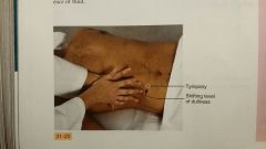
Normal Findings...Voluntary guarding occurs when a person is cold or ticklish. Muscle will relax on exhalation.
|
|
|
Abnormal Findings....
|
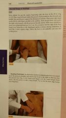
Hooking Technique - Palpate.
|
|
|

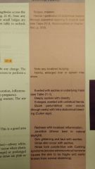
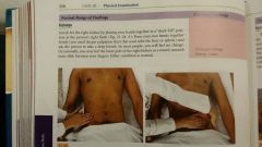
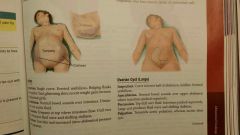
Spleen is NOT palpable. Must be enlarged 3 times normal size to be felt.
|
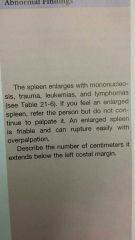
When enlarged the spleen slides out and bumps your fingertips. can be large enough to extend into lower quadrants. Note: Abnormal findings to the right.
|
|
|
Kidneys
|
Aorta
|
|
|
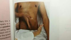
Rebound tenderness (Blumberg Sign)
|
Inspiratory Arrest (Murphy Sign)
|
|
|
|
Infant Normal Findings
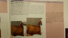
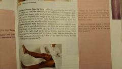
abdominal should be symmetric, although two bulges are common. Note umbilical hernia. usually appears 2 to 3 weeks abs is prominent. |
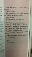
Diastasis recti - A separation of the rectus muscles with visible bulge along midline. Common in black infants. Note peristalsis. Auscultation, percussion and palpation. Note: Abdominal findings.
|
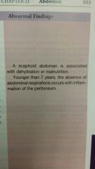
Abnormal Findings....
|
|
Abnormal Findings...
|
Abnormal Findings..?
|
|
|
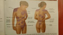
Common sites for referred abdominal pain
|
|
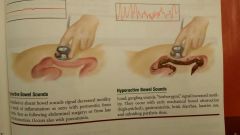
Bowel sounds
|
|
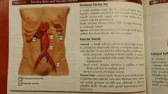
Friction Rubs and Vascular Sounds
|
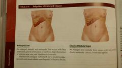
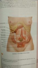
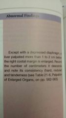
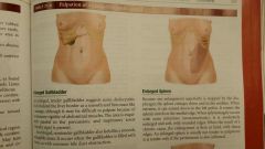
Palpation of Enlarged Organs
|
Continued....
|
|
Continued...
|
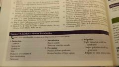
Checklist Abdominal Examination
|
|