![]()
![]()
![]()
Use LEFT and RIGHT arrow keys to navigate between flashcards;
Use UP and DOWN arrow keys to flip the card;
H to show hint;
A reads text to speech;
40 Cards in this Set
- Front
- Back
|
The organs and structures that make up the female reproductive system are? |
uterus ovaries fallopian tubes breasts |
|
|
Uterus |
|
|
|
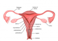
Female Reproductive System |
|
|
|
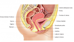
Position and relationships of the uterus |
• Usually anteverted and anteflexed • Lies supero-posterior to urinary bladder and utero-vesical pouch. • Lies anterior to the rectum, recto-uterine pouch• Inferior to the small bowel • Medial to the ovaries • Superior to the vagina. |
|
|
In which abdominal area is the uterus located? |
Hypogastric |
|
|
Functions of the uterus |
• Part of the pathway for sperm to reach uterine tubes. – Sperm enter via vagina- cervical canal – uterine body • Site for implantation of embryo and pregnancy.– If successful fertilisation of released ova results in development of embryo • Site of menstruation (monthly cycle) – Influenced by hormones oestrogen and progesterone |
|
|
Retroverted Uterus |
A retroverted uterus can be a genetic trait and occurs in 20% of women worldwide Remember that anatomical positions of organs are sometimes subject to slight change as a result of conditions |
|
|
The uterus is supported in the pelvis by: |
• Broad ligament • Utero-sacral ligament • Lateral cervical ligaments. • Round ligament. • Muscles of the perineum – especially Levator ani muscle |
|
|
Gross Anatomy of the Uterus |
The uterus can be divided into 4 sections:- • Fundus • Body • Cervix • Fallopian tubes (Salpinges) |
|
|
Microscopic anatomy |
3 layers of tissue make up the uterus • Perimetrium (outer)- serous layer = peritoneum • Myometrium (middle)- muscle • Endometrium (inner) – important for menstrual cycle – one layer is shed if implantation of ovum does not occur – If ovum implants, it thickens – affected by oestrogen and progesterone levels |
|
|
Blood Supply – the uterus is well supplied |
• Internal iliac arteries • Uterine arteries • Arcuate arteries • Radial arteries • Straight arterioles • Spiral arterioles |
|
|
Venous and Lymph drainage |
Venous - • Uterine veins • Internal iliac veins Lymph drainage - • Fundus to para-aortic nodes alongside ovarian arteries. • Body and cervix to int. and ext. iliac nodes • Lower vagina to inguinal nodes |
|
|
Nerve supply |
• Sympathetic and parasympathetic innervation from pelvic and hypogastric plexuses. • Smooth muscle of the myometrium only receives sympathetic innervation. • Uterine pain often felt in lower back or perineum. |
|
|
Location of the fallopian tubes and ovaries |
• The fallopian tubes are wider near their external ends (ampulla). • The ends have fimbriae– projections help to waft the released oocyte into fallopian tubes when stimulated by hormones |
|
|
What are the functions of the ovaries? |
A. Produce oestrogen B. Produce progesterone C. Produce secondary oocytes D. Produce oestrogen and secondary oocytes |
|
|
Ovaries - gross anatomy and function |
• Paired organs- resemble “unshelled almonds” • Either side of uterus, adjacent to fimbrae of fallopian tubes. • Each ovary has a hilus – the point of entry /exit for blood vessels and nerves. • Function:- • To produce secondary oocytes • To produce oestrogen and progesterone |
|
|
Blood, lymph, nerve supply and drainage of the ovaries |
• Arteries from abdominal aorta. • Right ovary drains to inferior vena cava • Left ovary drains to left renal vein. • Lymphatic drainage follows blood vessels to lateral aortic and pre-aortic lymph nodes. • Nerve supply – some conditions can irritate the obturator nerve |
|
|
Irradiation |
• In our professions we must be aware of the risks associated with gonad exposure through – Ionising radiation (Diagnostic Imaging) – Side effects of Radiotherapy • Somatic effects • Genetic effects |
|
|
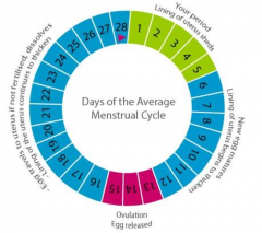
Average Menstrual Cycle |
|
|
|
At what stage of pregnancy is the foetus most sensitive to the effects of radiation? |
0-12 weeks |
|
|
Female Reproductive System Imaging |
• Ultrasound • Magnetic Resonance Imaging (MRI) • Computerised Tomography (CT) • Fluoroscopy – Hysterosalpinography |
|
|
Polycystic Ovary |
• Most common female endocrine disorder 14-44 years • Absent/irregular/heavy periods • Ovaries produce excessiveandrogens • Multiple immature follicles (NOT cysts as such) where growth has arrested – No ovulation |
|
|
Ovarian Carcinoma |
• Often initially diagnosed as irritable bowel • Ascites • Hereditary breast-ovariancancer syndrome • Ethnic origins – Ashkenazi Jews |
|
|
How many UK women were diagnosed with Ca Uterus in 2015? |
8500 |
|
|
Cervical Cancer Screening |
• Humanpapillomavirus link • 12th most common cancer in UK• Vaccines for teenage girls • PAPsmearsscreening – 42% reduction 1988-97 |
|
|
Ectopic Pregnancy |
• Foetusimplantoutside uterus • Can occur Fallopian tube (90%), cervix, ovary, abdomen |
|
|
Which is the modality of choice to locate a “lost” IUCD? |
Ultrasound |
|
|
The Breasts |
• The breasts are also known as the mammary glands. • They are present in both males and females and are modified sudoriferous glands |
|
|
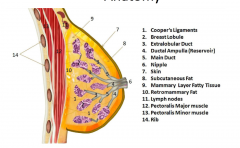
Breast Anatomy |
|
|
|
Breast Function |
• Function is to produce milk - lobes and ducts facilitate this process • They are responsive to certain hormones • They changes with monthly cycle and with age |
|
|
Anatomical position and structure |
Position • Over lie muscle - pectoralis major • Lies anterior to ribs (2nd-6th) Tissues • Glandular tissue (in lobes)– responsive to hormones • Adipose tissue (fat) is main tissue)lies between lobes • Fibrous tissue forms ligaments and strands to support the lobules |
|
|
Support, blood supply and drainage |
• Suspensory ligaments (Coopers ligaments) • Internal mammary arteries and thoracic branches of axillary artery, intercostal arteries. • Network of veins around the base of nippIe drain to axillary and internal mammary veins. |
|
|
Hormonal influences |
At puberty – ovarian and gonadotrophic hormones During pregnancy –oestrogen and progesterone prolactin, thyroid and adrenocortical hormones During and after childbirth - oxytocin |
|
|
Lymphatic drainage |
• Plexus of vessels (networks) in each breast. • One plexus lies on surface of pectoralis major • Each plexus drains to lymph nodes outside of breasts • Lateral aspect drains to large group of nodes in axillae • Medial aspect to nodes in internal mammary region (along sternum) • Some nodes above and below clavicle |
|
|
Drainage of lymph |
• Some lymph crosses over midline to contralateral nodes • Mainly the left breast ultimately drains to the thoracic duct (left lymphatic) and the right to the right lymphatic duct |
|
|
Clinical applications for you |
• You need to know why we screen for breast cancer • You need to know why patients have radiotherapy for breast cancer |
|
|
Approximately how many women are diagnosed with Breast Cancer in the UK each year? |
50,000 |
|
|
Incidence |
• 50,000 women are diagnosed each year with breast cancer and around 12,000 die of it • Family History – BRCA1, BRCA2 gene • Less than 1% of breast cancers occur in men (about 300 each year). |
|
|
How many men are diagnosed with Breast Cancer in the UK each year? |
300 |
|
|
Breast development and structural changes |
• Adolescent breast: greater amount of developing glandular tissue than fat. • Adult breast: less dense than adolescent and has a greater fat content. • Menopausal: Fatty or dense and fibrous, stretched suspensory ligaments |