Reading...
![]()
Play button
![]()
Play button
![]()
Use LEFT and RIGHT arrow keys to navigate between flashcards;
Use UP and DOWN arrow keys to flip the card;
H to show hint;
A reads text to speech;
163 Cards in this Set
- Front
- Back
|
2 Types of Neural Tissue Cells
|
Neurons
Neuroglia |
|
|
Cells that can send and receive signals
|
Neurons
|
|
|
Cells that support and protect neurons
|
Neuroglia
|
|
|
Neuroglia AKA
|
glial cells
|
|
|
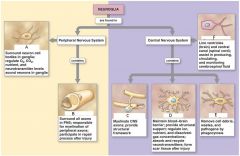
2 Anatomical Divisions of the Nervous System
|
Central Nervous System (CNS)
Peripheral Nervous System (PNS) |
|
|
System that consists of the spinal cord and brain
|
Central Nervous System
|
|
|
System that contains Neural Tissue, Connective Tissues & Blood Vessels
|
Central Nervous System
|
|
|
Functions of this system
processes and coordinates: a) sensory data: from inside & outside body b) Motor commands c) higher functions of the brain |
Central Nervous System
|
|
|
System which includes all neural tissue outside the CNS
|
Peripheral Nervous System
|
|
|
Functions of this System:
1) Deliver sensory info to CNS 2) Carry motor commands to peripheral tissues & systems |
Peripheral Nervous System
|
|
|
Bundles of axons with connective tissues and blood vessels
|
Nerves (peripheral nerves)
|
|
|
These carry sensory information and motor commands in PNS
|
Nerves
|
|
|
Nerves which connect to brain
|
cranial nerves
|
|
|
Nerves which attach to spinal cord
|
spinal nerves
|
|
|
2 Functional Divisions of the PNS
|
Afferent Division
Efferent Division |
|
|
Function of this division of the PNS:
carries sensory info from PNS receptors TO CNS |
Afferent Division
|
|
|
Function of this division of the PNS:
Carries motor commands FROM CNS to PNS muscles and glands |
Efferent Division
|
|
|
Afferent Division: These
a) detect changes or respond to stimuli b) neurons and specialized cells c)complex sensory organs (eg. eyes, ears) |
Receptors
|
|
|
Afferent Division: These
a) respond to efferent signals b) cells and organs |
Effectors
|
|
|
2 Systems in the efferent division of the PNS
|
1) Somatic Nervous System (SNS)
2) Autonomic Nervous System (ANS) |
|
|
This PNS system controls skeletal muscle contractions: voluntary and involuntary (reflexes)
|
Somatic Nervous Stystem
|
|
|
This PNS system:
controls subconscious actions: contractions of smooth/ cardiac muscle and glandular secretions |
Autonomic Nervous System
|
|
|
2 Divisions of the Autonomic Nervous System
|
1) Sympathetic division
2) Parasympathetic Division |
|
|
This division of the Autonomic Nervous System has a stimulating effect.
|
sympathetic division
|
|
|
This division of the Autonomic Nervous System has a relaxing effect.
|
parasympathetic division
|
|
|
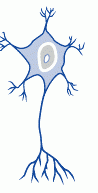
This type of neuron is common in the CNS
|
multipolar neuron
|
|
Name this Neuron
|
Multipolar Neuron
|
|
|
Type of neuron with:
a) cell body (soma) b) short, branched dendrites c) long, single axon |
multipolar neuron
|
|
|
Location of these in neuron
a) large nucleus and nucleolus b) perikaryon (cytoplasm) c) mitochondria (produce ATP) d) RER and ribosomes (produce neurotransmitters) e) Cytoskeleton |
Cell body
|
|
|
This part of the neuron cell body is made of neurofilaments, neurotubules and neurofibrils.
|
The cytoskeleton
|
|
|
Bundles of neurofilaments that provide support for dendrites and axon
|
Neurofibrils
|
|
|
Provide support for dendrites and axon
|
Neurofibrils
|
|
|
Dense areas of RER & ribosomes that make neural tissue appear gray (gray matter)
|
Nissl bodies
|
|
|
This part of the neuron receives info from other neurons and is 80-90% of the neurons surface
|
Dendritic spines
|
|
|
Long part of neuron that carries electrical signal (action potential) to target
**critical to function |
Axon
|
|
|
Structures of _________
a) axoplasm b) axolemma c) axon hillock d) initial segment |
axon structures
|
|
|
cytoplasm of the axon
|
axoplasm
|
|
|
These are located in which part of the neuron?
a) neurotubules b) neurofibrils c) enzymes d) organelles |
axoplasm
|
|
|
Specialized cell membrane which covers axoplasm
|
axolemma
|
|
|
Thick section of axon cell body which attaches initial segment
|
axon hillock
|
|
|
structure of axon which attaches to axon hillock
|
initial segment
|
|
|
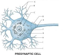
A) Dendritic Branches
B) Bissl Bodies (RER & free ribosomes) C) Mitochondrion D) Axon Hillock E) Initial Segment of Axon F) Golgi Apparatus G) Neurofilament H) Nucleus I) Nucleolus J) Dendrite |
|
|
Area where a neuron communicates w/ another cell
|
synapse
|
|
|
neuron that sends message
|
presynaptic cell
|
|
|
neuron that receives message
|
postsynaptic cell
|
|
|
the small gap that seperates the presynaptic membrane and the postsynaptic membrane
|
synaptic cleft
|
|
|
expanded area of axon of the presynaptic neuron
|
synaptic knob
|
|
|
Contains synaptic vesicles of neurotransmitters
|
synaptic knob
|
|
|
chemical messengers which
a) are released at presynaptic cleft b) affect receptors of postsynaptic membrane c) are broken down by enzymes d) are reassembled at synaptic knob |
neurotransmitters
|
|
|
2 Types of synapses
|
1) neuromuscular junction
2) neuroglandular junction |
|
|
synapse between neuron and muscle
|
neuromuscular junction
|
|
|
synapse between neuron and gland
|
neuroglandular junction
|
|
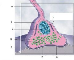
Structures of a typical synapse
|
A) Telodendrion
B) Synaptic Bulb C) Mitochondrion D) Synaptic Vesicles E) Precynaptic membrane F) Postsynaptic membrane G) Synaptic Cleft H) Endoplasmic Reticulum |
|
|
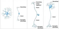
Four Structural Classifications of Neurons
|
Anaxonic neurons
Bipolar neurons Unipolar neurons Multipolar neurons |
|
|
Type of neuron found in brain and sense organs
|
Anaxonic neurons
|
|
|
Type of neuron found in special sensory organs (sight, smell, hearing)
|
Bipolar neurons
|
|
|
Type of neuron found in sensory neurons of PNS
|
Unipolar neurons
|
|
|
Neurons common in the CNS and
Include all skeletal muscle motor neurons |
Multipolar neurons
|
|
|
Name the neuron
Small All cell processes look alike |
Anaxonic Neurons
|
|
|
Name the Neuron
Are small One dendrite, one axon |
Bipolar Neurons
|
|
|
Name the Neuron
Have very long axons Fused dendrites and axon Cell body to one side |
Unipolar Neurons
|
|
|
Name the Neuron
Have very long axons Multiple dendrites, one axon |
Multipolar Neurons
|
|
Name the Neuron
|
Anaxonic
Bipolar Unipolar Multipolar |
|
|
3 Functional Classifications of Neurons
|
Sensory neurons
Motor neurons Interneurons |
|
|
Functions of which type of Neuron
1) Monitor internal environment (visceral sensory neurons) 2) Monitor effects of external environment (somatic sensory neurons) |
Sensory Neuron Functions
|
|
|
Structures of which type of neuron
1) Unipolar 2) Cell bodies grouped in sensory ganglia 3) Processes (afferent fibers) extend from sensory receptors to CNS |
Sensory Neuron Structures
|
|
|
Neuron that carries instructions from CNS to peripheral effectors Via efferent fibers (axons)
|
Motor Neurons
|
|
|
Two major efferent systems
|
Somatic nervous system (SNS
Autonomic (visceral) nervous system (ANS) |
|
|
This efferent system includes all somatic motor neurons that innervate skeletal muscles
|
Somatic nervous system
|
|
|
This efferent system:
visceral motor neurons innervate all other peripheral effectors (e.g., smooth muscle, cardiac muscle, glands, adipose tissue) |
Autonomic (visceral) nervous system
|
|
|
Most of these neurons are located in brain, spinal cord, and autonomic ganglia between sensory and motor neurons
|
Interneurons
|
|
|
These neurons are responsible for:
1) distribution of sensory information 2) coordination of motor activity |
Interneurons
|
|
|
These neurons are involved in higher functions
Memory, planning, learning |
Interneurons
|
|
|
Half the volume of the nervous system is made of?
|
Neuroglia
|
|
|
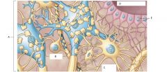
Four Types of Neuroglia in the CNS
|
Ependymal cells
Astrocytes Oligodendrocytes Microglia |
|
|
cells with highly branched processes; contact neuroglia directly
|
Ependymal cells
|
|
|
large cell bodies with many processes
|
Astrocytes
|
|
|
smaller cell bodies with fewer processes
|
Oligodendrocytes
|
|
|
smallest and least numerous neuroglia with many fine-branched processes
|
Microglia
|
|
|
These neuroglia cells form epithelium called ependyma
|
Ependymal cells
|
|
|
These neuroglia cells Line central canal of spinal cord and ventricles of brain
|
Ependymal cells
|
|
|
These neuroglia cells secrete cerebrospinal fluid (CSF)
|
Ependymal cells
|
|
|
These neuroglia cells have cilia or microvilli that circulate CSF
|
Ependymal cells
|
|
|
These neuroglia cells monitor CSF
|
Ependymal cells
|
|
|
These neuroglia cells contain stem cells for repair
|
Ependymal cells
|
|
|
These neuroglia cells Maintain blood–brain barrier (isolates CNS)
|
Astrocytes
|
|
|
These neuroglia cells Create three-dimensional framework for CNS
|
Astrocytes
|
|
|
These neuroglia cells Repair damaged neural tissue
|
Astrocytes
|
|
|
These neuroglia cells Guide neuron development
|
Astrocytes
|
|
|
These neuroglia cells Control interstitial environment
|
Astrocytes
|
|
|
Processes contact other neuron cell bodies
|
Oligodendrocytes
|
|
|
These neuroglia cells Wrap around axons to form myelin she
|
Oligodendrocytes
|
|
|
A) satellite cells
B) Schwann cells C) Oligodendrocytes D) Astrocytes E) Microglia F) Ependymal Cells |
|
Neuroglia in the CNS
|
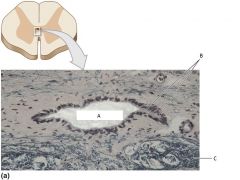
A) Central Canal
B) Ependymal Cells C) Gray Matter |
|
Neuroglia in the CNS
|
A) Gray Matter
B) Neurons C) Microglial Cell D) Central Canal E) Ependymal Cells |
|
|
Oligodendrocyte feature which:
1) increases speed of action potentials 2) insulates "_____" axons 3) makes nerves appear white |
myelination
|
|
|
myelinated segments of axon in Oligodendrocytes
|
internodes
|
|
|
Oligodendrocytes
gaps between internodes where axons may branch |
nodes (also called nodes of Ranvier)
|
|
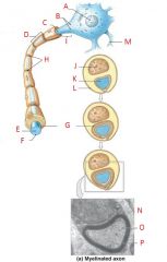
Schwann Cells and Peripheral Axons
|
A) nucleus
B) axon hilock C) Axon D) myelynated internode E) Axon F) Axolemma G) Myelin covering internode H) nodes I) initial segment (unmyelinated) J) Schwann cell of nucleus K) axon L) neurilemma M) dendrite N) neurilemma O) axon P) myelin |
|
|
Type of Neuroglia in the CNS
Myelination White matter Gray matter |
Oligodendrocytes
|
|
|
regions of CNS with many myelinated nerves
|
White matter
|
|
|
unmyelinated areas of CNS
|
Gray matter
|
|
|
Types of Neuroglia in the CNS
Migrate through neural tissue Clean up cellular debris, waste products, and pathogens |
Microglia
|
|
|
Masses of neuron cell bodies surrounded by neuroglia
found in the PNS |
Ganglia
|
|
|
Also called amphicytes
|
Satellite cells
|
|
|
Surround ganglia and regulate environment around neuron
|
Satellite cells
|
|
|
Also called neurilemmocytes
|
Schwann cells
|
|
|
Form myelin sheath (neurilemma) around peripheral axons
|
Schwann cells
|
|
|
____#____ Schwann cell sheaths ___#____ segment of axon and _________Schwann cells sheath entire axon
|
1
1 many |
|
|
These cells perform all communication, information processing, and control functions of the nervous system
|
neurons
|
|
|
These cells preserve physical and biochemical structure of neural tissue and are essential to survival and function of neurons
|
Neuroglia
|
|
|
All plasma (cell) membranes produce electrical signals by what?
|
ion movements
|
|
|
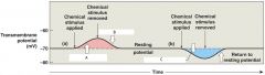
The transmembrane potential of resting cell
|
Resting potential
|
|
|
an electrical impulse
|
Action potential
|
|
|
Produced by graded potential
|
Action potential
|
|
|
Propagates along surface of axon to synapse
|
Action potential
|
|
|
Releases neurotransmitters at presynaptic membrane
Produces graded potentials in postsynaptic membrane |
Synaptic activity
|
|
|
Response (integration of stimuli) of postsynaptic cell
|
Information processing
|
|

Overview of Neural Activities
|
A) Resting potential
B) graded potential C) action potential D) synaptic activity E) information processing |
|
|
Three Requirements for Transmembrane Potential
|
1) Concentration gradient of ions (Na+, K+)
2) Selectively permeable through channels 3) Maintains charge difference across membrane (resting potential –70 mV) |
|
|
2 types of Passive Forces Acting Across the Membrane
|
Chemical gradients
Electrical gradients |
|
|
Name the gradient type
Concentration gradients of ions (Na+, K+) |
Chemical gradients
|
|
|
Name the gradient type
Separate charges of positive and negative ions Result in potential difference |
Electrical gradients
|
|
|
Check summary table 12-1 slide 50 Ch. 12
|
Check summary table 12-1 slide 50 Ch. 12
|
|
|
What happens in response to temporary changes in membrane permeability resulting from opening or closing specific membrane channels
|
Changes in Transmembrane Potential; Transmembrane potential rises or falls
|
|
|
Channels that are always open permeability changes with conditions
|
Passive channels (also called leak channels)
|
|
|
Channels that open and close in response to stimuli
at resting potential, most of these channels are closed |
Active channels (also called gated channels)
|
|
|
Three Classes of Gated Channels
|
Chemically gated channels
Voltage-gated channels Mechanically gated channels |
|
|
Channels that open in presence of specific chemicals (e.g., ACh) at a binding site found on neuron cell body and dendrites
|
Chemically gated channels
|
|
|
These channels:
1) respond to changes in transmembrane potential 2) Have activation gates (opens) and inactivation gates (closes) 3) Characteristic of excitable membrane 4) Found in neural axons, skeletal muscle sarcolemma, cardiac muscle |
Voltage-gated channels
|
|
|
These channels:
Respond to membrane distortion Found in sensory receptors (touch, pressure, vibration) |
Mechanically gated channels
|
|
|
Where are Chemically gated channels found?
|
on neuron cell body and dendrites
|
|
|
Where are Voltage-gated channels found?
|
in neural axons, skeletal muscle sarcolemma, cardiac muscle
|
|
|
Where are Mechanically gated channels found?
|
in sensory receptors (touch, pressure, vibration)
|
|
|
Also called local potentials
|
Graded Potentials
|
|
|
Name the type of potential
Changes in transmembrane potential that cannot spread far from site of stimulation |
Graded Potentials
|
|
|
Any stimulus that opens a gated channel produces what type of potential?
|
graded potential
|
|
|
Opening sodium channel produces graded potential:
resting membrane exposed to chemical sodium channel opens sodium ions enter the cell transmembrane potential rises What occurs? |
depolarization
|
|
|
A shift in transmembrane potential toward
0 mV |
Depolarization
|
|
|
Depolarization occurs when
movement of ______ through channel produces _________ depolarizes nearby ___________ (graded potential) change in potential is __________to stimulus |
Na+
local current plasma membrane proportional |
|
|
check slide 59 table 12-2
|
check slide 59 table 12-2
|
|
|
When the stimulus is removed, transmembrane potential returns to normal
|
Repolarization
|
|
|
Increasing the negativity of the resting potential
Result of opening a potassium channel Opposite effect of opening a sodium channel Positive ions move out, not into cell |
Hyperpolarization
|
|
|
A) depolarization
B) repolarization C) hyperpolarization |
|
|
Effects of graded potentials
1) At cell dendrites or cell bodies 2) At motor end plate |
1) trigger specific cell functions
2) releases ACh into synaptic cleft |
|
|
What is the All-or-none principle in Initiating Action Potential?
|
1) Action potential is either triggered, or not
2) If a stimulus exceeds threshold amount: the action potential is the same no matter how large the stimulus |
|
|
Four Steps in the Generation of Action Potentials
|
Step 1: Depolarization to threshold
Step 2: Activation of Na+ channels Step 3: Inactivation of Na+ channels, activation of K+ channels Step 4: Return to normal permeability |
|
|
During Step 2 (Activation of Na+ channels) of generation of action potentials, what happens?
|
Rapid depolarization
Na+ ions rush into cytoplasm Inner membrane changes from negative to positive |
|
|
How does Step 3 (Inactivation of Na+ channels, activation of K+ channels) of generation of action potentials happen?
|
At +30 mV
Inactivation gates close (Na+ channel inactivation) K+ channels open Repolarization begins |
|
|
In the Generation of Action Potentials, how does
Step 4: "Return to normal permeability" happen? |
K+ channels begin to close:
when membrane reaches normal resting potential (–70 mV) K+ channels finish closing: membrane is hyperpolarized to -90 mV transmembrane potential returns to resting level: action potential is over |
|
|
the amount of time it takes for an excitable membrane to be ready for a second stimulus once it returns to its resting state following excitation
|
refractory period
|
|
|
Name the time period:
Sodium channels open or inactivated No action potential possible |
Absolute refractory period
|
|
|
Name this time period:
Membrane potential almost normal Very large stimulus can initiate action potential |
Relative refractory period
|
|
|
_____________ of Action Potentials
Moves action potentials generated in axon hillock Along entire length of axon A series of repeated actions, not passive flow |
Propagation of Action potentials
|
|
|
Two methods of propagating action potentials
|
1) Continuous propagation: unmyelinated axons
2) Saltatory propagation: myelinated axons |
|
|
Name the type of propagation
1) Action potential along myelinated axon 2) faster and uses less energy than continuous propagation 3) Myelin insulates axon, prevents continuous propagation 4) Local current “jumps” from node to node 5) Depolarization occurs only at nodes |
Saltatory Propagation
|
|
|
Figure 12–16 Saltatory Propagation along a Myelinated Axon (Steps 1 and 2).
|
Figure 12–16 Saltatory Propagation along a Myelinated Axon (Steps 1 and 2).
|
|
|
What can effect action potential speed?
Larger equals what? |
Axon diameter
Larger diameter= lower resistance |
|
|
Any synapse that releases ACh is what type?
|
Cholinergic Synapses
|
|
|
4 Locations of Cholinergic Synapses
|
1) All neuromuscular junctions with skeletal muscle fibers
2) Many synapses in CNS 3) All neuron-to-neuron synapses in PNS 4) All neuromuscular and neuroglandular junctions of ANS parasympathetic division |
|
|
Events at a Cholinergic Synapse
1) ______ arrives, depolarizes __________ 2) ______ enter ________, trigger _________ of Ach 3) ACh binds to _____, depolarizes __________ membrane 4) AChE breaks ACh into _______and _________ |
1) Action potential, synaptic knob
2) Calcium ions, synaptic knob, exocytosis 3) receptors, postsynaptic membrane 4) acetate, choline |
|
|
Figure 12–17 slides 80-83
|
Figure 12–17 slides 80-83
|
|
|
Other than acetylcholine, name 4 important Neurotransmitters
|
Norepinephrine (NE)
Dopamine Serotonin Gamma aminobutyric acid (GABA) |