![]()
![]()
![]()
Use LEFT and RIGHT arrow keys to navigate between flashcards;
Use UP and DOWN arrow keys to flip the card;
H to show hint;
A reads text to speech;
35 Cards in this Set
- Front
- Back
|
4. Type of the epithelial lining of the digestive tract. |
Small Intestine Serosa •Serosa –VisceralPeritoneum •Simple squamousepithelial •Connectivetissue |
|
|
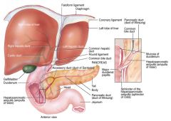
7. Functions of the gall bladder. |
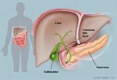
The gallbladder is a small pouch that sits just under the liver. The gallbladder stores bile produced by the liver. After meals, the gallbladder is empty and flat, like a deflated balloon. Before a meal, the gallbladder may be full of bile and about the size of a small pear.In response to signals, the gallbladder squeezes stored bile into the small intestine through a series of tubes called ducts. Bile helps digest fats so that they can be broken down by pancreatic lipase, but the gallbladder itself is not essential. Removing the gallbladder in an otherwise healthy individual typically causes no observable problems with health or digestion yet there may be a small risk of diarrhea and fat malabsorption. |
|
|
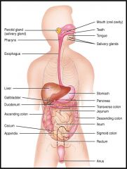
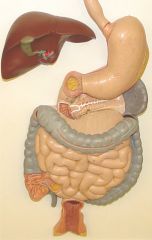
1. Knowthe structures/parts of the digestive system and digestive tract: a. Componentsof the digestive tract and accessory digestive organs. |
•Thegastrointestinal tract –Atube that extends from the mouth to the anus. Includes the: •Mouth •Pharynx •Esophagus •Stomach •Small intestine •Large intestine |
|
|
1. Know the structures/parts of the digestive system and digestive tract: b. accessory digestive organs. |
•Accessoryorgans –Salivary glands –Liver –Pancreas |
|
|
2. Define the following physiological terms: a. Gustation. |
the action or faculty of tasting |
|
|
2. Define the following physiological terms: b. Mastication. |
Mastication or chewing is the process by which food is crushed and ground by teeth. It is the first step of digestion, and it increases the surface area of foods to allow more efficient break down by enzymes. During the mastication process, the food is positioned by the cheek and tongue between the teeth for grinding. •Food masticated –Involuntary chewing reflex •stretch receptors in cheek, gums and tongue•Mixed with saliva •Food becomes a bolus |
|
|
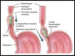
2. Define the following physiological terms: c. Deglutition. |
•Threestages 1.Boluspushed to back of oral cavity by tongue 2.Reflexbegins 1.Uvularises a.Closesnasopharynx 2.Tonguemoves to roof of mouth a.Closesoropharynx 3. Epilglottismoves down, larynx moves up a.Closes larynx 3.Peristalsis pushes food down esophagus
SWALLOWING(DEGLUTITION) •Reflex coordinated in medualla • Crainial nerves •Trigeminal •Facial •Glossopharyngeal •Hypoglossal •22muscles in •mouth, pharynx, and esophagus |
|
|
2. Define the following physiological terms: d. Ingestion. |
Ingestion is the consumption of a substance by an organism. In animals, it normally is accomplished by taking in the substance through the mouth into the gastrointestinal tract, such as through eating or drinking. |
|
|
2. Define the following physiological terms: e. Defecation |
the discharge of feces from the body. |
|
|
3. Anatomy and physiology of: a. Esophagus. |
•Musculartube •25cm (10 inches) long •Linedwith stratified squamous epithelium •Piercesthe diaphragm at hiatus –Upper esophageal sphincter –Lower esophageal sphincter •Belowdiaphragm •Opensduring swallowing •Allowsfood to pass into stomach |
|
|
3. Anatomy and physiology of: b. Stomach. |
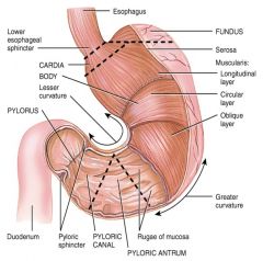
•Functions –Storage –Mechanicaland Chemical digestion •Layers –Serosa -outside –Muscularus -3 layers (only in stomach) •Longitudinal •Circular •Oblique Whydoes the stomach have 3 layers of muscle? –Mucosa-lining Rugae-folds |
|
|
3. Anatomy and physiology of: c. Small intestines |
•20feet long----1 inch in diameter •Largesurface area for majority of absorption •3Regions –Duodenum •10inches –Jejunum •8feet –Ileum •12feet –Ileocecal valve –Plica circularis •permanentfolds •Increasesurface area •cannotstretch out like rugae in stomach |
|
|
3. Anatomy and physiology of: d. Large intestines. |
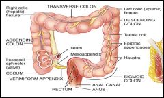
Functions –Formationof feces –Absorption •5feet long |
|
|
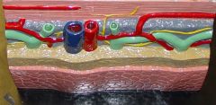
Lower Esophagus |
|
|
|
Small Intestine Layers |
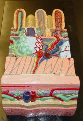
•Serosa •Muscularis •Submucosa •Mucosa |
|
|
5. Functions of the Liver. |
FUNCTIONS•Metabolism of –Carbohydrates –Lipids –Proteins•Detoxificationof blood •Excretion of bilirubin •Storageof vitamins andminerals•Productionof bile |
|
|
Functions of the Liver (Card 2) |
Functions of the Liver Card 2 1.Metabolism of nutrients a. Sugars, amino acids, lipids 1.Usedto build other molecules 2.Storedfor later use b.Substances built and released fromthe liver include 1. Plasma proteins 2.Glucose 3.Cholesterol 4. Bile |
|
|
Functions of the Liver (Card 3) |
Functions of the Liver (Card 3) 2.Detoxifies blood •Removesand breaks down •Hormones,toxins, bile pigments, drugs •ConvertsNH3 (ammonia)to urea •Alcohol •Cirrhosis- Scar tissue -Alcohol(3-4 drinks a day) -Hepatitis -Nonalcoholicfatty liver Disease - Fatty acid build upfrom obesity and diabetes |
|
|
Functions of the Liver (Card 4) |
Functions of the Liver (Card 4) 3. Excretion of bilirubin •Absorbedfrom heme ofbroken down red blood cells •Excretedwith bile 4. Storage Of Vitamins + Minerals • A, B12, D,E, K •Iron,copper 5. Synthesis of bile salts |
|
|
6. Composition and functions of Bile. |
•Composition –Water,Bile salts, Cholesterol •Functions –Emulsify fats –Absorption •Storage –Inconcentrated form in gallbladder |
|
|
6. Secretion of Bile |
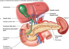
•Fats in duodenum •Stimulate release of CCK •CCK stimulates release of bile intoduodenum |
|
|
CCK |
Cholecystokinin (CCK or CCK-PZ; from Greek chole, "bile"; cysto, "sac"; kinin, "move"; hence, move the bile-sac (gallbladder)) is a peptide hormone of the gastrointestinal system responsible for stimulating the digestion of fat and protein. Cholecystokinin, previously called pancreozymin, is synthesized and secreted by enteroendocrine cells in the duodenum, the first segment of the small intestine. Its presence causes the release of digestive enzymes and bile from the pancreas and gallbladder, respectively, and also acts as a hunger suppressant. |
|
|
8. Types of motility seen in the digestive tract. a. Peristalsis |
rhythmic wave of smooth muscular contraction, used to propel materials (ex, squeezes food through GI tract) muscularis externacontracts to perform peristalsis and segmentation of materials in GI tract |
|
|
8. Types of motility seen in the digestive tract. b. Segmentation. |
irregular smooth muscle contraction that churns and fragments digestive materials (ex, "chops up" food in GI tract) muscularis externa contracts to perform peristalsis and segmentation of materials in GI tract |
|
|
8. Types of motility seen in the digestive tract. c. Churning. |
Turbulence or agitation (no definition in the notes) |
|
|
9. Know the site of secretions and functions of the following: a. Gastric enzymes |
Parietal Cells:
gastric-gland cells that secrete hydrochloric acid (HCl), which chemically digests food;also secretes intrinsic factor Chief Cells gastric-gland cell that secretes pepsinogen, which becomes pepsin and breaks down protein; (also secretes gastric lipase) (No notes on gastric enzymes) |
|
|
9. Know the site of secretions and functions of the following: a. intrinsic factor |
Intrinsic factor (IF), also known as gastric intrinsic factor(GIF), is a glycoprotein produced by the parietal cells of the stomach. It is necessary for the absorption of vitamin B12(cobalamin) later on in the small intestine. |
|
|
9. Know the site of secretions and functions of the following: Intestinal enzymes: a. Salivary amylase |
enzyme that digests carbohydrates secreted by parotid gland into saliva catabolizes (breaks down) complex carbohydrates; "breaks-down sugars" |
|
|
9. Know the site of secretions and functions of the following: b. Intestinal enzymes: Lipase |
•Activated by stomach acid •Breaks down Triglycerides into fatty acids and monoglycerides –Gastric lipase- •Works with lingual lipase •Digest 10%- 15% of dietary fats Protein digestion •Protease |
|
|
9. Know the site of secretions and functions of the following:. b. Intestinal enzymes: trypsin |
The enzyme called trypsin, present in pancreatic juice, is essential for efficient protein digestion. |
|
|
9. Know the site of secretions and functions of the following: c. Pancreatic enzymes. |
•Released into the duodenum of small intestine•Sodium Bicarbonate - pH 7.1 – 8.2 •Pancreatic amylase (Carb Enzymes) - Protein Enzymes •Trypsin , Chymotrypsin + Carboxypeptidase
•Lipase (Fat/Lipid Enzymes) |
|
|
Gastric Juice: HCL |
•pH 2 •Activates pepsinandlingual lipase •Denatures proteins •Convertsferric ions (Fe3+)to ferrous ions (Fe2+)–Fe2+ absorbed –hemoglobinsynthesis•Killsmicrobes |
|
|
10. Know the 3 regions and functions of the small intestine: a. Duodenum. |
The duodenum is the first and shortest segment of the small intestine. It receives partially digested food (known as chyme) from the stomach and plays a vital role in the chemical digestion of chyme in preparation for absorption in the small intestine. Many chemical secretions from the pancreas, liver and gallbladder mix with the chyme in the duodenum to facilitate chemical digestion. Located inferior to the stomach, the duodenum is a 10-12 inch (25-30 cm) long C-shaped, hollow tube. The duodenum is a part of the gastrointestinal (GI) tract, attached to the pyloric sphincter of the stomach on its superior end and to the jejunum of the small intestine on its inferior end. The pancreas, liver and gallbladder all deliver their digestive secretions into the duodenum through an orifice known as the ampulla of Vater, which is located roughly in the middle of the duodenum on the left side. http://www.innerbody.com/image_dige02/dige21.html |
|
|
10. Know the 3 regions and functions of the small intestine: b. Jejunum. |
The jejunum is the middle segment of the small intestine found between the duodenum and the ileum. Most of the nutrients present in food are absorbed by the jejunum before being passed on to the ileum for further absorption. The jejunum is a continuation of the small intestine following the duodenum. It begins at the duodenojejunal flexure, where the small intestine turns sharply toward the anterior direction. From the duodenojejunal flexure, the jejunum follows a convoluted path through the abdomen before continuing as the ileum. While the jejunum does not have an anatomical landmark to separate it from the ileum, it slowly changes its anatomical structure along its length as it transitions into the ileum. http://www.innerbody.com/image_endo03/dige21.html |
|
|
10. Know the 3 regions and functions of the small intestine: c. Ileum. |
The terminal ileum is the distal end of the small intestine that intersects with the large intestine. It contains the ileocecal sphincter, a smooth muscle sphincter that controls the flow of chyme into the large intestine. The terminal ileum is located on the right side of the abdominopelvic cavity in the umbilical and hypogastric regions. It is a tube about 1.25 to 1.5 inches (3 to 4 cm) long at the end of the ileum and terminates at the ileocecal sphincter. The ileocecal sphincter is a band of smooth muscle that controls the flow of chyme from the ileum into the cecum of the large intestine. |