Reading...
![]()
Play button
![]()
Play button
![]()
Use LEFT and RIGHT arrow keys to navigate between flashcards;
Use UP and DOWN arrow keys to flip the card;
H to show hint;
A reads text to speech;
127 Cards in this Set
- Front
- Back
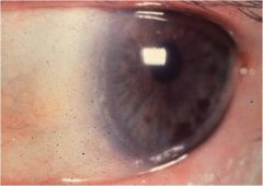
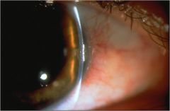
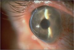
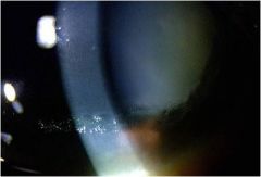
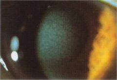
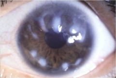
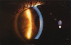
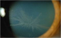
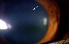
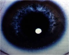
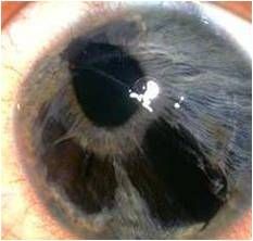
What disorder is this?
|
Arcus (Senilis)
|
|
|
Subjective for what disorder? =
-Asymptomatic -Very common (50% by age 50, 100% by age 80) -More common in African American |
Arcus Senilis
|
|
|
DDX for Arcus (Senilis)
|
DDX =
-Hyperlipidemia/high cholesterol -Not an unusual factor if over 40 to 50 -If under 40 consider other factors such as ----CV disease self or family ----CV risk factors |
|
|
Objective for what disorder? =
-Bilateral -1 to 2 mm white band in mid-peripery -Gradual onset. Inferior and Superior --> fills to complete -Cholesterol/lipid deposition at Boman's -Often hereditary factors |
Arcus (Senilis)
|
|
|
TX for Arcus (Senillis)?
Follow up for Arcus (Senilis)? |
-Definite referral if no medical evaluation w/i 2 yr and under 40
-Annual if no risk factor |
|
What disorder is this?
|
Arcus (Senilis)
|
|
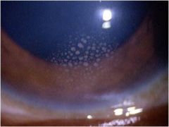
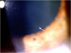
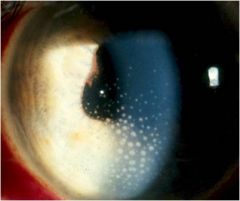
What disorder is this?
|
Limbal Girdle of Vogt
|
|
what disorder is this?
|
Limbal Girdle of Vogt
|
|
|
Subjective:
-Asymptomatic -Older patients 45+ -May be higher in women Objectice: -Bilateral -Narrow band of whitish crystalline-like opacity in nasal and temporal limbus area -degeneration of collagen fibers; not vascularized -Type I -- associated w/ early band keratopathy - see clear zone b/t limbus and opacity line -Type II -- simply peripheral corneal finding - no clear zone |
Limbal Girdle of Vogt
|
|
|
what is the DDX and TX for limbal Girdle of Vogt
|
DDX:
-Other peripheral corneal degenerations -Band keratopathy for Type I TX: -Nothing -- Type II -TX for Band keratopathy |
|
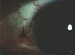
What is this disorder?
|
Dellen
|
|
|
What is this disorder?
|
Dellen
|
|
What is this disorder?
|
Dellen
|
|
What is this disorder
|
Dellen
|
|
|
Subjective:
-Asymptomatic (possibly) -Any age Objective: -focal, peripheral thinning near limbus -usually 0,5 to 1 mm, edges may be sloped or be steeply defined -may occupy up to 1/2 of corneal thickness but epithelium is intact -surrounding tissue may be clear or hazy -causes: secondary to dessication, poor wetting -often occurs adjacent to conjunctival mass (pinguecula, post-surgical edema) |
Dellen
|
|
|
What is the DDX and TX for Dellen?
|
DDX:
-other peripheral thinning disorders -identification/elimination of cause TX: -Lubrication -Therapeutic SCL |
|
What disorder is this?
|
Hassall-Henle Bodies
|
|
|
Subjective:
-Asymptomatic Objective: -Small, round thickenings of Descemet's membrane w/ overlying endothelial displacement in peripheral cornea = peripheral corneal guttata -AKA Descemet's wart's -Normal changes w/ aging -May see associated corneal edema |
Hassall-Henle Bodies
|
|
|
What is the DDX and TX for Hassall-Henle Bodies
|
DDX:
Fuchs (central) TX: None |
|
What is this disorder?
|
Hassall-Henle Bodies
|
|
What is this disorder
|
Hassall-Henle Bodies
|
|
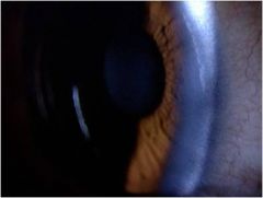
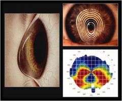
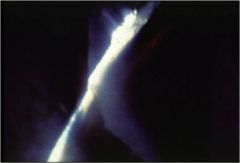
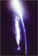
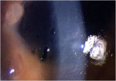
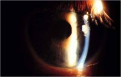
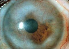
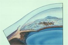
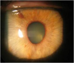
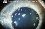
what is this disorder?
|
Pellucid Marginal Degeneration
|
|
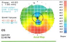
what disorder does this scan represent?
|
Pellucid Marginal Degeneration
|
|
What is this disorder called?
|
Pellucid Marginal Degeneration
|
|
|
Subjective:
-usually asymptomatic -decreased VA Objective: -Bilateral, inferior corneal thinning -Causes central cornea to bulge out above the thinning zone creating irregular ATR astigmatism -Hydrops and scarring can occur w/ progression -very similar to keratoconus and may be a variant |
Pellucid Marginal Degeneration
|
|
|
What is the DDX and TX for Pellucid Marginal Degeneration?
|
DDX:
-other marginal thinning TX: -Correct astigmatism w/ gas permeable CL, keratoplasty if necessary |
|
What disorder are these findings indicative of?
|
Pellucid Marginal Degeneration
|
|
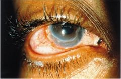
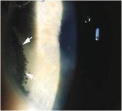
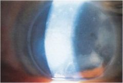
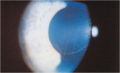
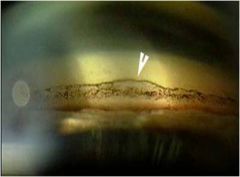
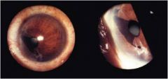
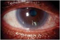
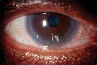
what disorder is seen on this eye?
|
Terrien's Marginal Degeneration
|
|
What disorder is seen?
|
Terrien's Marginal Degeneration
|
|
|
Subjective:
-May have associated pain, decreased VA Objective: -Rare, 75% males, any age, usually bilateral but asymmetric -Marginal stromal thinning opacification and superficial neovascularization -Begins as marginal opacificaton, usually superionasally (may look like arcus) -Lucid zone b/t tinning and limbus -central ege may have a yellw border of lipid -Distortion of cornea w/ irregular astigmatism -Minor trauma can cause ruptur |
Terrien's Marginal Degeneration
|
|
|
What is the DDX and TX for Terrien's Marginal Degeneration?
|
DDX:
-Other marginal thinning conditions -Inflammatory - esp fungal -Rheumatoid disease TX: -Steroid Keratoplasty, LV as necessary |
|
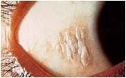
what is this disorder?
|
(Marginal) Furrow Degeneration
|
|
what is this disorder?
|
(Marginal) Furrow Degeneration
|
|
|
This disorder is idiopathic
Subjective: -bilateral tinning seen in or adjacent to Arcus -epithelium is intact -no neovascularization -VA's are not usually affected |
(Marginal) Furrow Degeneration
|
|
|
what is the DDX and TX for (Marginal) Furrow Degeneration?
|
DDX:
-Other peripheral thinning TX: -None |
|
|
This disorder is associated w/ systemic diseases
Subjective: -ring ulcer seen w/ acute rheumatoid arthritis, systemic lupus erythematous, leukemia, polyarteritis nodosa, tuberculosis |
(Marginal) Furrow Degeneration
|
|
|
What is the TX for systemic related (Marginal) Furrow Degeneration?
|
TX:
-treat underlying systemic cause |
|
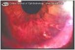
What is this disorder?
|
Mooren's Ulcer
|
|
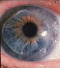
what is this disorder?
|
Mooren's Ulcer
|
|
|
Subjective:
-males may be more affected -2 types: ----Older pt - unilateral - usually responds to TX ----Younger pt -bilateral 25% - difficult to manage -Painful, photophobia -Nigerian individuals may have sever form - rapid progression/perforation Objective: -Begins w/ marginal infiltrate --> chronic, serpiginous limbal ulceration -3 to 12 month course, w/ remission. Mild trauma may perforate globe -Adjacent conjunctival injection/neovascularization -Etiology unclear; autoimmune rxn |
Mooren's Ulcer
|
|
|
what is the DDX and TX for Mooren's Ulcer?
|
DDX:
-other (non-inflammatory) degenerations -Infectious - no thinning -Fungal - HX of trauma (esp. vegetative) -systemic connective tissue disease TX: -Responds poorly - steroids may speed the perforation (so be careful if used) -Corneal specialists referral, conjunctival excision/corneal reconstriction |
|
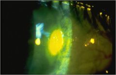
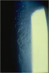
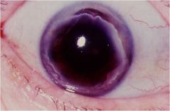
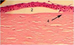
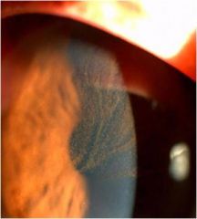
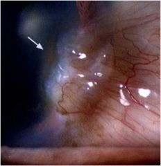
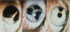
what is this disorder?
|
Posterior Crocodile Shagreen
|
|
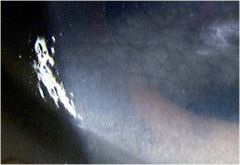
what is this a photo of?
|
Posterior Crocodile Shagreen
|
|
what is this disorder called?
|
Amyloid Degeneration
|
|
|
Subjective:
-Asymptomatic Objective: -Bilateral -Small, gray polygonal 'crocoile skin' patches of various sizes at Descemet's membrane |
Posterior Crocodile Shagreen
|
|
|
What is the DDX and TX for Posterior Crocodile Shagreen?
|
DDX:
-other posterior corneal degenerations and/or dystrophies TX: -None |
|
|
Subjective:
-degeneration in the area of Bowman's and epithelium -secondary to long standing disease (ie. trachoma, glaucoma, uveitis, bullous keratopathy) Objective: -fleshy mass; creates nodular surface -salmon pink to yellow-white -cornea may be vascularized |
Amyloid Degeneration
|
|
|
What is the DDX and TX for Amyloid degeneration?
|
DDX:
-from primary cause TX: -primary cause prevention (but its usually too late) -Keratectomy -Corneal transplant |
|
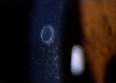
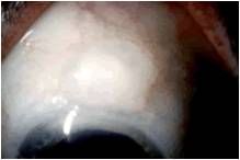
what is this disorder called?
|
Coats White Ring
|
|
what is this disorder called?
|
Coats White Ring
|
|
|
Subjective:
-Hx of foreign body -Asymptomatic Objective -Granular whitish oval ring in the cornea -may contain iron -thought to be lipid in nature |
Coats White Ring
|
|
|
what is the TX for Coats White Ring?
|
None
|
|
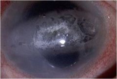
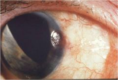
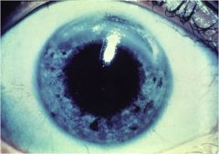
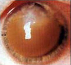
what is this condition called?
|
Band Keratopathy
|
|
what is this disorder called?
|
Band Keratopathy
|
|
what is this disorder called?
|
Band Keratopathy
|
|
|
Subjective:
-Asymptomatic earlery on; later decr. VA -May have RCE's in late stage Objective: -Calcium deposits w/i the interpalpebral fissure -White to yellow deposits at Bowman's and anterior stroma -May be secondary to ocular inflammation or systemic dieases: ----Chronic anterior uveitis, prolonged glaucoma, phthisis bulbi ----Hypercalcemia conditions: Sarcoid, Vit D toxicity, hyperparathyroid, metastatic carcinoma of the bones ----many other causes including autoimmune: gout, lupus, JRA |
Band Keratopathy
|
|
|
What is the TX for Band Keratopathy?
|
TX:
-primary causes - refer if systemic -ocular - lubrication or chelating agents (EDTA), scrape off band |
|
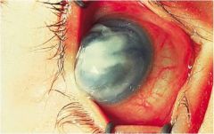
what is this disorder called?
|
Bullous Keratopathy
|
|
What is this picture showing?
|
Bullous Keratophathy
|
|
what is this pic showing?
|
Bullous keratopathy
|
|
what is this dysfunction
|
Bullous Keratopathy
|
|
|
Subjective:
-Acute -Painful Objective: -Caused by long term, prolonged corneal edema -Bubbling of the cornea, break down and reform, eventually scar |
Bullous Keratopathy
|
|
|
what is the DDX and TX for Bullous Keratopathy?
|
DDX:
-primary cause: Fuch's, keratoconus, etc... -post operative TX: -antiedema TX -early on - therapeutic CLS -late stages - symptomatology TX-poor prognosis -LV |
|
What is this disorder called?
|
Salzmann's Nodular Degeneration
|
|
|
Subjective:
-usually asymptomatic -may be more common in females Objective: -Non-inflammatory -Multiple bluish-white nodules, usually at the mid-periphery -Bilateral -Vision depends on location -Related to previous inflammation - esp. Phlyctenular disease |
Salzmann's Nodular Degeneration
|
|
|
What is the TX for Salzmann's Nodular Degeneration?
|
TX:
-Asymptomatic: monitor 3 to 6 months -w/ epithelial break down: therapeutic CLS, antibiotic qid -Keratoplasty, LV as necessary |
|
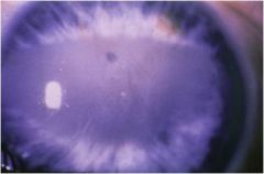
what is this condition called?
|
Xerophthalmia and Keratomalacia
|
|
what is this condition called?
|
Xerophthalmia
|
|
what is this condition called?
|
Keratomalacia
|
|
|
these two conditions are both related to vit A deficiency either by malnutrition or failure to absorb
|
Xerophthalmia and Keratomalacia
|
|
|
Subjective:
-Keratinization of the epithelium -Atrophy of conjunctival goblet cells -Corneal edema -Neovascularization -may see Bitot's spots |
Xerophthalmia
|
|
|
Subjective:
-Acute corneal tissue liquefaction -may see Bitot's spots |
Keratomalcia
|
|
|
what is the DDX and TX for Xerophthalmia and Keratomalcia?
|
DDX:
-other corneal degeneration TX: -Vit A supplementation - ocular changes may reverse |
|
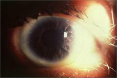
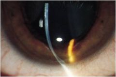
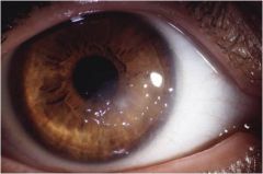
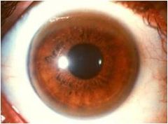
what is this disorder?
|
Kruckenberg's Spindle
|
|
|
what is this disorder called?
|
Kruckenberg's Spindle
|
|
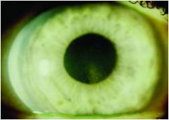
what is this disorder called?
|
Kruckenberg's Spindle
|
|

what is this disorder called?
|
Kruckenberg's Spindle
|
|
what is this disorder called?
|
Kruckenberg's Spindle
|
|
|
Objective:
-Brownish in color -Vertical spindle shaped pigment deposition on posterior cornea -Inferior 1/3 to 1/2 -Suggests old uveitis or pigment dispersion syndrome |
Kruckenberg's Spindle
|
|
what is this condition called?
|
Vortex Keratopathy
|
|
what is this condition called?
|
Vortex Keratopathy
|
|
|
Objective:
-Greyish or golden epithelial deposits -appear in a swirl pattern from a point below the pupil -occurs in pt w/ Fabry Disease (alpha-galactosidase A deficiencey, causing fat accumulation) and pt being treated w/ a varety of drugs, including but not limited to: ---Amiodarone (for antiarithmias) ---Hydroxycholorquine (for malaria?) ---Indomethacin (for gout) ---Tamoxifen (for breast cancer) |
Vortex Keratopathy
|
|
what is this called?
|
Arlt's Triangle
|
|
|
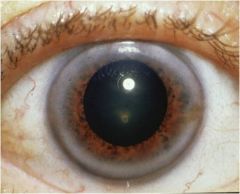
This corneal Pigmentation is:
-Brownish in color -Triangular shaped pigment at the 6 o'clock position on posterior cornea -pathognomonic for old uvea |
Arlt's Triangle
|
|
|
This corneal Pigmentation is:
-Brownish in color -Edematous haze in epithelium -Pathognomonic for EBMD, RCE |
Brawny cornea
|
|
what is this corneal disorder?
|
Brawny cornea
|
|
what is this corneal disorder?
|
Ferry's Ring
|
|
what is this corneal pigmentation disorder?
|
Fleisher's Ring
|
|
|
This Corneal pigmentation disorder is:
-Orangish-brown in color -Ferric deposition around a surgical filtration bleb -No pathologic indication |
Ferry's Ring
|
|
|
This Corneal pigmentation disorder is:
-Orangish in color -Ferric deposition at the base of keratoconic core -Pathognomonic for keratoconus |
Fleisher's Ring
|
|
|
This corneal pigmentation disorder is:
-Brownish in color -pigment granules forming a horizontal line on the inferior cornea -pathognomonic for pigmentary glaucoma |
Goar's line
|
|
|
This Corneal Pigmentation disorder is:
-Reddish-brown in color -Intracorneal or posterior corneal surface blood stain -Pathognomonic for: -----Hyphema-posterior stain -----Intracorneal bleeding neovascularization |
Hemosiderosis
|
|
What is this corneal pigmentation disorder?
|
Hemosiderosis
|
|
what is this corneal pigmentation disorder?
|
Hudson-Stahli Line
|
|
what is this corneal pigmentation disorder called?
|
Kaiser-Fleischer Ring
|
|
|
This corneal pigmentation is:
-Orangish-brown in color -deposition at Boman's -at area of upper and lower lid junction -more in males -frequency incr. w/ age -frequent site of RCE |
Hudson-Stahli Line
|
|
|
This corneal pigmentation is:
seen as: -faint segmental line -continuous line -line w/ a surrounding white opacity |
Hudson-Stahli Line
|
|
|
This corneal pigmentation disorder is:
-Orangish in color -posterior cornea -- anterior angle -best seen in gonioscopy, may be grossly visible -copper deposition -suggests for Wilson's hepaticolenticular disease |
Kaiser-Fleisher Ring
|
|
|
This corneal pigmentation is:
-seen in liver cirrhosis pt -basal ganglia degeneration -due to decreasedd ceruloplasmin w/ copper deposition into organs What is the disease called? |
Kaiser-Fleischer Ring
Wilson's Diseaes |
|
What is this disorder and what disease is associated with it?
|
Kaiser-Fleischer Ring
|
|
what is this corneal pigmentation disorder called?
|
Keratic Precipitates
|
|
what is this corneal pigmentation disorder called?
|
Keratomelanocytosis (Striate Melanokeratosis)
|
|
|
This corneal pigmentation disorder is:
-white or pigmented -Endothelial surface -Pathognomonic for: ---uveitis or trauma |
Keratic Precipitates
|
|
|
This corneal pigmentation disorder is:
-pigmented spokes radiating out into the cornea from the limbus -most in heavily pigmented individuals -most frequently at 4 and 8 o'clock -pathognomonic for: -----Truama -----Infectinon -----Focal toxic inflamm. of Staph |
Keratomelanocytosis (Striate Melanokeratosis)
|
|
|
This corneal pigmentation is:
-orangish in color -discoloration of midstroma -----interstitial keratitis -suggests syphilitic keratitis |
Salmon Patch
|
|
|
This corneal pigmentation is:
-pigment granules at Schwalbe's line -suggests pigmentary glaucoma |
Sampaolesi's Line
|
|
|
This corneal pigmentation is:
-Orangish brown in color -Ferric deposition at ptergyium leading edge |
Stocker's Line
|
|
|
what is this corneal pigmentation disorder?
|
Salmon Patch
|
|
what is this corneal pigmentation disorder?
|
Salmon Patch
|
|
what is this corneal pigmentation disorder called?
|
Sampaolesi's Line
|
|
what is this corneal pigmentation disorder?
|
Stocker's Line
|
|
what is this syndrome called?
|
ICE (Iridocorneal Endothelial Syndrome)
|
|
|
This syndrome is;
1. Typically unilateral 2. More often seen in females 3. Consists of three overlapping disorders ---Chandler syndrome ---Cogan-Reese syndrome ---Progressive Iris Atrophy 4. The common link b/t the three forms in an abnormal corneal endothelium 5. The endothelium can proliferate and migrate into the angle and onto the iris surface 6. The migration into the angle can cause synechial angel closure leading to GLAUCOMA |
ICE syndrome
|
|
|
Objective for this corneal ulcer is:
-Corectopia (malposition of the pupil) -Pseudopolycoria (multiple "pupils") in a previously normal iris -iris atrophy -corneal endothelial abnormalities ----a hammered appearance similar to Fuch's ----Best viewed w/ specular reflection ----Corneal edema due to endothelial defects |
ICE syndrome
|
|
what is this syndrome called?
|
ICE syndrome
|
|
what is this syndrome called?
|
ICE syndrome
|
|
what is this irido-corneal endothelial syndrome called?
|
Chandlers Syndrome (ICE)
|
|
what is this irido-corneal enothelial syndrome called?
|
Cogan-Reese syndrome (ICE)
|
|
what is this irido-corneal epithelial syndrome disorder called
|
Progressive Iris Atrophy (ICE)
|
|

fill in the boxes for Chandler's syndrome
ICE Disorder IOP Corneal edema Corectopia Glaucoma prevalence % |

DISORDER Chandler's Syndrome
IOP Normal to high CORNEAL EDEMA Moderate to severe CORECTOPIA Mild to moderate Glaucoma prevalence ~5% |
|
|
fill in the boxes
ICE Disorder Cogan-Reese IOP Corneal edema Corectopia Glaucoma prevalence % |
Disorder CoganReese
IOP Usually high Corneal edema Mild-moder Corectopia Modera-sev Glaucoma prevalence %~50% |
|
|
fill in the boxes for Progressive Iris Atrophy
ICE Disorder IOP Corneal edema Corectopia Glaucoma prevalence % |
ICE Disorder ProgIrisAtroph
IOP Usually high Corneal edema Mild-moder Corectopia Moder-Sever Glaucoma prevalence % ~37% |
|
what is this disorder called
|
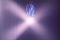
Filamentary Keratitis
|
|
what is this disorder caused by dry eye called
|
filamentary keratitis
|
|

what is this disorder caused by Sjogren's called
|
filamentary keratitis
|
|
what is this disorder called
|
filamentary keratitis
|
|
what is this corneal disorder called
|
filamentary keratitis
|