![]()
![]()
![]()
Use LEFT and RIGHT arrow keys to navigate between flashcards;
Use UP and DOWN arrow keys to flip the card;
H to show hint;
A reads text to speech;
85 Cards in this Set
- Front
- Back
|
drug interaction sites by % |
GPCR's 45% |
|
|
number of genes in the human genome
|
about 19,000 |
|
|
types of methods for detecting protein-protein interactions
|
Molecular biology methods: yeast two hybrid screening
biophysical methods: fluorescence-based technology |
|
|
For FRET, these two peaks must overlap
|
donor fluorescence and acceptor absorption
|
|
|
FRET stands for
|
Fluorescence resonance energy transfer
-or- Forster resonance energy transfer |
|
|
definition of FRET
|
nonradiative energy transfer from an excited molecule to another nearby molecule
|
|
|
formulas for FRET
|
E = 1/(1+r/R0) = (# quanta transferred)/(# quanta absorbed by D)
|
|
|
types of FRET donor-acceptor pairs
|
fluorsecent proteins
organic dyes chelates of lanthanides (Eu, TR-FRET) |
|
|
advantage of using organic dyes for FRET pairs
|
improved photo and pH stability
|
|
|
spectroscopy definition
|
study of light and its interaction with matter
|
|
|
spectrometry definition
|
measurement of a light spectrum
|
|
|
analytical uses of spectroscopy
|
quantitation of drugs for QC
monitor reactions of biomolecules qualitative ID of unknown drugs by characteristic absorption or emission bands |
|
|
relating equation, wavelength and frequency
|
(lambda)×(nu) = c
|
|
|
light energy and frequency relating equation
|
E = (h)(nu)
|
|
|
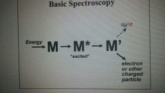
basic spectroscopy diagram
|
|
|
|
Atomic Emission Spectroscopy definition
|
quantitative optical emission from excited atoms to determine analyte concentration
|
|
|
atomic emission spectroscopy - what happens to the sample |
molecules/atoms in solution are aspirated into excitation region where they are desolvated, vaporized, or atomized |
|
|
types of spectroscopy and their uses
|
emission - elemental analysis |
|
|
types of absorption spectroscopy and what they measure
|
UV/VIS - electronic transitions |
|
|
formula for total energy from its components
|
ET = ER + EV + EE
|
|
|
formula for energy change when transitions are allowed
|
delta-E = E* - E0 = hc/lambda
|
|
|
spectral regions of light (cm)
|
radio >10 |
|
|
How atomic emission spectroscopy works |
Atoms are excited to higher energy levels, then relax, emitting light of characteristic wavelengths |
|
|
What does a polychromator do |
counts photons |
|
|
How absorbance is used for quantitation |
If light passes through a substance that absorbs light, intensity (I0) will be reduced to I |
|
|
Equation used to relate absorbance to concentration |
Beer-Lambert Law: A = epsilon*b*c
epsilon = molar absorptivity b = path length |
|
|
How to determine molar absorptiity for new compound |
Use calibration curve |
|
|
Basic components of absorption spectrometry instrumentation |
Source, monochromator, detector |
|
|
Source types for absorption spectrometry |
Incandescent (limited to visible spectral range) Luminous gas (ex: neon light, higher intensity and wavelength range) |
|
|
Monochrometer types for absorption spectrometry |
Filters (like colored glass, must be selected for narrow wavelength range)
Dispersion devices ( prisms/gratings, provide wider and more selectable wavelength range) |
|
|
types of photodetectors for absorption spectroscopy |
phototube - converts light into electrical current
photomultiplier - more sensitive because dynode series amplifies electrical current |
|
|
What does absorbance mean? |
molecules absorb specific wavelengths of light that promote them to excited state; excited state is quantized representing unique configurations of electrons, and vibrational and rotational modes |
|
|
UV/Vis most useful for/least useful for |
Most - analyte concentration
Least - qualitative (ID of compounds) |
|
|
effect of conjugation on wavelength |
Increased wavelength (b/c decreased energy) |
|
|
Efffect of lone pairs on wavelength |
more easily delocalized l.p. ==> higher wavelength |
|
|
effect of pH on wavelength |
depends - stability of product determines effect |
|
|
Bathochromic |
shift to longer wavelength |
|
|
Hypsochromic |
shift to shorter wavelength |
|
|
Hyperchromic |
Shift to greater absorbance |
|
|
Hypochromic |
Shift to lower absorbance |
|
|
UV/Vis spectrometry applications
|
pH titrations
Qualitative information (related molecular features) Detectors for chromatography |
|
|
Reason why UV/Vis is poor for qualitative studies |
solution spectra (esp. in water) are broad, nondescript bands |
|
|
Colorimetry |
analysis performed in visible spectral region |
|
|
visible spectral region in angstroms |
4000 - 7000 |
|
|
Typical colorimetry analyte will be.. |
highly conjugated |
|
|
Example of colorimetry |
Phenolphthalein analysis |
|
|
Fluorescence definition |
emission of light from a molecule that has previously absorbed light energy |
|
|
Chemiluminescence definition |
emission of light from a molecule that has previously absorbed light energy from a chemical reaction |
|
|
Stages of fluorescent emission |
1 - excitation - photon absorbed 2 - excited state lifetime 3 - fluorescent emission |
|
|
Stokes shift |
difference between absorption and emission wavelength |
|
|
reason why emission wavelength is higher (from stokes shift) |
energy dissipation during excited state lifetime; lower energy => higher wavelength |
|
|
How to minimize background fluorescence |
Select filters that reduce transmission of E2 relative to E1
Select probes that absorb and emit at longer wavelengths |
|
|
Fluorescence quantum yield definition |
ratio of number of molecules fluorescing to number excited |
|
|
Factors influencing fluorescence quantum yield |
excitation wavelength lifetime of state temperature pH solvent |
|
|
Molecular factors favoring fluorescence |
planar conjugated sterically uncrowded EDG's fused rings rigid chelation to metals |
|
|
fluorescence probes |
chromophores, localize to specific region of specimen and respond to specific stimulus |
|
|
Which is more sensitive for quantification, fluorescence or absorbance spectrometry? |
Fluorescence |
|
|
Chemiluminescence difference from fluorescence spectroscopy |
energy for absorption provided by chemical reaction rather than light
No source or primary filter needed |
|
|
Example of chemiluminescence labeling reagent |
Luminol |
|
|
What is radioactivity? |
spontaneous emission of electromagnetic radiation or atomic fragments from an unstable nucleus |
|
|
Half life definition |
time for isotope to decay to something else (which may or may not also be radioactive) |
|
|
alpha decay |
(4He2+) Helium nucleus emitted, reduces atomic # by 2 and mass # by 4
Not dangerous |
|
|
beta-minus decay |
neutron spontaneously converts to proton and emits electron
atomic number increases by 1 no change in mass number
stopped by minimal barrier - not particularly dangerous |
|
|
beta-plus decay |
*antimatter*
Proton spontaneously converts to neutron and emits positron |
|
|
How to measure radioactivity energy |
megaelectronvolts (MeV) |
|
|
Specific activity definition |
number of curies (decays per second) per unit mass |
|
|
Scintillation definition |
radiation converted to UV/visible radiation by collision with scintillator |
|
|
gamma radiation |
emission of high-energy photon, usually along with other decays
very dangerous |
|
|
Rate of radioactive decay |
first order units = curies (3.7e10 decays/s) |
|
|
Beta liquid scintillation counting process |
Radioactive sample added to scintillation cocktail
beta particles emitted, excites solvent
solvent energy transferred to fluor |
|
|
(Symbolic) steps in beta scinitillation |
1. S + E-beta --> S* 2. S* + PPO --> PPO* 3. PPO* + POPOP --> PPO + POPOP* 4. POPOP* --> POPOP + hv (long wavelength) |
|
|
Methods of two-color FRET |
Sensitized emission
Donor dequenching
Fluorescence lifetime imaging (FLIM)
|
|
|
Sensitized emission (for FRET) definition |
D excitation/A emission
Issues: spectral bleedthrough contamination |
|
|
Donor dequenching (for FRET) definition |
measuring the intensities of an identical donor fluorophore in the absence and presence of the acceptor |
|
|
FLIM (for FRET) definition |
measuring the reduction in the D lifetime that results from quenching in the presence of an acceptor |
|
|
Limitation of FRET, how to address these |
donor and acceptor are prompt fluorophores with short half-lives
background fluorescence
Solution: TR-FRET (time resolved FRET) |
|
|
TR-FRET basics |
uses long-lived fluorophores (lanthanides)
uses time-resolved detection (delay between excitation and emission detection) |
|
|
Why are lanthanides good for TR-FRET |
very long Stokes shifts
long emission half-lives (usec to msec) |
|
|
Common mechanism of BRET |
Coelenterazine oxidized by luciferise through dioxetane intermediate |
|
|
Example types of cell viability assays and their advantages |
ATP detection assay (most sensitive, least steps, quickest, small interference
Tetrazolium or resazurin reduction (cheap, adequate performace)
Fluorogenic protease (less cytotoxic, multiplexing) |
|
|
Important factors to remember for any cell viability assay |
Use tightly controlled, consistent source of cells
perform appropriate characterization of assay conditions (reagent concentration, incubation time) |
|
|
Distribution coefficient in chromatography |
concentration of component in stationary phase/concentration of component in mobile phase |
|
|
Theoretical plate equation |
N = L/H |
|
|
Van Deemter Equation |
H = A + B/u +Cu
A = Eddy diffusion B = Longitudinal Diffusion C = Mass transfer coefficient u = linear velocity |
|
|
Eddy diffusion |
multiple path effect |