![]()
![]()
![]()
Use LEFT and RIGHT arrow keys to navigate between flashcards;
Use UP and DOWN arrow keys to flip the card;
H to show hint;
A reads text to speech;
22 Cards in this Set
- Front
- Back
|
A variation of red blood cell size is known as |
Anisocytosis
|
|
|
- Appear smaller than normal mature erythrocyte. - More dense staining than normal mature erythrocyte. - No central zone pallor. - Associated with immune-mediated hemolytic anemias.
|
Spherocytes |
|
|
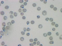
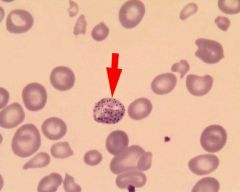
Heinz Bodies
Round structure representing denatured hemoglobin. Up to 5% normal in cats. |
|
|
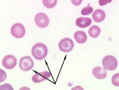
Target cells |
|
|
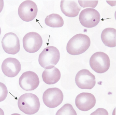
Howell Jolly Bodies
Basophilic nuclear remnants seen in young erythrocytes in response to anemia; removed by the spleen. |
|
|
Name the erythroid maturation series in proper order. |
1. Rubriblast 2. Prorubricyte 3. Rubricyte 4. Metarubricyte 5. Reticulocyte 6. Erythrocyte |
|
|
Describe canine RBC shape |
Discocytic, biconcave disc. Central pallor due to thin layer of hemoglobin. Shape facilitates better transporting of oxygen. |
|
|
Signs of regeneration |
1. Polychromasia 2. Anisocytosis 3. Nucleated RBCs 4. Howell Jolly Bodies |
|
|
Rouleaux
A groping of erythrocytes in stacks; seen with increased fibrinogen or globulin. |
|
|
Agglutination
An antibody coats the erythrocyte causing bridging and clumping. |
|
|
Basophilic Stippling
Presence of small, dark-blue bodies within the erythrocyte. Seen in lead poisoning. |
|
|
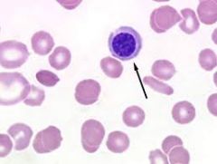
Nucleated RBC
Cells released into circulation early during anemia. |
|
|
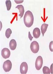
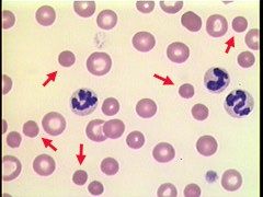
Schistocyte
RBC fragments; usually from shearing of the red cell by intravascular trauma; DIC |
|
|
Spherocytes |
|
|
What is the difference between feline and canine RBCs? |
Cat red blood cells are smaller than canine red blood cells and they have limited central pallor. |
|
|
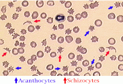
Acanthocytes: Irregular, spiculated red cells with few unevenly distributed surface projections of various length and diameter. Schistocyte: RBC fragments; usually from shearing of the red cell by intravascular trauma. DIC.
|
|
|
RBCs with a smaller than normal diameter and decreased MCV; seen in iron deficiency. Akitas may have this type of cells. |
Microcytes |
|
|
RBCs that are larger than normal with an increased MCV. Poodles may have this type of cells. |
Macrocytes |
|
|
RBC that exhibit a bluish tint when stained with Wright's stain; organelles remain in cytoplasm indicating young cells. |
Polychromasia |
|
|
RBC changes from disc shape to spheres with projections; result of pH change due to slow drying of blood films. |
Crenation |
|
|
What are the three basic classes of etiological anemia? |
1. Blood loss. 2. Hemolysis. 3. Inadequate production.
|
|
|
What is macrocytosis? |
Increase of the MCV seen in young RBCs. |