Reading...
![]()
Play button
![]()
Play button
![]()
Use LEFT and RIGHT arrow keys to navigate between flashcards;
Use UP and DOWN arrow keys to flip the card;
H to show hint;
A reads text to speech;
56 Cards in this Set
- Front
- Back
|
CNI? What is it called when this n is severed? What bone do these nn go through?
|
Olfactory nerve. Anosmia. Pass through the cribriform plate of the ethmoidal bone (both terms mean sieve).
|
|
|
CNII? Describe how it courses in to the brain.
|
Optic nerve. Photoreceptor cells send signals to retinal cells --> to optic n --> optic chasm --> to optic tracts --> to lateral geniculate nucleus of the thalamus --> primary visual cortex located in the occipital lobe of cerebrum.
|
|
|
Which CNII nerve fibers will decussate at the optic chasm? How can knowing this be helpful diagnostically?
|
Medial optic fibers. If an injury is at the optic n vs the optic tract, pt will experience different symptoms.
|
|
|
CNII enters the cranium via ? which is in the ? bone.
|
CNII enters the cranium via OPTIC CANAL which is in the SPHENOID bone.
|
|
|
What is immediately posterior to the optic chasm? How is the useful diagnostically?
|
The pituitary gland is immediately posterior to the optic chasm and if there's something wrong with it, there might also be something wrong visually.
|
|
|
CNVIII? Serves what?
|
Vestibulocochlear N. Strictly sensory nn that serves the cochlear and vestibular organ.
|
|
|
What two nn come together to form CNVIII? Where does it emerge from the cranium?
|
Vestibular and Cochlear nn form CNVIII. It emerges from the Internal Acoustic Meatus (N123)
|
|
|
The receptors of the inner ear (cochlea and vestibule) have cell bodies that are located where?
|
The cell bodies are located in ganglia in the inner ear.
|
|
|
Damage to CNVIII can result in:
|
Hearing and or balance issues
|
|
|
Where do the Oculomotor neurons (CNIII) originate?
|
They originate in the midbrain at the oculomotor nucleus.
|
|
|
Describe the course of the CNIII.
|
Originates in midbrain oculomotor nuclease --> passes through cavernous sinus (N103) --> through superior orbital fissure (N4) --> into orbital cavity where it innervates muscles.
|
|

Label related structures and muscles innervated by CNIII
|
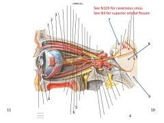
1. Superior Rectus
2. Levator palpebrae superioris muscle 3. Superior oblique muslce 4. Inferior oblique muscle 5. Medial rectus m 6. Inferior rectus m 7. Midbrain 8. Pons 9. Medulla |
|
|
Damage to CNIII presents with what symptoms.
|
Oculumotor n damage presents with ptosis - drooping of the upper eyelid (i.e. levator palpebrae superioris); and the eye deviates laterally bc of the unopposed action of the lateral rectus muscle that is innervated by the Abducent CNVI
|
|
|
CNIV innervates what muscle?
|
The superior oblique muscle that travels through a trochlea - hence called the Trochlear N
|
|
|
Describe where CNIV originates and its progression toward its insertion.
|
Originates in the trochlear nucleus of the mid brain --> emerges from the DORSAL aspect of the brain stem --> through cavernous sinus --> through superior orbital fissure --> innervates superior oblique
|
|
|
What is the CNVI?
|
The Abducent
|
|
|
Describe the course of the CNVI.
|
Originates in the abducent nucleus of the brain stem --> emerges at the junction bn the pons and medulla --> passes through cavernous sinus --> pierces optical cavity via the orbital fissure --> innervates the lateral rectus m.
|
|
|
Describe the eye movement caused by CNVI
|
The ABducent nerve ABducts the eye.
|
|
|
If you see a pt with one eye deviating toward the nose, where might there be an injury (2 things)
|
Trauma to the base of the skull or an aneurism in the cavernous sinus area.
|
|
|
CNXI? What does it innervate? (N126)
|
Accessory Nerve. SCM and Trap
|
|
|
Describe the course of CNXI (N126)
|
CNXI nerve roots emerge at several locations along the brain stem and coalesce as the ascend and pass through the foremen magnum. The single n now loops around and comes down through the jugular foremen to innervate the SCM and trap.
|
|
|
CNXII is called what an innervates what?
|
Hypoglossal N - innervates the Extrinsic m of the tongue.
|
|
|
What are the extrinsic muscles of the tongue? (there are four but CNXII only innervates 3)
|
1. Genioglossus
2. Hyoglossus 3. Styloglossus 4. Geniohyoid m* Not innervated by CNXII. |
|
|
Describe the course of the CNXII to the extrinsic tongue muscles.
|
Originates in the hypoglossal nucleus in the medulla. Emerges and passes thru the hypoglossal canal. Runs anterior to the carotid and IJV and runs along the base of the hyoglossus before innervating the extrinsic muscles.
|
|
|
CNV1 is? What kind of nerve is it? What tissues does it supply?
|
Ophthalmic Nerve of the trigeminal. It is a purely sensory nerve that receives sensory input from the upper aspects of the face (eye lid, lacrimal gland, anterior portion of the nose, scalp).
|
|
|
Describe the course of CNV1
|
Since sensory only it starts at peripheral tissues (e.g. forehead, eyelid, lacrimal gland) --> orbital cavity --> through superior orbital fissure --> to cell bodies in the trigeminal ganglion --> to pons.
|
|
|
What is CNV2? What kind of n is it and what tissues does it serve?
|
CNV2 is the Maxillary N. It is a purely sensory n that serves the upper lip/teeth, palate, cheak, etc.
|
|
|
Describe the course of CNV2
|
Since it is purely sensory, it starts at the mid face-ish --> enters the cranium via the foremen rotundum (N44) --> to cell bodies in the trigeminal nerve ganglion --> to the pons.
|
|
|
What is CNV3? What kind of n is it and what tissues does it serve?
|
CNV3 is the Mandibular division of the Trigeminal. It is both a motor and sensory neuron that supplies the lower jaw.
|
|
|
Describe the course of the CNV3
|
The main CNV3 trunk enters the cranial cavity via the foremen ovale. Sensory fibers have nuclei in the trigeminal gangion --> central processes pass to the brainstem (pons).
|
|
|
CN VII? Is it S, M or SM?
|
Facial N. Its SM
|
|
|
Describe the path of the CNVII
|
It exits the caudal boarder of the pons (bn the pons and medulla) --> enters the petrous division of temporal bone via Internal Acoustic Meatus --> exits petrous via Stylomastoid Foramen --> pass through parotid --> innervates m. of facial expression.
|
|
|
Where do the sensory neurons arise in the facial nerve? Where do they go? What is the n called once it gets close to the tongue?
|
The geniculate ganglion neurons go to the anterior 2/3 of the tongue. Called the Chorda Tympani.
|
|
|
Facial n innervates this accessory mastication muscle:
|
Buccinator via Buccal Branches
|
|
|
This branch of the facial n innervates the Occipital Belly or Occipitofrontalis Muscle
|
The posterior auricular branch.
|
|
|
Which n innervates the stapedius and auricular mm?
|
Facial n
|
|
|
CNIX? Is S, M, SM?
|
Glossopharyngeal N is a Sensory and Motor neuron.
|
|
|
Describe the path of CNIX
|
3-4 roots come off the side of the medulla and enters the jugular foramen
|
|
|
Describe the somatic sensory component of the glossopharyngeal n. Hint: there are three major structures.
|
1. Posterior 1/3 of tongue
2. Pharynx (tonsils, soft palate, auditory tubules) 3. Carotid Body and Sinus - monitors lack of O2 and monitors pressure. |
|
|
What is the only motor component of the Glossopharyngeal N? What does damage to this branch result in?
|
Stylopharyngeous m - damage to this branch causes diminished gag reflex.
|
|
|
CNX? Is it S, M, SM
|
Vagus is both SM
|
|
|
Describe the path of CNX
|
Emerges from the Medulla and exits the cranium through the jugular foramen.
|
|
|
In general, the Autonomic Nervous System innervate what?
|
Cardiac m, smooth m, and glands.
|
|
|
What is the function of the ANS (3)?
Is it subdivided into parasympathetic and sympathetic divisions? |
1. Emergency (fight or flight)
2. Maintain homeostatis (rest & digest) 3. Reproduction Yes, subdivided into para/sympathetic divisions. |
|
|
Recall how the para/sympathetic neurons are set up (see back for diagram). Describe the respective lengths of both.
|
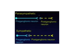
Has a preganglion and postganglion neuron. Parasymp has a long preganglion and a short postganglion.
|
|
|
What happens at ganglion?
|
Where the "baton exchange" occurs. Where the presynaptic nerve synapses with the postsynaptic neuron.
|
|
|
What are the four parasympathetic ganglion of the head?
|
SPOC Ganglion (Think Star Trek):
1. Submandibular 2. Pterygopalatine 3. Otic 4. Ciliary |
|
|
What CN innervates the smooth muscle of the eye leading to pupil constriction and thickening of the lens (accommodation)? Is this para- or sympathetic?
|
Oculomotor (CNIII). Parasympathetic
|
|
|
Describe the path of the parasympathetic portion of the oculomoter neuron
|
Originates in the midbrain at the EDINGER-WESTPHAL nucleus (pre-ganglion) --> travels with occulomoter n through superior orbital fissure --> ciliary ganglion --> short ciliary n (post-ganglion) --> around globe --> sphincter pupillae m (pupil constriction) and ciliary m (lens accommodation for near vision - reading a book during RR).
|
|
|
If you have an injury along the parasympathetic n of the occulomoter, what will happen?
|
You'll have a huge an enlarged pupil and pt will have loss of accommodation for near vision.
|
|
|
Describe the path of the n that leads to tears. First, what cranial nerve is this?
|
CNVII - Facial.
Starts at Superior Salivatory Nucleus --> emerges with Facial N --> (branch point) --> greater petrosal n --> deep petrosal --> Nerve (Vidian) of Pterygoid Canal --> Pterygopalatine Ganglion --> continues to travel with trigeminal/zygomatical temoral n (CNV) --> lacrimal nn --> lacrimal gland. |
|
|
What do pts complain about with damage to the parasympathic n fibers of CNVII?
|
Dry eyes.
|
|
|
What innervates the sublinguinal and submandibular gland? (N132)
|
CNVII - Facial N (parasympathetic)
|
|
|
Describe the parasympathetic nerve fiber track (N132) to the salivary glands.
|
Originates at the superior salivatory nucleus --> emerges with facial n --> (branch point) --> chorda tympani --> merges with lingual n (branch of CNV3) greater petrosal --> submandibular ganglion --> submandibular and sublinguinal gland
|
|
|
Recall the Bell's Palsy example she used in class and how damage to different areas causes different symptoms. Draw the nerves involved and describe the symptoms.
|
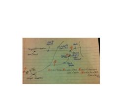
Damage at 1:
a) Facial m paralysis Damage at 2: a) and b) loss of taste c) loss of salivation Damage at 3: a) b) c) and d) hyperacusis Damage at 4: a) b) c) d) and e) loss of tast (palate) 6) loss of lacrimation **Note - my drawing is a bit off - post submandibular ganlion nerve fibers do not innervate taste bud on tongue. |
|
|
Describe the Otic Ganglion Schema (N124)
|
Originates at the Inferior salivary nucleus in brain stem --> emerges with CNIX (glossal pharyngeal) --> thru tympanic cavity/N --> lesser petrosal --> OTIC Ganglion --> auriculotemporal n --> parotid gland
|