![]()
![]()
![]()
Use LEFT and RIGHT arrow keys to navigate between flashcards;
Use UP and DOWN arrow keys to flip the card;
H to show hint;
A reads text to speech;
84 Cards in this Set
- Front
- Back
|
Aritfact |
structure of appearance that is not natural/ fuzzy image due to manipulation |
|
|
Cable |
EKG machine cable 10 cables |
|
|
Calibration |
measurement of its variation |
|
|
Galvanometer |
measurement by electromagnetic action |
|
|
Graph Paper |
small box = 0.04 large box =.20 h=time v=volt |
|
|
Ground Lead |
contrivance for guiding cable |
|
|
Isoeletric Line |
base line above is positive deflection below is negative deflection |
|
|
Lead |
electrocardiographic cable connects with thewelectrons placed at a particular point of your heart |
|
|
Pericarditis |
inflation of the 2 layers sac-like membrane of the heart |
|
|
Sensor |
respond to physcial stumil electrodes |
|
|
Stylus |
putting ink on paper as it prints pencil / pen |
|
|
Ventricular Hypertrophy |
increase in volume or organ produced by enlargement of exciting cells. |
|
|
Normal Sinus
|

|
|
|
Sinus Tachycardia
|
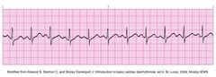
file:///Users/s08034274/Desktop/EAf1KUxBtLDE4H-dj8yyyQ_m.jpg |
|
|
Sinus Bradycardia
|
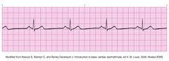
file:///Users/s08034274/Desktop/e5iEKKKXxOFnSqcgAvDMdA_m.jpg |
|
|
Atrial Flutter
|
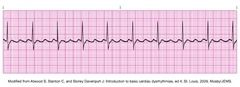
file:///Users/s08034274/Desktop/k.mO7Yhg0Td7JewXV8YQQw_m.jpg |
|
|
Atrial Fibrillation
|
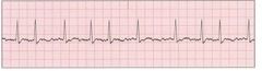
file:///Users/s08034274/Desktop/-fsPEKh5lIXGGRYQL-Sowg_m.jpg |
|
|
T wave
|
represents ventricular repolarization
|
|
|
depolarization
|
the most important electrical event in the heart, the state of cellular stimulation in the heart that causes it to contract
|
|
|
isoelectrical line
|
represents no eletrical activity
|
|
|
QRS complex
|
represents ventricular depolarization and the resulting ventricular contraction
|
|
|
SA node
|
the pacemaker of the heart
|
|
|
P wave
|
represents atrial repolarization with resulting atrial contraction
|
|
|
pathway for conduction thru the heart
|
SA node, AV node, bundle of His, bundle branches, Purkinje fibers
|
|
|
PR interval
|
PR interval the beginning of atrial depolarization to the beginning of ventricular depolarization, the time it takes the impulse to travel from the SA node to the AV node, lasts about 0.12-0.20 seconds
|
|
|
ST segment
|
ST segment the period of time between ventricular depolarization and the beginning of ventricular repolarization
|
|
|
blood pressure
|
blood pressure The amount of force exerted against the walls of an artery by the blood
|
|
|
ekg stand for |
electrocardiogram
|
|
|
When we do an EKG we are looking at the heart from :
|
12 different points of view or angles
|
|
|
Name the limb leads.
|
RL,LL,LA,RA |
|
|
Name the chest or precordial leads:
|
V1, V2, V3, V4, V5, V6
|
|
|
A 6 second strip in Lead II is called a
|
rhythm strip
|
|
|
What education should you give to your patient before starting the EKG?
|
It is a non-invasive test that won't hurt, have to lie still, clothing should be removed from the waist up, no jewelry, Any lotion needs to be removed with alcohol pads
|
|
|
Where is V1 placed?
|
Where 4th intercostal space on the right
|
|
|
Where is V2 placed?
|
4th intercostal space on the left
|
|
|
Where is V3 placed?
|
Between V2 and V4
|
|
|
Where is V4 placed?
|
Mid-clavicular line on the left side, 5th intercostal space
|
|
|
Where is V5 placed?
|
Between V4 and V6
|
|
|
Where is V6 placed?
|
Mid-axillary line on the left side, 5th intercostal space
|
|
|
Sinus Bradycardia
|
SR < 60 bpm
|
|
|
Sinus Tachycardia
|
SR > 100 bpm
|
|
|
Sinus Arrhythmia
|
Sinus Arrhythmia irregular regular sinus rhythm between 60-100 bpmvaries with respirationinhale = fasterexhale = slower
|
|
|
The large squares on the EKG paper are equal to...
|
.20 seconds |
|
|
The _____ switch controls the gain or amplitude on the EKG.
|
sensitivity
|
|
|
When preparing for lead placement you should first care for...
|
skin preparation
|
|
|
Electrode site should be
|
clean, smooth, dry
|
|
|
An ECG tracing measures amount of voltage and ____ it takes for the voltage to travel throughout the heart...
|
time |
|
|
The glucose tolerance test is a ___ test
|
finger stick |
|
|
How much of a second is a small square
|
0.04 |
|
|
STRESS TESTING
|
MEASURES THE EFFICIENCY OF THE HEART DURING A PREDETERMINED AMOUNT OF EXERCISE
|
|
|
HOLTER MONITOR
|
A SMALL PORTABLE EKG MONITOR USUALLY WORN BY A PATIENT FOR 24 HOURS TO DETECT CARDIAC ARRHYTHMIAS
|
|
|
FIBRILLATION
|
AN EXTREMELY RAPID AND INCOMPLETE CONTRACTION OF THE HEART MUSCLE
|
|
|
WANDERING BASELINE
|
ARTIFACT THAT INDICATES WHEN THE STYLUS MOVES AWAY FORM THE CENTER OF THE PAPER
|
|
|
Ventricular Tachycardia
|
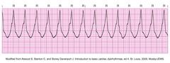
file:///Users/s08034274/Desktop/-OTDS1a6.ta6Dnfje8JBlw_m.jpg |
|
|
First Degree Heart Block
|
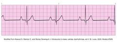
file:///Users/s08034274/Desktop/Vw2mGDyayAhgLGqJimrjNA_m.jpg |
|
|
Second Degree Heart Block
|
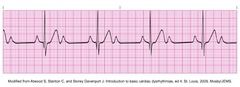
file:///Users/s08034274/Desktop/o5mgtypnIJOI-Uiu3c-cMQ_m.jpg |
|
|
Third Degree Heart Block
|
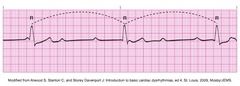
file:///Users/s08034274/Desktop/fYd7gCiATHE3GLBNLycJwQ_m.jpg |
|
|
When the SA node is stimulated, what mechanical action happens in the heart?
|
Atria contract
|
|
|
What has to happen for the ventricles to contract?
|
What has to happen for the ventricles to contract? Electrical conduction has to pass through the AV node, down the Bundle of His, through the right and left bundle branches, into the purkinje fibers. Once they reach the Purkinje fibers, then the ventricles will contract.
|
|
|
Name the 2 types of EKG machines:
|
Single channel and multichannel
|
|
|
What education should you give to your patient before starting the EKG?
|
It is a non-invasive test that won't hurt, have to lie still, clothing should be removed from the waist up, no jewelry, Any lotion needs to be removed with alcohol pads
|
|
|
How should the patient be positioned when performing an EKG?
|
supine position, arms at the sides
|
|
|
supraventricular rhythms are
|
above the AV NODE
|
|
|
Heart blocks are at
|
at the AV NODE
|
|
|
ventricular rhythms are
|
below the AV node
|
|
|
Premature Beats
|
QRS occurs (early) before next expected beatPremature Atrial Contraction (PAC) Premature Junctional Contraction (PJC) Premature Ventricular Contraction (PVC)
|
|
|
PVCs have the following features:
|
Broad QRS complex (≥ 120 ms) w/ abnormal morphology Premature: occurs earlier than expected for the next sinus impulse Discordant ST segment & T wave changesUsually followed by a full compensatory pauseRetrograde capture of the atria may or may not occur
|
|
|
Ventricular Rhythms
|
Wide QRSIdioventricular RhythmVentricular TachycardiaVentricular Fibrillation
|
|
|
ALTERNATING CURRENT
|
ARTIFACT THAT INDICATES ELECTRICAL INTERFERENCE
|
|
|
ANEURYSMECTOMY
|
SURGICAL REMOVAL OF AN ANEURYSM
|
|
|
ANGIOGRAPHY
|
THE PROCESS OF X-RAYING THE BLOOD VESSELS
|
|
|
ARRHYTHMIA
|
IRREGULAR HEART ACTION
|
|
|
ARTIFACT
|
UNWANTED, ERRATIC MOVEMENTS OF THE STYLUS ON THE PAPER
|
|
|
ARTIFACTS
|
UNWANTED, MOVEMENT OF THE STYLUS ON THE PAPER
|
|
|
AUGMENTED LEADS
|
COMBINED LIMB LEADS
|
|
|
BASELINE
|
THE HEART AT REST
|
|
|
CARDIAC CATHETERIZATION
|
A DIAGNOSTIC PROCEDURE WHICH DETERMINES OXYGEN CONTENT OF THE BLOOD AND MEASURES BLOOD FLOW PRESSURE
|
|
|
CARDIAC CYCLE
|
ONE COMPLETE HEART BEAT
|
|
|
CARDIAC EVENT MONITOR
|
A RECORDING DEVICE THAT CAN RECORD CARDIAC ACTIVITY FOR UP TO 30 DAYS
|
|
|
DEPOLARIZATION
|
CONTRACTION OF THE HEART
|
|
|
ELECTRODES
|
TEN SENSORS ON THE PATIENTS ARMS, LEGS AND CHEST
|
|
|
ISCHEMIA
|
DECREASED AMOUNT OF BLOOD SUPPLY TO THE HEART MUSCLE
|
|
|
LEAD WIRES
|
ATTACHED TO ELECTRODES TO RECORD THE PATIENTS ELECTRICITY
|
|
|
ST SEGMENT
|
TIME INTERVAL FROM THE END OF VENTRICULAR CONTRACTION TO THE BEGINNING OF VENTRICULAR RELAXATION
|