Reading...
![]()
Play button
![]()
Play button
![]()
Use LEFT and RIGHT arrow keys to navigate between flashcards;
Use UP and DOWN arrow keys to flip the card;
H to show hint;
A reads text to speech;
40 Cards in this Set
- Front
- Back
|
Describe where/how the pupillary sphincter and dilator muscles reside - also how they're innervated.
|
Pupillary are proximal to the pupil and are radially arranged. Sympathetic opens the pupil.
Sphincter are distal to the pupil and are concentric. Parasympathetic - constriction closes the pupil |
|
|
What are the extraocular muslces attached to?
|
Sclera
|
|
|
Sicne the cornea does not have a blood supply, how does it receive nutrients and protection from infection?
|
Tears and aqueous humor.
|
|
|
What are the five layers of the cornea?
|
1. Epithelium
2. Bowman's Membrane 3. Stroma 4. Descemet's Membrane 5. Endothelium |
|
|
What are the two primary functions of the cornea's endothelium layers? What purpose does its innervation have?
|
1. Block the passage of foreign material
2. Provide a smooth surface that absorbs oxygen and cell nutrients from tears. It's innervated with pain receptors |
|
|
What layer exists between the epithelium and stroma? What is it composed of and what happens if it's stratched?
|
1. Bowman's membrane. Composed primarily of collagen. If scratched, can form a scar.
|
|
|
What percentage of the cornea's thickness does the stroma contribute? What percentage of the focusing power? What percentage of water/collagen?
|
1. 90% of cornea's thickness
2. 80% of the eyes focusing power 3. 78% water and 16% collagen |
|
|
Where does Descemet's membrane belong? What does it do? What happens if injured?
|
It is a thin but strong sheet of tissue composed of collagen fibers just below the stroma. It protects. It's regenerated after injury.
|
|
|
What are the two principle jobs of the cornea's endothelium? What happens if the endothelium cells are damaged?
|
1. Keeps cornea clear and pump excess fluid out of the stroma.
2. If damaged, it is lost forever 3. If not functioning properly, corneal edema, blindness, |
|
|
What is the most common refreactvie error? Is the eye too long or short? Where does the image focus with respect to the retina? IS this near or farsightedness?
|
Myopia. The eye is too long. The image focuses in front of the retina. This is nearsightedness
|
|
|
What are the three parts of the Uvea
|
1. Choroid
2. Ciliary body 3. Iris |
|
|
What are the three components of the Choroid?
|
1. Vessel layer
2. Chorocapillary layer 3. Bruch's membrane |
|
|
Upon what layer of the choroid does the retinal pigmented epithelia rest?
|
Bruch's membrane
|
|
|
The ciliary body is an expansion of what? What three regions does it contact?
|
Expansion of the stroma of the choroid near the lens. It is in contact with the vitreous body, sclera, and posterior chamber/lens
|
|
|
The ciliary body has what that project to the lens. What are these projections called?
|
Ciliary processes. Zonula fibers
|
|
|
What fluid is the ciliary body responsible for absorbing?
|
Aqueous humor is drained from the anterior chamber via trabecular meshwork.
|
|
|
Describe the anterior aspect of the Iris. What does it contain?
|
It is made up of the vascular, loose CT with interspersed melanocytes. The number of melanocytes determines eye color.
|
|
|
The posterior surface of the iris is lined with what? What is the point?
|
Pigmented epithelium. IT helps to reduce scatter.
|
|
|
Upon which layer does the two muscles masses rest upon in the iris (i.e. the sphincter pupillae and the concentric pupillae)?
|
pigmented epithelium
|
|
|
What produces the aqueous humor? Describe the flow of the aqueous humor.
|
Ciliary process. It passes from the post to anterior chamber between the iris and the lens. It is drained via trabecular meshwork. From there it passes into the canal of Schlemm.
|
|
|
There are two types of Gluacoma. What are they and which is the most common?
|
Primary Open Angle Glaucoma and Angle Closure Glaucoma. Open angle is the most common.
|
|
|
Glaucoma is damage to the nerve. This can happen in two ways:
|
1. Increased pressure
2. Normal pressure - "Normal tension glaucoma" |
|
|
What does "secondary glaucoma" refer to?
|
Any case in which another disease causes or contributes to increased eye pressure.
|
|
|
Describe Open-angle glaucoma.
|
It is caused by to an obstruction in the drainage system of the eye.
|
|
|
What is normal pressure of the eye? What is normal cup to optic disc ratio?
|
12-22 mmHg; 0-1. Higher is worse.
|
|
|
Describe angle-closure glaucoma.
|
It is the acute form of the disease where there is poor access to the drainage system in the eye.
|
|
|
Describe the three structural components of the lens
|
1. Capsule - ECM surrounding lens
2. Epithelium - anterior surface of lens 3. Lens fiber - body of the lens |
|
|
The lens has lower refractive power than cornea (~ ? D) However it can do this:
|
10 D
Accommodate |
|
|
Describe accommodation. What happens when some tries to focus on something near?
|
The ciliary muscles CONSTRICT to relax the fibers on the lens - cause the lens to thicken. The opposite happens when trying to focus on far object.
|
|
|
What are the two major components of vitreous body?
|
1. Type 2 collagen
2. Hyaluronic acid |
|
|
The retina has two regions derived from different layers of the optic cup during embryogenesis:
|
1. Neural or sensory retina
2. Retinal pigment epithelium (RPE) |
|
Name these layers
|
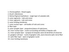
a
|
|
|
The fovea has a high concentration of rods or cones?
|
Cones
|
|
|
In regard to a photoreceptor, what is in the inner segment and what is in the outer segment?
|
Inner: Organelles for protein synthesis and E production
Outer: Flattened membrane discs with photosensitive visual pigments. |
|
|
Describe the distribution of rods and cones
|
Rods are most numerous except at fovea. They are very sensitive to light
Cones are most numerous at fovea and have membrane discs. Responsible for high acuity & color vision |
|
|
Is there a blood supply to the fovea? Why or why not?
|
No. The vascularture is in the inner layers and since the fovea doesn't have an inner, it does not have a blood supply.
|
|
|
What are the major functions of the RPE:
1. With respect to light 2. With respect to nutrients 3. With respect to retinal 4. With respect to outer segment 5. With respect to structural integrity of retina |
1. It absorbs scatter light
2. Transports nutrients and ions bn photoreceptors & choriocapillaris 3. Reisomerizes all-trans retinal 4. Renews outer segment ~10%/d 5. Secretion of growth factors for maintenance and structural integrity of retina |
|
|
What two vessels supply the retina?
|
1. Outer retina is supplied by choriocapillaries
2. Inner is supplied by central retinal artery. |
|
|
The central retinal artery is a branch of which artery?
|
The opthalmic artery
|
|
|
Impaired foveal specialization is a hallmark of diseases such as: (2)
|
1. Aniridia
2. Albinism. |