![]()
![]()
![]()
Use LEFT and RIGHT arrow keys to navigate between flashcards;
Use UP and DOWN arrow keys to flip the card;
H to show hint;
A reads text to speech;
66 Cards in this Set
- Front
- Back
|
What are the three body systems which facilitate movement? |
Skeletal system: bones that act as levers and provide structure for muscles to pull Muscular system: muscles deliver force to move one bone in relation to another Nervous system: delivers signals to muscles which cause the to contract and create movement |
|
|
What are the three types of muscles? |
Smooth muscles: in organs, involuntary constriction, not striated, spindle shape, each fibre has single nucleus Cardiac Mucle: In heart, branching, lightly striated, intercalated, single nuclues per fibre Skeletal muscle: connecting to bone, heavily striated, run in parallel tracts, multinucleated |
|
|
What are the function of skeletons? |
-provide support and protection for body organs -provide surface for muscle attachment therefore facilitating movement |
|
|
Describe the two different types of skeletons |
Endoskeletons: internal skeletons which typically consist of numerous bones Exoskeletons: external skeletons which are comprised of connected segments |
|
|
Bones are connected to other bones by ___________ an bones are connected to muscles by __________. |
ligaments, tendons |
|
|
What are synovial joints? What are joint's function? |
capsules that surround the areas where two bones connect. Joints maintain structural stability by allowing certain movement but restricting others. |
|
|
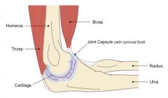
What are the three main components of synovial joints? What is the function of each? |
Joint capsule: seals the joint, gives stability, and restricts range. Cartilage: Lining on bone surface that absorbs shock, distributes load, and facilitates smoother movement. Synovial fluid: provides oxygen and nutrients to cartilage and reduces friction by lubricating the bones |
|
|
What are the six main types of synovial joints? |
PBS CHiP Pivot joint, Ball and socket joint, Saddle joint Condyloid joint, Hinge joint, Plane joint |
|
|
What kind of joint is the human elbow joint? what does it connect? What movement does it allow? |
The elbow joint is a hinge joint that connects the humerus and the radius+ulna. It can move in one direction with flexion and extension only, with only a small amount of possible rotation |
|
|
Draw and annotate the elbow joint |
|
|
|
What are muscles? How do they work in antagonistic pairs? |
-contracting tissue that provide force to move bones. -they connect at the point of origin and the point of insertion (the static and the moving bone) -they work in antagonistic pairs. E.g. when one contracts, the other relaxes, allowing opposing movements. e.g bicep (flexion) and tricep (extension) |
|
|
What example of antagonistic muscles allow insects like the grass hopper to jump? |
the flexor tibiae muscle and extensor tibiae muscle, which connect the femur and the tibia When the flexor contracts, it brings the tibia and femur closer together, and when the extensor muscle contracts, it extends the leg, allowing the insect to jump |
|
|
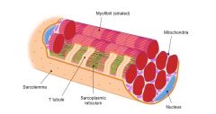
Outline the structure of skeletal muscles |
-skeletal muscles consist of tightly packed muscular bundles (fascicles) surrounded by perimysium. -each fascicle contains muscle fibres (multiple muscle cells fused together) -each muscle fibre contains tubular myofibrils that are responsible for contraction -each myofibril is made up of repeating contractile units called sarcomeres |
|
|
What specialized features do muscle fibres have which facilitate muscle contraction? |
-multinucleated (acd cell fusion)
-many mitochondria (high ATP consumption) -sarcoplasmic reticulum stores calcium ions for muscular contractions -contian myofibrils made up of thin actin and thick myosin myofilaments -sarcholema surrounds the entire muscle fibre and has invaginations called T tubules |
|
|
Draw a muscle fibre. |
|
|
|
What is the function of myosin and actin within the sarcomere? |
They move in relation to each other, causing the lengthening and shortening of the sarcomere |
|
|
What are Z lines? |
dense protein disks that hold myofilaments in place from. actin radiates from the Z disks and it also helps keep myosin in place |
|
|
What is the visual result of the sarcomere? |
A striped pattern along the length of muscle fibres -the center of the sarcomere is dark due to actin and myosin overlap (A band) -the peripheries appear lighter due to actin only (I band) -A slightly lighter region in the middle of the A band may appear due to myosin only (H zone) |
|
|
When drawing the sarcomere what should you keep aware of? |
-myosin is thicker than actin -myosin filaments have protruding heads -striated banding pattern should be named (e.g. A band, H zone, I band, Z disk) |
|
|
What are the four steps of muscle contraction? |
-depolarization and calcium ion release -actin and myosin cross-bridge formation -sliding mechanism of actin and myosin filaments -sarcomere shortening (muscle contractions) |
|
|
Explain the first stem of muscle contraction: depolarization and calcium ion release |
-motor neuron releases acetylcholine into the motor end plate acd action potential -acetylcholine triggers depolarization in sarcolemma which spreads through muscle fibre via T tubules -depolarization triggers Ca2+ release from sarcoplasmic reticulum |
|
|
Explain the second step of muscle contraction: Actin and mysoin cross-bridge formation |
-the binding sites on actin for myosin heads are covered by troponin and tropomyosin (troponin is the little heads, tropomyosin is the ribbon which covers binding sites) -Ca2+ binds to troponin and moves the tropomyosin, exposing binding sites -myosin heads form a cross bridge with actin filaments |
|
|
Explain the third step of muscle contraction: sliding mechanism of actin and myosin |
-ATP binds to myosin head, breaking cross-bridge btwn actin and myosin -ATP hydrolysis causes myosin heads to change position and swivel, moving to the next actin binding site -myosin binds to new actin site and returns to original configuration -this reorientation drags actin along myosin: sliding mechanism |
|
|
Explain the fourth step of muscle contraction: sarcomere shortening |
-repeated reorientation of myosin heads drags actin along myosin -drags Z lines closer together acd actin anchorage -I band gets shorter/non existant -individual sarcomeres become shorter in length and the whole muscle fibre contracts |
|
|
How can you tell from a micrograph that a saromere is contracting? |
-I bands get smaller -H zones get smaller -A bands stay the same length -Z lines get closer together |
|
|
Describe slow twitch fibres |
-muscular endurance, contract slowly, don't fatigue easily -many mitochondria, use oxygen, aerobic -red acd many blood vessels -prevalent in endurance athletes |
|
|
Describe fast twitch fibres |
-strength, contract fast, fatigue easily -anaerobic, less mitochondria, -lighter acd less blood vessels -prevalent in strength athletes |
|
|
What is urea? How and why is it produced? |
A waste product from the metabolism of amino acids. AA cannot be stored in the body, so via deamination they become ammonia, urea, or uri acid. |
|
|
What is deamination |
The removal of the amine group |
|
|
What do animals use to excrete nitrogenous wastes from the blood stream. |
It depends. Some animals (all mammals), use the kidneys. Insects use the Malipighian tubules. |
|
|
If an animal's ancestor used uric acid to excrete nitrogenous waste, what would they use to excrete nitrogenous waste? |
Likely, uric acid. This is because animals cannot undergo an entirely new physiology even though they qualify as a new species. |
|
|
What are the advantages and disadvantages of excreting nitrogenous wastes as ammonia? |
Pro: requires little energy to produce Con: so toxic that it must be diluted and removed quickly by using a lot of water e.g. Fish |
|
|
What are the advantages and disadvantages of excreting nitrogenous wastes as urea? |
Pro: less nrg than uric acid. toxic, but only at abnormally high levels. Con: requires more nrg than ammonia, requires some water for dilution and removal e.g. mammals |
|
|
What are the advantages and disadvantages of excreting nitrogenous wastes as uric acid? |
Pro: insoluble in aq solutions like blood and cytoplasm. can be stored in specialized structures within some eggs. require little or no water for dilution and excretion. Con: requires lots of energy to produce acd complexity e.g. birds |
|
|
How did the fishes evolutionary history effect its form of nitrogenous waste? |
Ammonia acd unlimited supply of water and nrg efficiency |
|
|
How did mammals' evolutionary history effect its form of nitrogenous waste? |
urea acd less water accessibility than fish. urea isn't as toxic so mammals can cope as long as kidneys work. can be stored temporarily in urinary bladder. |
|
|
How did birds' evolutionary history effect its form of nitrogenous waste? |
because of self-contained egg. needed waste that was insoluble and could be stored (unlike ammonia). uric acid can be stored in developing egg while bird develops. adult birds use urea so they won't have to find water as frequently. |
|
|
How do insects excrete nitrogenous waste? |
-insects have open circulatory system, bathing organs in blood -body cavities have Malpighian tubules in pools of blood -tubes have distal end and proximal end -components of blood enter at distal end and go through selective reabsorption, leaving uric acid, excess water, and Na+, K+. and Cl- behind -wastes dumped into gut, excreted as feces |
|
|
How does blood in the renal artery compare to blood in the renal vein? |
-differ in levels of water, salt ions, and urea -acd filtering action of nephrons |
|
|
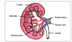
What is urine? How is it produced? |
Urine is the fluid produced by the kidneys. it consists of water and dissolved waste products from the blood stream. Urine is produced by the nephrons and collects in the renal pelvis, which drains it into a tube called the ureter. This connects to the urinary bladder. |
|
|
Draw and annotate a diagram of the kidney |
|
|
|
What are nephrons? How many are there in a kidney and what do they consist of? |
-Filtering units of kidneys. -1.25 million -capillary bed (glomerulus) filters some substances from blood -Bowman's capsule surrounds glomerulus -tubule extends from Bowman's capsule which is proximal convoluted tubule, loop of Henle, and distal convoluted tubule -2nd capillary bed (peritubular capillary bed) surrounds 3 part tuble |
|
|
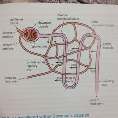
Draw and label a diagram of the nephron |
|
|
|
What is ultrafiltration? Where does it occur? |
Ultrafiltration: various substances are filtered through the glomerulus (and its fenestrations) under unusually high blood pressure in this capillary bed. It occurs in the Bowman's capsule. |
|
|
What is the function of the basement membrane, efferent and afferent arteriole, and capillary fenestrations? |
basement membrane: prevents large molecules from becoming a part of the filtrate efferent and afferent arteriole: afferent is wider diametre, efferent narrower, causing high pressure fenestrations: allow larger molecules out when they open under high pressure |
|
|
Why is reabsorption important? Where does it occur? |
After ultrafiltration, many of the substances in the filtrate are indispensible and need to be reabsorbed (water, salt ions, glucose). This occurs via the proximal convoluted tubule and into the peritubular capillary bed |
|
|
How does the structure of the proximal convoluted tubule facilitate reabsorption? |
-microvilli: increase SA for reabsorption -one cell thick: for quick diffusion into capillaries -mitochondria: for active transport |
|
|
How are salt ions reabsorbed? |
-majority (but not Na+, CL-, and K+) leave filtrate via reabsorption -actively transported into tubule cells and then into intercellular fluid outside tubule -ions taken into peritubular capillary bed |
|
|
How is water reabsorbed? |
-movement of salt ions induces water to follow same route via osmosis -under normal circumstances most water stays in filtrate until body can decide how much water needs to be kept |
|
|
How is glucose reabsorbed? |
-all glucose is reabsorbed in healthy nephron -via active transport -cannot be facillitated diffusion because gradient would disappear 50% through |
|
|
What is osmoregulation? |
Body's response mechanism that attempt to maintain homeostatic levels of water. any regulatory mechanism that affects water balance in an animal's body. |
|
|
What physiological factors determine how much water is excreted in urine? |
-total recent volume of water ingested recently -respiration rate (via excercise and temperature) -ventilation (via exercise acd exhalation) |
|
|
What occurs after reabsorption in the proximal convoluted tubule? discuss the permeability of the ascending and descending loop of Henle. |
-filtrate travels down descending loop of Henle. -segment is permeable to water and impermeable to salt -filtrate enters ascending where it is impermeable to water and permeable to salt ions. -salt ions are pumped out inter intercellular fluid |
|
|
The medulla is _________. Why? |
hypertonic. acd loop of Henle descending into medulla. causes some water to move out via osmosis, but most stays in the ascending loop and goes into the distal convoluted tubule (still pretty hypotonic). |
|
|
Why is water reabsorbed in the collecting ducts? |
-water content is still high in "urine" (hypotonic) -interstitial fluid in medulla is hypertonic -if no reabsorbtion occured, we'd have to drink a lot of water to make up for water loss |
|
|
The collecting duct is differentially permeable to water. Why? |
Acd ADH (antidiuretic hormone). If present, it makes the collecting duct permeable to water and water moves by osmosis into the hypertonic medulla interstitial fluid. From there water enters the peritubular capillary bed. If it isn't present, collecting duct is impermeable to water and it stays in the collecting duct. |
|
|
by what and to where is ADH secreted? |
What: secreted by the posterior lobe of the pituitary gland where: circulates in the blood stream and acts on target tissue of the kidney collecting duct |
|
|
Why does the loop of Henle differ in some animals? |
there is a positive correlation between loop of Henle length and animal's need to conserve water. e.g. kangaroo rats take in very little water (only from food) and recycle most water by having a very long loop of Henle that produces a large hypertonic area for water re absorption via ADH and collecting duct |
|
|
What is the blood like in the renal vein compared to the renal artery? |
-less urea -less salt ions (Na+, K+, Cl-, etc.) -less water -nearly same amount of glucose -nearly same amount of protein -no change in blood cells |
|
|
What are osmoregulators? What are osmoconformers? |
osmoregulators: have a different internal solute conc. compared to environment. they can osmoregulate and expend a great deal of energy on these mechanisms osmoconformers: water moves in and out freely due to osmotic balance. solute conc. is the same as the environent. must be very basic and restricted to living in environments that are iso-osmotically matched. |
|
|
What is haemodialysis? |
-mechanism to remove waste from blood after kidney failure -patient's blood moves through tube w/ a semi-permeable membrane into a dialysis solution -urea is small enough to move through -dialysis has no urea, so urea moves out via concentration gradient -must be repeated every 1-3 days, takes a few hours |
|
|
What is a kidney transplant? |
-taking a kidney from a suitable donor (tissues match) and putting it in a recipient -people can live normally with one kidney, so a close family member can donate one -after recieving it, patient must recieve immune-suppressing drugs for the rest of their life. |
|
|
What might the composition of urine tell you about your health? |
glucose: there should be no glucose in urine in healthy individual blood cells: blood cells are too big to fit in fenestrations, so this is a sign of kidney malfunction, infection, or renal tube bleeding Proteins: too large to leave fenestratins, should not be found Drugs: leaves body via kidneys, so people may be tested for them (e.g. sports teams) |
|
|
What are the symptoms of dehydration? |
-sleepiness -constipation -dry mouth and skin -dizziness and headache |
|
|
What are the symptoms of overhydration? |
-change in behaviour/confusion -blurred vision -muscle cramps -nausea and vomiting |
|
|
Why is water so important? |
It is the solvent of life (the solvent component in blood and cytoplasm) |