Reading...
![]()
Play button
![]()
Play button
![]()
Use LEFT and RIGHT arrow keys to navigate between flashcards;
Use UP and DOWN arrow keys to flip the card;
H to show hint;
A reads text to speech;
26 Cards in this Set
- Front
- Back
|
Blank
|
Blank
|
|
|
Epithelial types and location - squamous
|
lining surfaces utilizing simple passive diffusion -
Lines blood vessels (as endothelium) and the pleural, peritoneal and other serous cavities (as mesothelium) Comprises the parietal layer of Bowman’s capsule, the thin loop of Henle, etc. |
|
|
Epithelium types and location - cuboidal
|
Found in secretive or absorptive tissue.
Lines distal tubules in the kidney, follicles in the thyroid gland, the surface of the ovary, etc. Simple cuboidal lines the ducts of sweat glands. |
|
|
Epithelium types and location - columnar
|
Lines the stomach, intestine and the excretory ducts of many glands
|
|
|
Epithelium types and location - pseudostratified
|
Ciliated epithelium found in airways of the nose and lungs, in the uterus and fallopian tubes where cilia propel the ovum.
|
|
|
Describe the structures of epithelial cells and give examples of sites - simple
|
One cell thick, every cell in contact with underlying basement membrane. Found where absorption and filtration occur.
|
|
|
Describe the structures of epithelial cells and give examples of sites - stratified.
|
Found where body linings have to withstand mechanical or chemical insult.
Keratinized found in skin and the lining of the esophagus. Transitional found in the bladder, ureter and urethra. |
|
|
Desribe the apical modification of epithelia -
|
Microvilli - Extension of the membrane, Brush border, Absorption
Stereocilia - Long microvilli Cilium (pl. cilia) - 9+2 microtubules, Movement Kinocilium - 9+0 microtubules, Sensory Flagellum - 9+2 microtubules, Movement |
|
|
Describe the basolateral modification of epithelia
|
Basal folds - increase reabsorbtive area
Junctions - tight, intermediate and desmosomes. Hemidesmosome - half a desmosome connecting to basement membrane. Gap junctions - allow flow of ions |
|
|
Tight Junctions
|
Zonula Occludens
Location: Apicolateral Ligand: Occludins |
|
|
Desmosomes
|
Macula Adherens
Location: Midlateral Ligans: CAMs, Desmoplakins |
|
|
Intermediate Junctions
|
Zonula Adherens
Location: Apicolateral Ligand: Vinculin |
|
|
Hemidesmosomes
|
Focal Adhesion
Location: Basolateral Ligand: Integrin/Laminin |
|
|
Gap Junctions
|
Zonula Communicans
Location: Lateral Connexon made by 6 Connexins |
|
|
Describe the classes of glands.
|
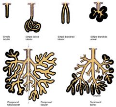
Simple gland: the duct does not branch
Compound gland: the duct branches Shape of secretory unit defines whether the gland is classified as acinar or alveolar (sac or flasklike) or tubule (either straight, coiled or branched) |
|
|
Describe the types of glandular secretions
|
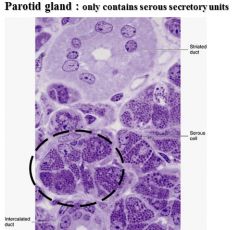
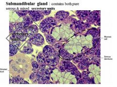
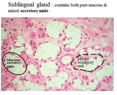
May be classified as mucous, serous or mixed
Mucus is a viscous material produced by mucous glands that usually protects or lubricates Serous secretions (from serous glands) are watery and often rich in enzymes |
|
|
Parotid Glands
|
|
|
|
Submandibular Glands
|
|
|
|
Sublingual Gland
|
|
|
|
Cilia and Chronic Sinusitis, Recurrent Ear Infections and Bronchiectasis
|
Cilia damage or mutation is common from Chronic Sinusitis. A loss of ciliated cells and an increasing number of nonciliated columnar cells with microvilli, ciliary disorientation, metaplasia and extrusion of epithelial cells. Compound cilia and Short cilia were often seen in infected mucosa indicating ciliogenesis.
|
|
|
Kartagener's Syndrome
|
Primary ciliary dyskinesia (PCD), also known as immotile ciliary syndrome. A defect in the action of the cilia lining the respiratory tract (lower and upper, sinuses, Eustachian tube, middle ear) and fallopian tube. Any impairment can result in poor mucociliary clearance, with subsequent upper and lower respiratory infection. Also responsible for situs inversus (inversion of organs) and sterility in males.
|
|
|
Polycystic Kidney Disease
|
Characterized by the presence of multiple cysts in both kidneys. The cysts are numerous and are fluid-filled resulting in massive enlargement of the kidneys. The disease can also damage the liver, pancreas, and in some rare cases, the heart and brain.
|
|
|
Junctional complexes as target of pathonogenic agents.
|
They are the target of pathogens seeking to gain entry into individual cells and tissues and that mutations in the genes that encode them lead to significant tissue and organ defects in metabolic diseases and cancers.
|
|
|
Bullous Vesicular Lesions
|
Bullous pemphigoid, also referred to as BP, is an acute or chronic autoimmune skin disease, involving the formation of blisters, more appropriately known as bullae, at the space between the skin layers epidermis and dermis. The bullae are formed by an immune reaction, initiated by the formation of IgG[1] autoantibodies targeting hemidesmosomes. It can also rarely involve the mucous membranes. Results in a separation along the dermal-epidermal junction and eventually stretch bullae.
|
|
|
Epithelial Metaplasia
|
When cells are faced with physiological or pathological stresses they respond by adapting in several ways, possibly changing into another, more protected type of epithelium.
|
|
|
Epithelial Carcinoma
|
Tumors can arise from epithelial tissue - carcinoma.
Benign: Chondroma, Lipoma Malignant: Carcinoma, Sarcoma |