Reading...
![]()
Play button
![]()
Play button
![]()
Use LEFT and RIGHT arrow keys to navigate between flashcards;
Use UP and DOWN arrow keys to flip the card;
H to show hint;
A reads text to speech;
7 Cards in this Set
- Front
- Back
The zona orbicularis is oscopic landmark for access to which structures? 1-Iliopsoas; 2-Pectineus ;3-Sartorius
4-Adductor brevis; 5-Rectus femoris |
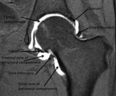
zona orbicularis is the oscopic landmark for access to the iliopsoas. Arthroscopic release of the iliopsoas is performed for tx of an internal snapping hip,caused by the iliopsoas snapping over the iliopectineal eminence or the femoral head, zona orbicularis is a ring of capsular tissue, hip arthoscopy Identification of the zona orbicularis can guide the surgeon to the iliopsoas tendon that is immediately deep to this structure.Ans1
|
|
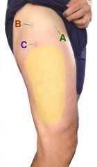
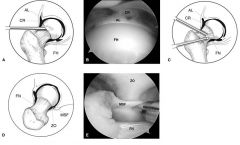
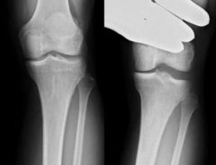
Hx: 29yo M undergeoes hip oscopy using the 3 portals Fig A. Postop he develops numbness in the distribution shown in yellow. This complication was most likely caused by ? 1-Inj to the Pudendal N->Portal A; 2-Inj to the Fem N -> Portal B; 3-Inj to the Lat Fem Cutaneous N -> Portal A; 4-Inj to the Common Peroneal N from Portal C
5. Injury to the a sensory branches of the sciatic nerve from Portal B |
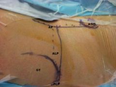
LFCN is@ >est risk of inj w/ placement of the ant portal, Ans 1: Transient N inj -> groin (pudendal N) is 2^ traction against the perineal post, Ans 2: ant portal that is too far med risks inj to the fem N, Ans 4: Transient N inj affecting the dorsum of the ft (peroneal) 2^ traction, Ans 5: The sciatic N does not have sensory branches innervating the distribution in Fig A.Ans3
|
|
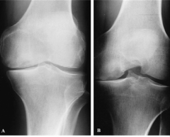
Which of the following xrays views is most sensitive for detecting knee jt OA changes? 1-NWB AP; 2- WB AP; 3- NWB PA in 45 deg flex;4-WB PA in 45 deg flex 5-Merchant
|
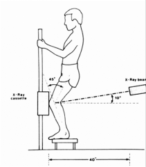
45 deg PA flex WB view is the best for demonstrating subtle joint-space loss, especially in the lateral compartment, earliest loss of cartilage occurs in the 30 to 60 deg flexion zone which is easily overlooked on x-rays in full extension. WB views are always preferrable when evaluating for arthritis.Ans4
|
|
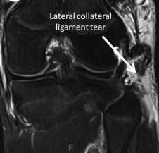
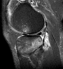
Hx:27yo soccer player injures his knee during a game. PE-Lachman is 1A, laxity to varus stress w/ knee flex @ 30 deg. Dial test = incr ER @ 30 deg, but not at 90 deg in comparison to the contralateral leg. Which structure(s) are torn? 1-ACL; 2-LCL; 3-ACL & LCL; 4-LCL & PLC; 5-PCL &PLC
|
LCL is part of the posterolateral corner, but can be injured in isolation or along with the rest of the posterolateral corner. An isolated LCL tear= flex knee @ 30 deg and applying varus stress. posterolateral corner = dial test, ER the affected tibia. A PLC injury = incr'd ER @ 30 deg, while a combined PLC/PCL injury = incr'd ER @ 30 & 90 deg, Grade 1A indicates less than 5mm of translation and a firm endpoint indicating that the ACL is intact.Ans4
|
|
In relation to the femoral insertion of the popliteus, the femoral attachment of the LCL is? 1-pos & prox
2-pos & distal; 3-ant & prox; 4-ant & distl; 5-directly superficial |
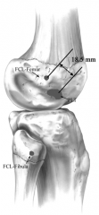
LCL's femoral attachment is posterior and proximal compared to the popliteus femoral insertion with the average distance between their femoral attachments being 18.5 mm.Ans1
|
|

Which of the following most accurately describes the primary role of satellite cells? 1-To act as an intermediary in the cell-signalling pathway for bone remodeling; 2-To regenerate skeletal muscle p/ mus inj; 3-To regen periosteum p/ periosteal damage in a child; 4-To bind chemotherapeutic ligands in the tx of lymphoma of bone; 5-To express high amounts of sonic hedgehog surface protein
|
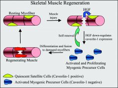
primary role of satellite cells is to regenerate skeletal muscle after muscle injury, 1: The osteoblast acts as an intermediary in the cell-signalling pathway for bone remodeling. 3: Satellite cells are not involved in the regeneration of periosteum, 4: Satellite cells play no role in binding chemotherapeutic drugs, 5: Sonic hedgehog surface protein is involved in limb bud generation.Ans2
|
|
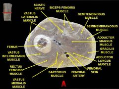
Concomitant flexion of the hip and extension of the knee is most likely to result in an injury to which structure? 1-Sartorius; 2-Rectus femoris; 3-Adductor magnus; 4-Biceps Femoris; 5-Tensor fascia lata
|
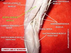
Flex of the hip & ext of the knee most likely would injure the hamstring. The hamstring is composed of the semimembranosus, semitendinosus, and biceps femoris and all 3 components originate at the ischial tuberosity, rectus fem is 1 of 4 of quad, vastas-med, lat, inter; biceps femoris- in the back of thigh as a hamstring.Ans4
|