Reading...
![]()
Play button
![]()
Play button
![]()
Use LEFT and RIGHT arrow keys to navigate between flashcards;
Use UP and DOWN arrow keys to flip the card;
H to show hint;
A reads text to speech;
57 Cards in this Set
- Front
- Back
|
What are the scolex, neck and strobila portions of a tapeworm?
|
scolex - head
neck - new segments are added here strobila - reproductive system is located here |
|
|
What are the main anatomical points of the scolex of a tapeworm?
|
may contain:
multiple acetabulem hooks bothria - pits or grooves bothridia - muscular projections that physically hold onto host |
|
|
What are the main anatomical points of the strobila of a tapeworm?
|
linear series of reproductive organs (hermaphroditic) each referred to proglottid
can be monozoic - one set of proglottid polyzoic - multiple sets of proglottids |
|
|
What is strobilation?
|
the process of new proglottids that arise at the anterior end of the strobila
|
|
|
What are the three types of proglottids?
|
immature - immature reproductive organs
mature - mature reproductive organs gravid - shelled-embryo containing proglottids |
|
|
What types of copulation can occur with tapeworms?
|
inter-proglottid
intra-proglottid inter-worm |
|
|
What is the significance of Hymeolepis proglottids?
|
proglottids shed and disintegrate en route through intestines of host (humans) and the only thing left in feces
|
|
|
What is the significance of Teania & Dipylidium proglottids?
|
proglottids shed, but remain intact in feces
|
|
|
What is the significance of Diphyllobothrium proglottids?
|
Proglottids are retained and eggs are released from proglottids
|
|
|
Where are the hosts of tapeworms?
|
EVERY VERTEBRATE CLASS
|
|
|
What two diseases arise from infection of Taenia solium?
|
Taeniasis - adult tapeworm infection
Cysticercosis - larvae infection in flesh to be consumed and infected |
|
|
What are the definitive and intermediate hosts of Taenia solium?
|
definitive - human
intermediate - pig |
|
|
How are humans infected with Taenia solium?
|
1) eating undercooked pork of which contains cysticerci (larval stage of adult tapeworm)
2) cysticerci developed in humans by consuming eggs accidentally from human feces |
|
|
Where is Taenia solium usually found, geographically?
|
Latin America, Africa, India, SE Asia, China, Slavic countries
|
|
|
What is the structure of a Taenia solium scolex?
|
4 acetabula
crowned rostellum (hooked ring around primary oral sucker) |
|
|
What is the lifecycle of the adult Taenia solium (and Taenia saginata for that matter)?
|
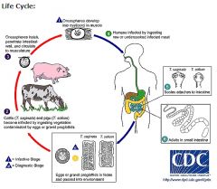
Taeniasis is the infection of humans with the adult tapeworm of Taenia saginata or Taenia solium. Humans are the only definitive hosts for T. saginata and T. solium.
Eggs or gravid proglottids are passed with feces 1; the eggs can survive for days to months in the environment. Cattle (T. saginata) and pigs (T. solium) become infected by ingesting vegetation contaminated with eggs or gravid proglottids 2. In the animal's intestine, the oncospheres hatch 3, invade the intestinal wall, and migrate to the striated muscles, where they develop into cysticerci. A cysticercus can survive for several years in the animal. Humans become infected by ingesting raw or undercooked infected meat 4. In the human intestine, the cysticercus develops over 2 months into an adult tapeworm, which can survive for years. The adult tapeworms attach to the small intestine by their scolex 5 and reside in the small intestine 6. Length of adult worms is usually 5 m or less for T. saginata (however it may reach up to 25 m) and 2 to 7 m for T. solium. The adults produce proglottids which mature, become gravid, detach from the tapeworm, and migrate to the anus or are passed in the stool (approximately 6 per day). T. saginata adults usually have 1,000 to 2,000 proglottids, while T. solium adults have an average of 1,000 proglottids. The eggs contained in the gravid proglottids are released after the proglottids are passed with the feces. T. saginata may produce up to 100,000 and T. solium may produce 50,000 eggs per proglottid respectively. (pic) |
|
|
What are mild symptoms of a T. solium infection?
|
asymptomatic
hunger pains abdominal discomfort chronic indigestion increased WBC |
|
|
What is tx for T. solium?
|
Praziquantel (veterinary use, but also used in humans) - preferred
Niclosamide (human use, but regulated by the CDC) |
|
|
Cysticercosis
|
egg ingestion that causes Cysticerci infection of Taenia solium in the muscles of the host
|
|
|
What is the path of infection for cysticercosis?
|
1) Humans ingests eggs present in human feces (contaminated hands, eating utensils, oral sex)
2) Eggs hatch in SI (stimulated by bile salts) 3) Oncosphere penetrates wall of the SI, gaining entrance into systemic circulation via the hepatic portal vein. 4) Cysticerci (<10 mm max dia at maturity) prefers subcutaneous tissues: Between muscle bundles Eye Brain |
|
|
How long does it take to get cysticercosis from ingestion of an egg?
|
~ 2 months
|
|
|
What do cysticercus look like?
|
semitransparent, opalescent white
elongate oval shaped 0.6 - 1.8 cm bladder filled w/ fluid aka bladder worms |
|
|
What are cysticercosis symptoms?
|
CNS symptoms
eye symptoms atrophy and pain when contracting infected muscle |
|
|
What does the cyst of a cysticercosis do when the cysticercus is killed by an immune response?
|
The cyst calcifies and makes a calcified cyst, which is where "crunchy pork" comes from.
|
|
|
What is the portion of the lifecycle of a Taenia solium that causes Cysticercosis?
|
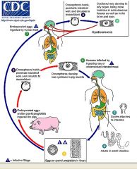
Cysticercosis is an infection of both humans and pigs with the larval stages of the parasitic cestode, Taenia solium.
This infection is caused by ingestion of eggs shed in the feces of a human tapeworm carrier 1. Pigs and humans become infected by ingesting eggs or gravid proglottids 2, 7. Humans are infected either by ingestion of food contaminated with feces, or by autoinfection. In the latter case, a human infected with adult T. solium can ingest eggs produced by that tapeworm, either through fecal contamination or, possibly, from proglottids carried into the stomach by reverse peristalsis. Once eggs are ingested, oncospheres hatch in the intestine 3, 8 invade the intestinal wall, and migrate to striated muscles, as well as the brain, liver, and other tissues, where they develop into cysticerci 9. In humans, cysts can cause serious sequellae if they localize in the brain, resulting in neurocysticercosis. The parasite life cycle is completed, resulting in human tapeworm infection, when humans ingest undercooked pork containing cysticerci 4. Cysts evaginate and attach to the small intestine by their scolex 5. Adult tapeworms develop, (up to 2 to 7 m in length and produce less than 1000 proglottids, each with approximately 50,000 eggs) and reside in the small intestine for years 6. |
|
|
what is nematode related ecdysis?
|
epidermal cuticle is shed four times to reach the adult morphology. cuticle shedding is called ecdysis
|
|
|
What is special about nematode sexuality?
|
sexually dimorphic and (smaller) males have paired copulatory spicules that bite into female vagina during mating
|
|
|
What are the two morphological forms of nematodes that are infectious?
|
filariform - infectious larvae
rhiabditiform - free-living stage |
|
|
What species of nematode are relevant to human infection?
|
Ascaris lumbricoides - largest nematode worm, found in areas of poor sanitation
|
|
|
*Where are Ascaris lumbricoides found geographically?
|
temperate and tropical areas - rural areas of SE United States*
|
|
|
How long does it take for Ascaris lumbricoides to undergo a complete lifecycle?
|
4 mos
|
|
|
What is the lifecycle of an Ascaris lumbricoides?
|
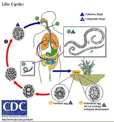
Adult worms 1live in the lumen of the small intestine.
A female may produce approximately 200,000 eggs per day, which are passed with the feces 2. Unfertilized eggs may be ingested but are not infective. Fertile eggs embryonate and become infective after 18 days to several weeks 3, depending on the environmental conditions (optimum: moist, warm, shaded soil). After infective eggs are swallowed 4, the larvae hatch 5, invade the intestinal mucosa, and are carried via the portal, then systemic circulation to the lungs 6. The larvae mature further in the lungs (10 to 14 days), penetrate the alveolar walls, ascend the bronchial tree to the throat, and are swallowed 7. Upon reaching the small intestine, they develop into adult worms 1. Between 2 and 3 months are required from ingestion of the infective eggs to oviposition by the adult female. Adult worms can live 1 to 2 years. (pic) |
|
|
What are Ascaris lumbricoides infection symptoms?
|
alveolar destruction -> asthma, pneumonia
hepatopancreatiatits loose stools (similar to loose stools) fever malaise abdominal distension tenderness vomiting alveolar hemorrhage |
|
|
Tx for Ascaris lumbricoides
|
Mebendazole for all roundworms stimulates worm migration
Endoscopy if necessary |
|
|
What does Wuchereria bancrofti cause?
|
elephantiasis
|
|
|
Where are Wuchereria bancrofti geographically found?
|
tropics and subtropics
|
|
|
What is special about Wuchereria bancrofti infectious third-stage filarial larvae?
|
These are the infectious stage of W. bacncrofti delivered by mosquitoes to a human in a bloodmeal
|
|
|
What are the hosts for Wuchereria bancrofti?
|
intermediate - Culcine or Anopheline mosquito
definitive - human |
|
|
What is the lifecycle of the Wuchereria bancrofti?
|
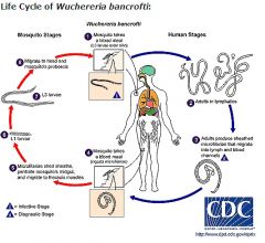
Different species of mosquitoes are vectors of W. bancrofti filariasis depending on geographical distribution.
During a blood meal, an infected mosquito introduces third-stage filarial larvae onto the skin of the human host, where they penetrate into the bite wound 1. They develop in adults that commonly reside in the lymphatics 2. The female worms measure 80 to 100 mm in length and 0.24 to 0.30 mm in diameter, while the males measure about 40 mm by .1 mm. Adults produce microfilariae measuring 244 to 296 μm by 7.5 to 10 μm, which are sheathed and have nocturnal periodicity, except the South Pacific microfilariae which have the absence of marked periodicity. The microfilariae migrate into lymph and blood channels moving actively through lymph and blood 3. A mosquito ingests the microfilariae during a blood meal 4. After ingestion, the microfilariae lose their sheaths and some of them work their way through the wall of the proventriculus and cardiac portion of the mosquito's midgut and reach the thoracic muscles 5. There the microfilariae develop into first-stage larvae 6 and subsequently into third-stage infective larvae 7. The third-stage infective larvae migrate through the hemocoel to the mosquito's prosbocis 8 and can infect another human when the mosquito takes a blood meal 1. (pic) |
|
|
How long will microfilariae wait to be picked up by an appropriate mosquito intermediate host?
|
1 - 1.5 years is possible
|
|
|
What symptoms are present with W. bancrofti infection?
|
1) filarial/elephantoid fever begins with chill -> elevated temp for 1-2 days followed by 2-5 day fever resolution
2) lymphangititis (lymph vessel blockage and subsequent edema) 3) secondary to (2) edematous sites damage blood vessels and then keritanize so they remain enlarged 4) cough, wheezing 5) weight loss 6) lymphadenopathy |
|
|
induration
|
woody hardening of lymphangititis compromised external tissue in order to resist further stretching - elephantiasis
|
|
|
What is a secondary complication to elephantiasis limbs?
|
bacterial and fungal infections
|
|
|
What demographic of people are the most susceptible to W. bancrofti infection?
|
human males
|
|
|
Tx of W bancrofti?
|
1) Hetrazan (DEC trade name) with an unknown mechanism
2) Mectizan 3) surgical |
|
|
What disease does Trichinella spiralis cause?
|
Trichinosis aka Trichinellosis
|
|
|
What are the routes of infection of Trichinella spiralis?
|
many carnivores and omnivores get T. spiralis from eating raw or undercooked pork (Trichinellosis) or eating eggs results in encysted larvae in human muscles (Trichinosis)
|
|
|
What are the two general phases of the T. spiralis lifecycle?
|
1) Enteral phase - occurs in intestine and is generally transitory
2) Parenteral phase - occurs outside of intestine and is marked by larval migration and excystment |
|
|
What are the two general subcycles of the T. spiralis lifecycle?
|
1) sylvatic cycle - no human involvement
2) domestic cycle - occurs with human intervention by feeding pigs raw infected scraps, human infection by ingestion of undercooked pork and bear meat |
|
|
What is the relative infection rate of T. spiralis in the U.S. compared to the rest of the world?
|
U.S. is 3x more infected than the rest of the world.
|
|
|
What is the complete lifecycle of the T. spiralis?
|
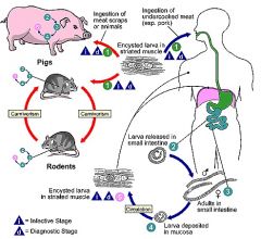
Trichinellosis is acquired by ingesting meat containing cysts (encysted larvae) 1 of Trichinella.
After exposure to gastric acid and pepsin, the larvae are released 2 from the cysts and invade the small bowel mucosa where they develop into adult worms 3 (female 2.2 mm in length, males 1.2 mm; life span in the small bowel: 4 weeks). After 1 week, the females release larvae 4 that migrate to the striated muscles where they encyst 5. Trichinella pseudospiralis, however, does not encyst. Encystment is completed in 4 to 5 weeks and the encysted larvae may remain viable for several years. Ingestion of the encysted larvae perpetuates the cycle. Rats and rodents are primarily responsible for maintaining the endemicity of this infection. Carnivorous/omnivorous animals, such as pigs or bears, feed on infected rodents or meat from other animals. Different animal hosts are implicated in the life cycle of the different species of Trichinella. Humans are accidentally infected when eating improperly processed meat of these carnivorous animals (or eating food contaminated with such meat). (pic) |
|
|
What are the specific steps of the Enteral Phase of the T. spiralis lifecycle?
|
1) Ingestion of infected raw or undercooked meat that contains the 1st stage larvae in skeletal muscle (1mm X 35 um)
2) Parasite freed of host tissue by digestive chemicals 3) Larvae penetrates several columnar epithelial cells lining the microvilli of the SI = intramulticellular parasite 4) Molts 4X in 28 hrs 5) Adult molt occurs 30 hrs post infection in SI 6) Female sheds 1st stage larvae (80 X 7 um) 5-6 days post-infection 7) Newborn have a stylet to penetrate SI wall to enter a blood vessel or lacteal in microvilli |
|
|
What are the specific steps of the Parenteral Phase of the T. spiralis lifecycle?
|
1) 1st stage larvae enter general circulation to become distributed throughout the body
2) Larvae uses esophageal stylet to burrow through tissues until skeletal muscle is found 3) Muscle cell converted to nurse cell over 9 d period 4) By day 12 the nurse cell is providing nutrition and removing waste 5) Larva-nurse cell complex is mature (max growth) by day 20 post infection 6) Viable for years: any species of mammal can be a reservoir for infection 7) Transmission by carnivores and scavengers |
|
|
What are the symptoms associated with T. spiralis infection?
|
it is proportional to the number of larvae penetrating muscle cells
fever eiosinophilia /\ IgE muscle inflammation circumorbital edema (swelling around eyelides) CNS symptoms diarrhea (5 - 15 times a day) |
|
|
What is the differential symptom for T. spiralis, trichinosis infections?
|
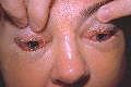
circumorbital edema
(pic) |
|
|
How can T. spiralis be killed?
|
freezing 20 days @ -15C
quickfreeze 24h @ -37C heat up to 140F (microwaves won't do it) |
|
|
Tx for T. spiralis infections?
|
corticosteroids for inflammation
Mebendazole for best prognosis with all nematodes Albendazole is better tolerated |