Reading...
![]()
Play button
![]()
Play button
![]()
Use LEFT and RIGHT arrow keys to navigate between flashcards;
Use UP and DOWN arrow keys to flip the card;
H to show hint;
A reads text to speech;
146 Cards in this Set
- Front
- Back
|
form & fxn
|
Food breakdown
- HCl - Bile Nutrient & water absorption - Microvilli Protective/ Immunologic functions - Barrier - GALT, mucosal lymphocytes & plasma cells Elimination of wastes |
|
|
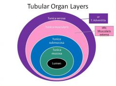
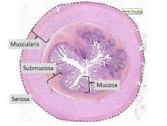
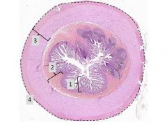
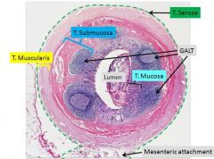
tubular organ layers
|
|
|
|
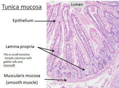
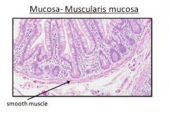
tunica mucosa
|
Components:
- Mucosal epithelium - Lamina propria - Muscularis mucosa Innermost layer |
|
|
|
|
|
mucosa - lamina propria
- Loose CT - Blood vessels - Lymphatics - Lymphocytes - Plasma cells |
|
|
|
|
|
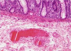
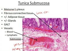
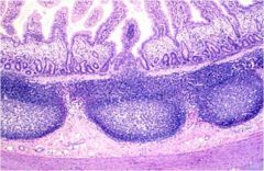
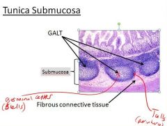
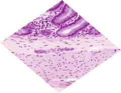
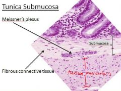
tunica submucosa
|
Meissner’s plexus
Fibrous connective tissue +/- Adipose tissue +/- Glands GALT Vessels: - Blood - Lymphatic |
|
|
|
|
|
|
|
|
|
|
|
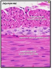
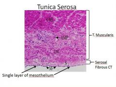
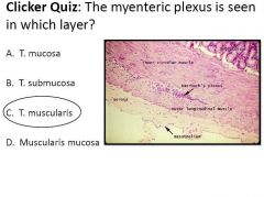
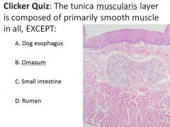
tunica muscularis
|
Smooth muscle (2 layers)
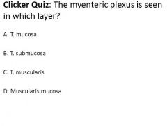
Inner circular Outer longitudinal ** Esophagus: may contain skeletal muscle, smooth muscle, or both Vessels Myenteric plexus (a.k.a. Auerbach’s plexus) b/w inner circular & outer longitudinal layers |
|
|
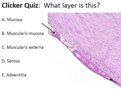
know the appearance and layers of tunica muscularis
|
|
|
|
|
|
|
|
|
|
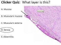
tunica adventitia
|
Similar to T. Serosa- BUT NO mesothelial layer
- Esophagus - Distal colon & Rectum Adventitia is in areas where you are not in a cavity (on ext surface of the cervical esophagus) Same as serosa, just where you find it & no mesothelial layer |
|
|
|
|
|
|
|
|
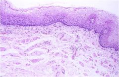
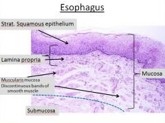
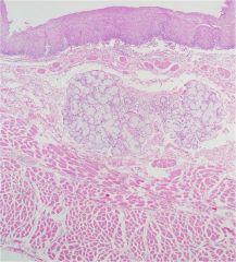
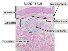
esophagus - tunica mucosa
|
Stratified squamous epithelium
- Variably keratinized - Non-absorptive - Protective Muscularis mucosa - Absent cranially in some species (pig, dog) - May be discontinuous |
|
|
esophagus - tunica submucosa glands
|
Glands
- secretions help ingesta pass - distribution varies between species Most domestic animals: - Glands in the cranial third (cervical) Dog: - Mucous glands, entire length Pigs: - more abundant in the cranial half - do not extend into caudal half |
|
|
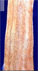
dog esophagus
tunica submucosa with dilated glands |
|
|
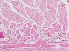
all spp have some component of ______ muscle in the esophagus
|
skeletal
(may or may not have smooth muscle) |
|
|
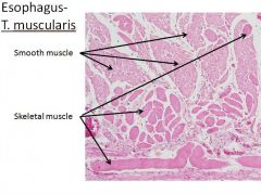
esophagus - tunica muscularis
|
Types:
- Skeletal muscle - Smooth - Mixed Dog- mostly skeletal muscle, changes next to stomach to mixed (caudal to diaphragm) Cat & Horse- Switches from skeletal to smooth in caudal 1/2 - 1/3 of esophagus Ruminants- muscle layer of esophagus is entirely skeletal mm. |
|
|
Inner circular muscle layers of esophagus become thick near
|
the cardia
(esp prominent in the horse) |
|
|
esophagus - tunica adventitia/serosa
|
T. adventitia
Cervical and anterior mediastinum: contains vessels and connective tissue that blends with the surrounding CT T. serosa covered by mesothelium: Thoracic pleura & just caudal to diaphragm |
|
|
|
|
|
|
|
|
|
|
|
|
|
|
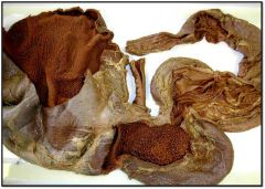
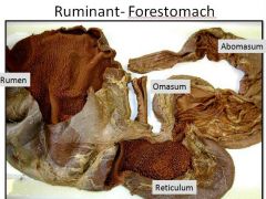
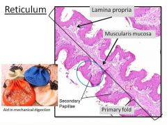
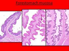
fxns of ruminant forestomach
|
Fermentation
Mixing Absorption Acidifaction |
|
|
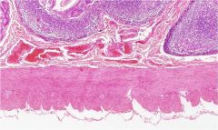
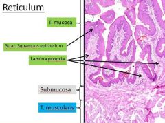
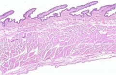
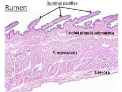
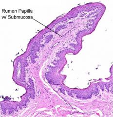
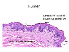
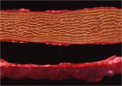
layers of ruminant forestomach
|
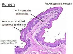
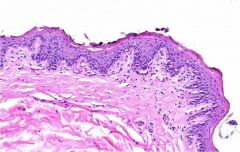
T. mucosa
Keratinized stratified squamous epithelium - Absorptive capabilities (water, fatty acids, nutrients) - Lack glands Mucosal papillae and folds - Increased surface area - Some contain smooth muscle T. submucosa T. muscularis T. serosa |
|
|
|
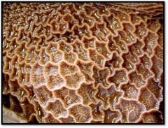
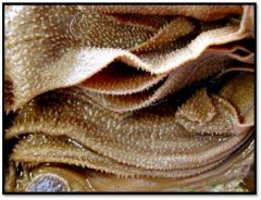
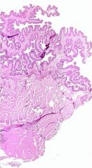
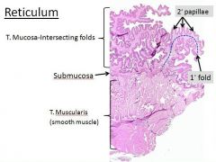
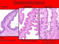
reticulum
|
|
|
reticulum
|
|
|
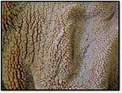
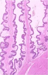
rumen
|
|
|
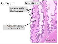
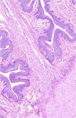
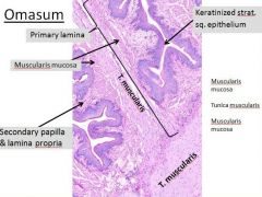
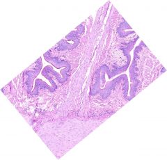
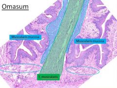
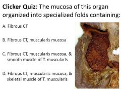
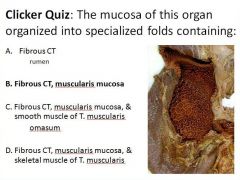
omasum
|
|

|
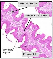
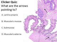
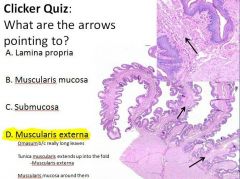
reticulum
T. Mucosa: Honeycomb- Intersecting long primary folds (laminae) Muscularis mucosa- within upper segment of 1◦ fold (aka. long fold) 2◦ papilla (aka. Conical papilla) |
|
|
|
|
|
|
|
|
|
|
|
|
|
|
what effect does a high grain or acidic diet have on ruminal papilla?
short grain diet? |
High grain or acidic diet
- Shorter papilla Short grain - Longer papilla |
|
|
|
|
|
|
|
|
|
|
|
|
|
|
|
|
|
|
|
|
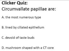
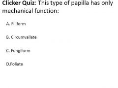
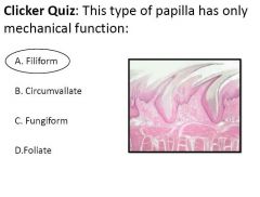
d. mushroom shaped with CT core
|
|
|
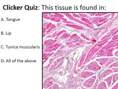
The T. muscularis of the cow esophagus is composed of:
a. Skeletal muscle b. Smooth muscle c. Mixed |
a. skeletal muscle
|
|
|
|
|
|
|
|

|
|
|
|

|
|
|
A. Dog esophagus
|
|
|
|
|
|
|
|
|
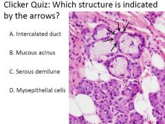
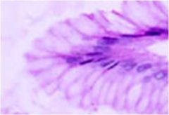
C. Serous demilune
|
|
|
|
|
|
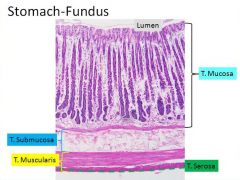
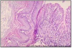
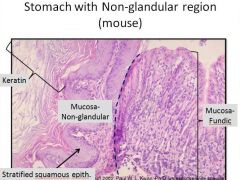
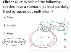
stomach
glandular vs non glandular regions |
Glandular
- Dog - Cats - Pigs (except pars esophagea) - Abomasum of ruminants Non-glandular region - (Stratified squamous epithelium, keratinized) - Horse - Rodents (Rats, mouse, gerbil, etc.) |
|
|
glandular stomach
T. mucosa |
Columnar epithelium- mucous cells
Lamina propria - Glands: cardiac, fundic, pyloric Gastric pits- (“foveolae”) - tubular structures that connect to invaginations of epithelium from surface |
|
|
glandular stomach
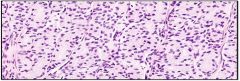
T. muscularis |
Smooth muscle:
- Circular - Longitudinal - Oblique layer |
|
|
glandular stomach
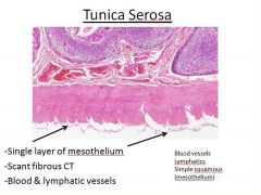
T. serosa |
T. Serosa
- CT - Vessels - Mesothelium |
|
|
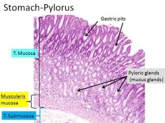
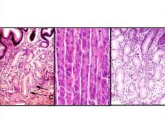
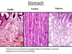
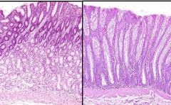
how do you tell apart cardia and pylorus?
|
Pylorus and cardia look similar
Pylorus is thicker Both have mucus glands |
|
|
gastric glands
cardia and pylorus |
Branched tubular mucous glands
Function: protect the lining of the stomach Gastric pits of cardia- more shallow than pyloric glands Cell types: - Mucous gland cells- basally located nuclei and pale abundant pale grey foamy cytoplasm - Few scattered parietal cells (in cardia) |
|
|
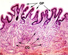
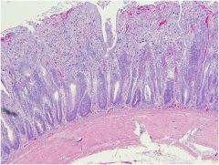
stomach - cardia
GP= Gastric pits CG= cardiac glands = Mucus glands in lamina propria Simple columnar mucus epithelium |
|
|
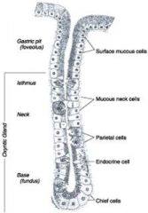
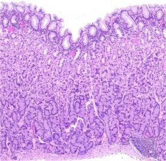
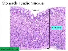
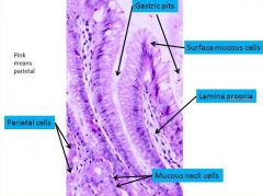
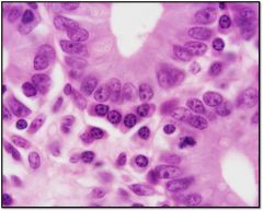
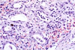
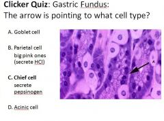
gastric glands - fundus and body
|
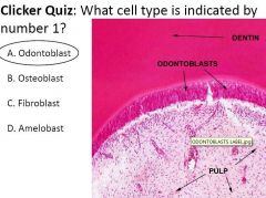
1. Surface mucous cells
2. Mucous neck cells 3. Parietal cells 4. Chief cells 2-4 only located in fundus |
|
|
|
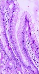
gastric glands - fundus/body
Surface mucous cells: - Cover the surface and line the gastric pits - Columnar cells with basally located oval nuclei and abundant apical cytoplasm containing tiny mucous droplets. - SECRETE: mucus |
|
|
gastric glands - fundus/body
Surface mucous cells: - Cover the surface and line the gastric pits - Columnar cells with basally located oval nuclei and abundant apical cytoplasm containing tiny mucous droplets. - SECRETE: mucus |
|
|
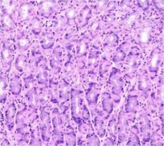
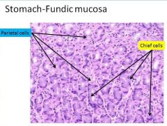
gastric glands - fundus/body
Parietal cells (aka. oxyntic cells): - Found between the mucous neck cells and chief cells - Large, polygonal cells with eosinophilic cytoplasm and central round nuclei - SECRETE: HCl, gastric intrinsic factor |
|
|
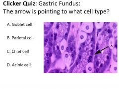
gastric glands - fundus/body
Chief cells: - Located deeper in the fundic glands - Eosinophilic apical cytoplasm (zymogen granules) and basophilic basal cytoplasm (rER) - SECRETE: pepsinogen, trypsinogen, renin (ruminant), gastric intrinsic factor (some species) |
|
|
know the strx of the fundic mucosa
|
|
|
|
|
|
|
|
|
|
|
|
|
|
|
|
|
|
|
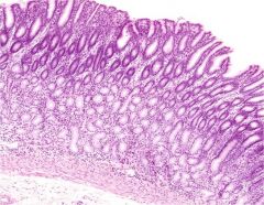
stomach - pyloric mucosa
|
|
|
|
|
|
|
|
|
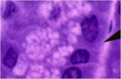
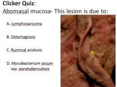
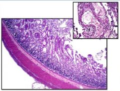
B. Ostertagiosis
|
|
|
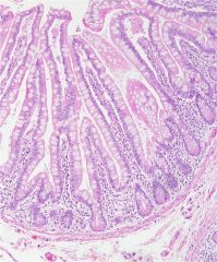
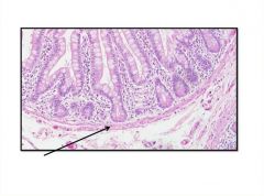
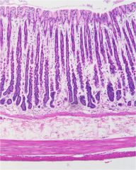
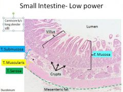
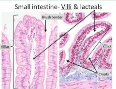
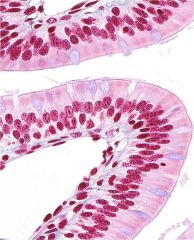
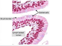
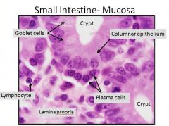
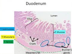
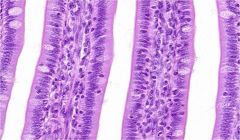
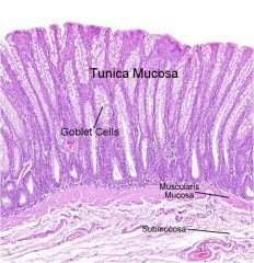
small intestine
T. mucosa |
Enterocytes: Simple columnar epithelium + microvilli
Goblet cells: secrete mucus (lubrication/protection) Increase in number as move aborad Villi: surface mucosal projections lined by epithelium Crypts of Lieberkuhn: space between villi rapidly proliferating cells Lymph lacteals Immune cells (lymphocytes, plasma cells, globular leukocytes) Endocrine cells (not going to see this) |
|
|
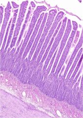
villi are present only in ______
how do they appear in carnivores vs. ruminants |
small intestine
Long-slender in carnivores Shorter- thicker in ruminants |
|
|
what 2 things increase the surface area for absorption in the small intestine
|
villi
microvilli |
|
|
|
|
|
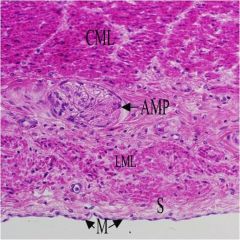
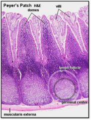
small intestine
T. submucosa/ T. muscularis/ T. serosa |
T. Submucosa
GALT/Peyer’s patches T. Muscularis Smooth muscle: inner circular layer & outer longitudinal layer (2 layers oriented perpendicular to each other) T. Serosa Mesothelium |
|
|
|
|
|
|
|
|
|
|
|
|
|
|
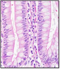
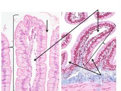
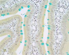
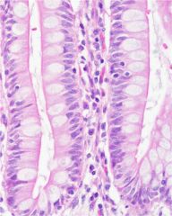
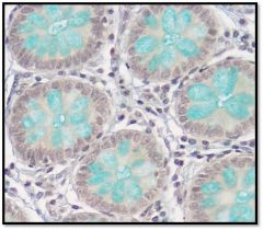
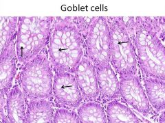
small intestine - mucosa
goblet cells - Columnar epith. - Loose CT - Vessels - Lymphocytes - Plasma cells |
|
|
|
|
|
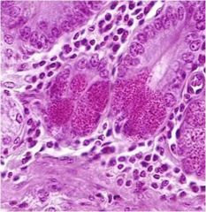
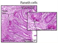
paneth cells
Clusters at base of crypts Cells= pyramid shaped with intensely eosinophilic cytoplasmic granules & basilar nuclei Secrete protein- includes lysozyme (bactericidal) Prominent in horse, also seen in ruminants |
|
|
|
|
|
enteroendocrine cells
|
Triangular, argentaffin cells
Secrete: - Serotonin - Motilin - Vasoactive intestinal polypeptide (VIP) - Somatostatin - Gastrin |
|
|
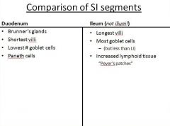
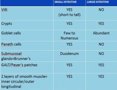
comparison of SI segments
|
|
|
|
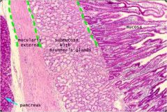
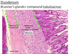
Brunner’s glands- mixed mucous and serous
|
|

|
|
|
|
|
|
|
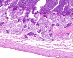
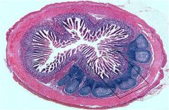
ileum
|
|
|
ileum
|
|
|

|
|
|
jejunum
|
|
|
ileum
|
|
|
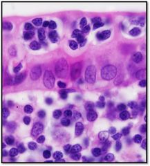
M cells
|
Epithelial cells (lack microvilli)
Found in epithelium over mucosal lymphoid tissue (Peyer’s patches) Function= Mucosal immunity- take up antigen/organisms for transepithelial delivery to antigen presenting cells |
|
|
ileum
notice crypts and lamina propria |
|
|
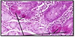
parvo effect on crypt cells
notice crypt at bottom center is beginning to regenerate |
|
|
parvo effect on tissue
|
|
|
|
|
|
|
|
|
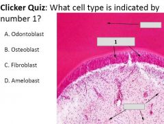
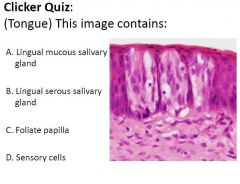
D. sensory cells
|
|
|
|
|
|
D. All of the above
|
|
|
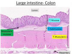
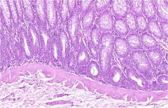
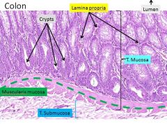
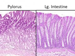
large intestine
|
|
|
large intestine
(describe it) |
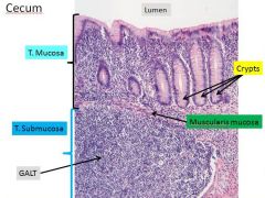
consists of cecum & colon
Mucosa: - NO villi, only crypts - Many goblet cells - No Paneth cells Submucosa and muscle wall- similar to rest of tract Function= Absorb H2O - Lubrication- mucus (pass out waste) |
|

|
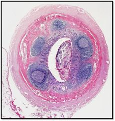
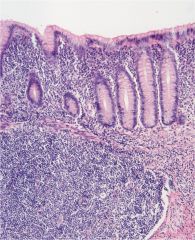
large intestine - cecum
prominent lymphoid tissue |
|
|
|
|
|
|
|
|

|
|
|
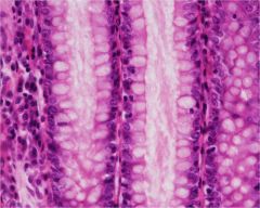
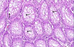
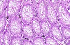
colon
(tons of goblets = large intestine) |
|
|
|
|
|
|
|
|
|
|
|
lamina propria
lymphocytes, plasma cells, capillaries |
|
|
how do the large and small intestine differ?
|
|
|
|
Peyers patches are found throughout intestine but are most prominent where?
|
in ileum and cecum
|
|
|
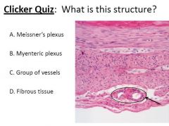
C. Group of vessels
|
|
|
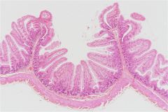
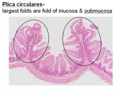
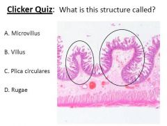
C. Plica circulares
|
|
|
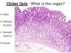
C. Stomach (pylorus)
|
|
|
|
|
|
B. Goblet cells
|
|
|
C. Has hypsodont teeth
|
|
|
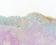
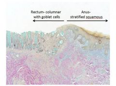
rectum vs. anus
|
Rectum=
Last part of colon Enlarged T. muscularis forms the anal sphincter Derived from= hindgut Anus- Derived from= surface ectoderm Abrupt transition: simple columnar with goblet cells - stratified squamous epithelium |
|
|
|
|
|
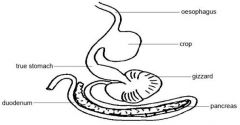
avian digestive tract
|
Oral cavity
- no teeth - Beak Esophagus - large diameter - Most birds (not all) have a crop Stomachs - Proventriculus= glandular stomach - Ventriculus= muscular stomach (a.k.a. Gizzard). It contains small stones that facilitate grinding of foodstuffs Small Intestine - similar to mammals - segments are not as histologically distinct Large intestine - short colon (Short villi extend into the lumen of the colon, unlike mammals.) - paired ceca (important sites for fermentation) Cloaca - common opening of the digestive, reproductive and urinary systems |
|
|
know the layout of the avian digestive tract
|
|
|
|
crop
diverticulum of esophagus Strat squam to columnar epi |
|
|
esophagus/ crop layers
|
T. Mucosa
- Non-keratinized stratified squamous to columnar - Mucous glands (lacking in crop) T. Submucosa T. Muscularis - Entirely smooth muscle T. Adventitia |
|
|
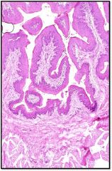
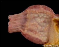
Proventriculus (glandular stomach)
Glands= Secrete HCl and pepsinogen |
|
|
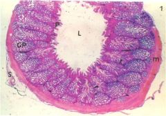
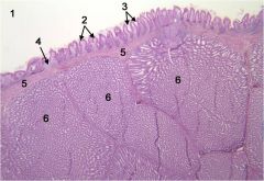
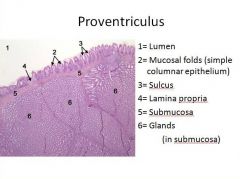
proventriculus (mucosal folds look like villi)
T. Mucosa - Folds surrounding opening of glands - Simple columnar epithelium - Lamina propria - Muscularis mucosa T. Submucosa - Glands T. Muscularis= thin - Inner longitudinal - Middle circular - Outer longitudinal (like stomach of mammals) T. Serosa= typical |
|
|
|
|
|
|
|
|

|
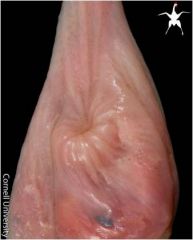
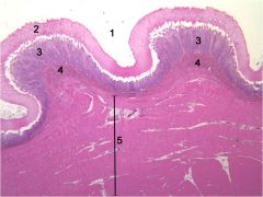
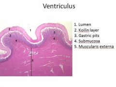
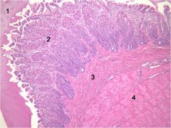
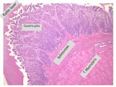
ventriculus
T. Mucosa - Cuboidal to columnar epithelium - Glands - “Koilin” over surface (a.k.a. pellicle) T. Submucosa T. Muscularis T. Serosa |
|
|
|
|
|
|
|
|
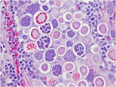
chicken coccidiosis
|
|
|
|
chicken coccidiosis
|
|
|
chicken coccidiosis
|