![]()
![]()
![]()
Use LEFT and RIGHT arrow keys to navigate between flashcards;
Use UP and DOWN arrow keys to flip the card;
H to show hint;
A reads text to speech;
70 Cards in this Set
- Front
- Back
- 3rd side (hint)
|
Tooth eruption |
The process by which developing teeth: -emerge through the soft tissue of the jaws and the mucosa -enter the oral cavity - contact the opposing teeth -function in mastication |
|
|
|
Tooth eruption |
A complex and multistep process |
|
|
|
The eruptive process |
Is not rapid and it is not continuous |
|
|
|
Tooth eruption |
Ends only when the tooth is lost |
|
|
|
3 phases of tooth eruption |
1. Preeruptive phase 2. Prefunctional eruptive phase 3. Functional eruptive phase (posteruptive phase) |
|
|
|
Preeruptive phase |
Includes all movements of primary and permanent tooth crowns from the time of their initiation and formation to the time of crown completion |
|
|
|
Preeruptive phase |
Developing crowns move within the jaws as the face and jaws grow --all movements occur within the crypts of the bone and before root formation begins - this phase ends when root formation begins |
|
|
|
Preeruptive movements of crowns anterior teeth |
Permanent anterior teeth develop lingual to the incisal level of the primary teeth |
|
|
|
As primary teeth erupt |

...The permanent teeth reposition themselves -lingual to the apical third of the primary roots |
|
|
|
Anterior teeth |

|
|
|
|
Preeruptive movements of crowns posterior teeth |
Permanent premolars begin to develop near the occlusal area of the primary molars |
|
|
|
As primary molars erupt |

.. The permanent premolars gradually move -to a position within the roots of the primary molars |
|
|
|
Preeruptive phase |
Maxillary permanent molars and mandibular permanent molars are accessional teeth They do not replace a primary tooth |
|
|
|
Maxillary molars |
Develop within the maxillary tuberosity, during the preeruptive phase - their occlusal surfaces are slanted distally (away from the midline) |
|
|
|
The mandibular molars |
Develop in the ramus of the mandible - their occlusal surfaces are slanted mesially (toward the midline) |
|
|
|
Prefunctional eruptive phase |
Begins with the initiation of root formation Ends when the teeth reach occlusal contact |
|
|
|
4 phases of eruption |
1. Root formation 2. Incisal or occlusal movement 3. Emergence 4. Occlusal contact |
|
|
|
Root formation |
Proliferation of epithelial root sheath stimulates formation of root dentin and pulp tissue -more space is required for the developing root |
|
|
|
Incisal or occlusal movement |
-Developing tooth needs more room -eruption pathway begins to develop -reduced enamel epithelium fuses with oral epithelium |
|
|
|
Emergence |
Crown tip penetrates through fused epithelial layers and into the oral cavity |
|
|
|
Occlusal contact |
- movement occurs until contact is made with the opposing tooth -size of the clinical crown grows larger (size of anatomical crown is constant) -gingival attachment migrates apically |
|
|
|
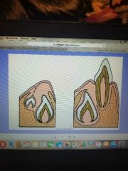
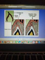
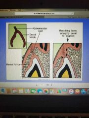
Eruption pathway |
The area of the dental follicle degenerates to form a pathway for the crown towards the oral mucosa Blood vessels decrease, nerve fibers break apart |
|
|
|
Eruption pathway |
The degenerated area develops into an inverted triangular shape Fibers in this area called the gubernacular cord, help guide the crown along the pathway |
|
|
|
Gubernacular cord |
|
|
|
|
Development of eruption pathway |
Eruption pathway must be accompanied by resorption of the bony crypt and remodeling of alveolar bone |
|
|
|
Development of eruption pathway |
1. Circulating monocytes accumulate in the area and become osteoclasts to resorb surrounding bone -they also resorb roots of primary teeth |
|
|
|
Development of eruption pathway |
2. Macrophages release hydrolytic enzymes that break up blood vessels and nerves |
|
|
|
Osteoblasts |
Produce new bone to accommodate the new position of the crown in development of eruption pathway |
|
|
|
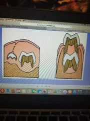
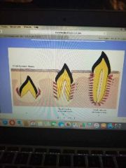
Reduced enamel epithelium |
Surrounds the crown of the tooth As the tooth moves along the eruption pathway, the REE fuses with the oral epithelium *This is called the reduced epithelial layer |
|
|
|
REE |
The tooth tip penetrates through the reduced epithelial layer The organic cuticle layer still remains on the tooth surface |
|
|
|
Reduced epithelial layer |

As the tooth emerges into the oral cavity, the epithelial attachment shifts farther down onto the crown |
|
|
|
Junctional epithelium |
Reduced epithelial layer continues to migrate down the crown until the entire crown has erupted into the oral cavity It then becomes known as this. |
|
|
|
PDL fiber development |
As the tooth emerges, fiber bundles attach themselves to the tooth surface The first fibers formed are those in the cervical area Fibroblasts produce the collagen fibers |
|
|
|
Initial fiber bundles |
|
|
|
|
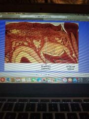
Alveolar bony crypt |
The height of this increases as the tooth emerges because the root is continuing to form |
|
|
|
PDL fibers |
The direction of the collagen fiber bundles changes as the tooth erupts |
|
|
|
Collagen fibers |
These have a high turnover rate (24 hours) at this stage to accommodate the tooth eruption and constant rearrangement |
|
|
|
Occlusion |
When the tooth reaches here, the orientation of the fiber bundles becomes complete |
|
|
|
Apical fibers |
Develop at the end of the root along with bony trabeculae |
|
|
|
Redirection of fiber bundles |

|
|
|
|
Functional eruptive phase |
(Post eruptive phase) Begins when the tooth comes into occlusal contact and continues for as long as the tooth is present in the mouth |
|
|
|
Functional eruptive phase |
At the beginning of this phase , root completion is still occurring |
|
|
|
Permanent teeth |
Takea about 2-3 years after initial emergence for completion of root formation |
|
|
|
Deciduous teeth |
Takes about 1-1.5 years for completion of root formation |
|
|
|
Occlusal contact results in changes |
- Principal fibers of the PDL arrange themselves in their preferred directions. - Principal fibers increase in size. -blood vessels and nerves become more organized in the interstitial spaces -mineral density of the alveolar bone increases |
|
|
|
Tooth movement during posteruptive phase |
The functional eruptive phase continues for as long as the tooth is present It compensates for loss of the enamel surface due to abrasion and attrition It can be physiological or pathological |
|
|
|
Physiological movement |
1. Occlusal wear 2. Mesial drift 3. Perioral forces |
|
|
|
Occlusal wear |
Occlusion changes with attrition and abrasion Cementum is usually deposited at the apex of the root to compensate for loss of occlusal surfaces |
|
|
|
Perioral forces |
Cheek/tongue movements or habit |
|
|
|
Pathological movement |
1. Mesial drift 2. Excessive parafunction 3. Periodontal disease 4. Bulimia |
|
|
|
Mesial drift |
As a result of tooth loss |
|
|
|
Excessive parafunction |
Grinding/bruxism |
|
|
|
Periodontal disease |
May result in tooth movement |
|
|
|
Bulimia |
Excessive loss of enamel |
|
|
|
4 theories of causes of tooth eruption |
1. Root growth 2. Vascular pressure 3. Bone growth 4. Ligament traction |
|
|
|
Vascular pressure |
Increase in tissue fluid pressure in the periapical region moves the tooth |
|
|
|
Bone growth |
Selective resorption and deposition of bone causes the tooth to move |
|
|
|
Ligament traction |
Cells and fibers of the PDL pull the tooth into occlusion |
|
|
|
Eruption of primary teeth |
The 6/4 rule - for every 6 months of age, 4 teeth erupt -6 mos. - 4 teeth -12 mos- 8 teeth -30 mos - 20 teeth (full complement) |
|
|
|
Order of emergence |
Central, lateral, 1st molar, canine, 2nd molar Sane pattern for max and mand teeth Mandibular centrals are usually the first teeth to erupt at about 6 mos of age |
|
|
|
Permanent teeth |
Mandibular centrals and max and mand molars erupt at about 6-7 years of age |
|
|
|
Mandibular sequence |

(Central. 1st molar), lateral, canine, 1st premolar, 2nd premolar, 2nd molar, 3rd molar |
|
|
|
Maxillary sequence |
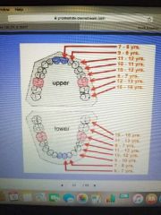
- 1st molar, central, lateral, 1st premolar, 2nd premolar, canine, 2nd molar, 3rd molar |
|
|
|
Tooth eruption- primary dentition |
2-8 years old |
|
|
|
Mixed dentition |
8-12 years old |
|
|
|
Permanent dentition |
12+ years old |
|
|
|
Successional teeth |
The permanent teeth that replace the primary teeth (20) |
|
|
|
Accesional teeth |
Permanent teeth that do not replace a primary tooth (total of 12) |
|
|
|
Leeway space |
Accounts for the difference in size between the primary teeth and the permanent teeth |
|
|
|
Leeway space |
Maxillary arch- 1.3mm Mandibular arch- 3.1mm |
|