![]()
![]()
![]()
Use LEFT and RIGHT arrow keys to navigate between flashcards;
Use UP and DOWN arrow keys to flip the card;
H to show hint;
A reads text to speech;
50 Cards in this Set
- Front
- Back
|
Will appear as a white area around the crown |
enamel
|
|
|
the grayish-white area that is semi-radiopaque and forms the bulk of the tooth
|
dentin
|
|
|
this boundry is distinct due the different densities and opacities
|
DEJ
|
|
|
radiolucent (appears radiographically as a dark area)
|
pulp
|
|
|
normally you DON'T see this on an xray (it is very thin and has the same radiodensity and dentin)
|
cementum
|
|
|
thin, dark line that circumscribes the tooth)
|
periodontal ligament space
|
|
|
dense bone that surrounds the root; appears as a white line
|
lamina dura
|
|
|
continuous with the lamina dura that is the coronal most part of the alveolar process; appears radiopaque |
alveolar crest
|
|
|
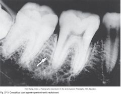
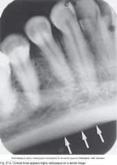
dense outer layer of bone; appears radiopaque; examples are inferior border of mandible, lamina dura of alveolus, alveolar crest
|
cortical bone ("compact bone")
|
|
|
composed of trabeculae (radiopaque) and marrow spaces (radiolucent); located between the lamina dura of adjacent teeth and multirooted teeth
|
cancellous bone ("spongy" bone) |
|
|
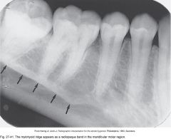
mylohyoid ridge |
|
|
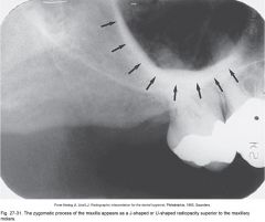
zygomatic process, appears as a J or U shaped radiopaque line above max molars |
|
|
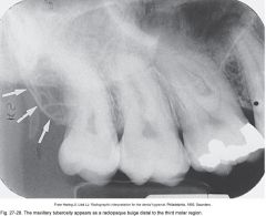
maxillary tuberosity |
|
|
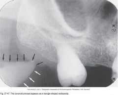
coronoid process |
|
|
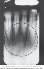
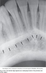
mental fossa above mental ridge in ant. mand. apex of teeth. Dark vertical lines are nutrient canals |
|
|
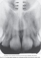
nasal septum |
|
|
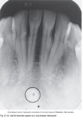
Genial tubercles |
|

|
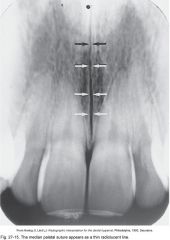
median palatal suture |
|
|
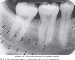
mandibular canal |
|
|
cancellous bone- spongy bone |
|
|
cortical bone |
|
|
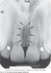
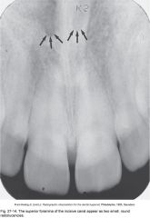
incisive foramen |
|
|
superior foramena of the incisive canal appear as 2 round radiolucensies |
|
|
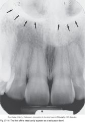
floor of nasal cavity |
|
|
median palatal suture |
|
|
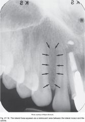
lateral fossa between lateral incisor and canine. |
|
|
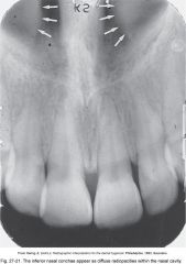
nasal cavity |
|
|
inferior nasal conchae |
|
|
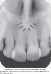
anterior nasal spine. V shape at floor of midline of nasal cavity |
|
|
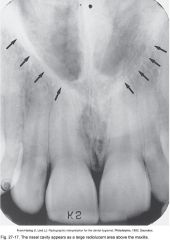
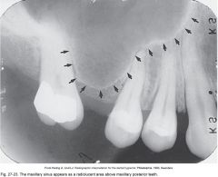
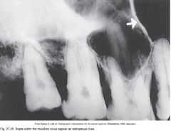
maxillary sinus |
|
|
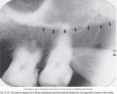
zygoma |
|
|
septa of maxillary sinus |
|
|
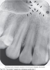
inverted Y, maxillary sinus and nasal sinus |
|
|
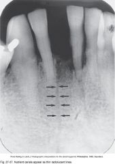
nutrient canals |
|
|
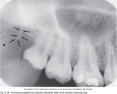
Hamulus. Most patients you will not go back this far. You can also see the coronoid process. |
|
|
mental ridge |
|
|
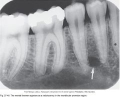
mental foramen. Btwn first and second premolars, |
|
|
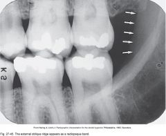
external oblique ridge |
|
|
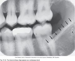
Internal oblique ridge |
|
|
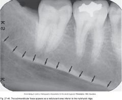
submandibular fossa inferior to the mylohyoid ridge |
|
|
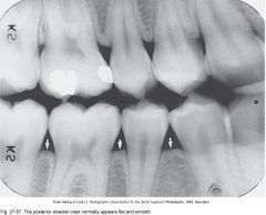
posterior alveolar ridge |
|
|
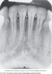
anterior alveolar crest |
|
|
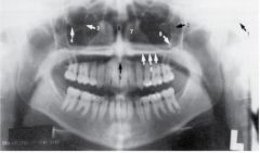
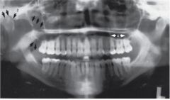
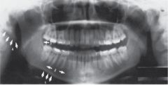
Figure 29-02. Normal anatomic landmarks of the maxilla and surrounding structures seen on panoramic images: 1, external auditory meatus; 2, pterygomaxillary fissure; 3, infraorbital foramen; 4, orbit; 5, anterior nasal spine; 6, nasal septum; 7, nasal conchae; 8, hard palate; 9, zygomatic process of maxilla. |
|
|
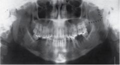
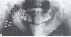
1, glenoid fossa; 2, articular eminence; 3, maxillary tuberosity; 4, maxillary sinus; 5, zygoma |
|
|
lateral pterygoid plate |
|
|
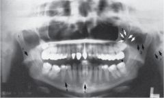
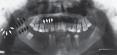
: 1, condyle; 2, coronoid notch; 3, coronoid process; 4, mandibular foramen; 5, mental foramen; 6, genial tubercles; 7, styloid process. |
|
|
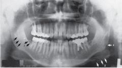
1, mandibular canal; 2, hyoid; 3, internal oblique ridge; 4, angle of mandible. |
|
|
1, inferior border of mandible; 2, submandibular fossa; 3, external oblique ridge; 4, soft tissue of ear. |
|
|
1, palatoglossal air space; 2, nasopharyngeal air space; 3, glossopharyngeal air space. |
|
|
Soft tissues seen on panoramic images: 1, tongue; 2, soft palate and uvula; 3, ear. |