![]()
![]()
![]()
Use LEFT and RIGHT arrow keys to navigate between flashcards;
Use UP and DOWN arrow keys to flip the card;
H to show hint;
A reads text to speech;
54 Cards in this Set
- Front
- Back
|
In order to accurately position the patient for the hip radiographs, one must localize two bony points on the pelvis. These two reference points are: |
(1) Superior margin of the symphis(SMS) (3)Anterior superior iliac spine(ASIS) |
|
|
How many degree should the feet and lower limb be internally rotated for an AP pelvis radiograph? |
15 to 20 degrees |
|
|
The central ray for an AP pelvis is directed perpendicular to the center of the IR. The central ray entrance point will be about? |
2 inches superior to the pubic symphysis |
|
|
Which of the following will be shown in "profile" if the lower limbs are in correct position for an AP pelvis? |
Greater trochanters |
|
|
Which of the following methods will demonstrate the femoral necks in the AP oblique projection? |
Modified Cleaves |
|
|
For the AP oblique femoral necks (modified cleaves method) the central ray is directed? |
0 degrees |
|
|
How much should the thighs be abducted for the AP oblique projections of the femoral necks( modified cleaves method)? |
45 degrees |
|
|
Where does the central ray enter the patient for the AP hip? |
2 1/2 inches(6.4 cm) distal on a line draw perpendicular to the midpoint of a line between ASIS and pubic symphysis |
|
|
How many degrees is the lower limb and footed rotated internally for an AP hip? |
15-20 degrees |
|
|
Which of the following methods will demonstrate the hip in a lateral projection? |
Lauenstein,Hickey |
|
|
Which of the following methods demonstrate the hip in an axiolateral |
Danelius-Miller |
|
|
Unless contradicted, the lower limb and leg should be internally rotated for an axiolateral projections of the hip(Danelius-Miller). How many degrees of rotation are required? |
15-20 degrees |
|
|
Which of the following devices are necessary to perform an axiolateral projection of the hip (Danelius-Miller)?
(1)sandbags(2)leg support device(3)vertical IR holder? |
1,2 and 3 |
|
|
Which of the following describes the position of the IR for the axiolateral projection of the hip (Danelius-Miller)?
(1)parallel with the long axis of the femoral neck (2)its upper border in the crease above the iliac crest (3)perpendicular to the long axis of the femur? |
1 and 2 |
|
|
Where is the IR centered for an AP pelvis?
|
midway between the ASIS and the pubic symphysis
|
|
|
Where is the central ray directed for the AP oblique projection (modified Cleaves) of the femoral necks?
|
1inch superior to the pubic symphysis
|
|
|
The angle of the SI joints is ____ degrees relative to the midsagittal plane
|
25 to 30 degrees
|
|
|
The body is placed at what angle for the AP oblique projection (Judet-method) of the acetabulum?
|
45 degrees
|
|
|
What is the central-ray entrance point for the AP oblique projection (Judet-method) of the acetabulum?
|
2 inches inferior to the ASIS
|
|
|
The internal oblique position of the AP oblique projection (Judet method) demonstrates the?
|
iliopubic column and posterior rim of acetabulum
|
|
|
The external oblique position of the AP oblique projection (Judet method) demonstrates the?
|
ilioischialcolumn and anterior rim of acetabulum
|
|
|
The AP axial projection Inlet (Bridgeman method) requires the central ray be directed:
|
40 degrees caudad
|
|
|
The hip bone is composed of which of the following:(1) ilium(2) pubis(3) ischium |
All the above |
|
|
The neck of the femur projects anteriorly at an approximate angle of ? |
15-20 degrees |
|
|
The hip joint is a ___ joint: |
synovial—ball-and-socket |
|
|
Which of the following best describes the female pelvis?(1) heavy bones(2) oval inlet(3) wide outlet |
(2) Oval inlet and (3) Wide outlet |
|
|
Flattening of the femoral head due to a vascular interruption is known as? |
Legg-Calvé-Perthes disease |
|
|
What percentage of each bone forms acetabulum? |
2/5 ilium, 2/5 ischium, 1/5 pubis |
|
|
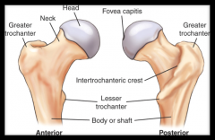
The area of the proximal femur where the ligamentum fovea inserts is called? |
Fovea capitis |
|
|
On which bone would we find ASIS? |
Ilium |
|
|
On which bone would you find ala? |
Ilium |
|
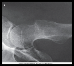
Refer to the image. What projection(method) is demonstrated? |
Axiolateral( DaneliusMiller) Method |
|

Examine this AP oblique (Judet)image of the right hip obtained with the patient positioned for the internaloblique. What patient position isdepicted in this image? |
45 degrees LPO |
|
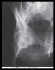
Examine this AP oblique (Judet)image of the right hip obtained with the patient positioned for the internaloblique. What is the anatomy ofinterest? |
posterioracetabular rim and iliopubiccolumn |
|
|
Proximal Femur |
|
|
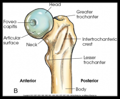
Anterior and Posterior View of Femur |
|
|
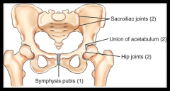
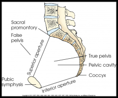
Joints of the Pelvis |
|
|
Sacroiliac(2) |
Class: Synovial Mobility: Amphiarthrodial Movement: Slight |
|
|
Hip(2) |
Class: Synovial Mobility: Diarthrodial Movement: Freely |
|
|
Symphysis Pubis |
Class: Cartilaginous Mobility: Amphiarthrodial Movement: Slight |
|
|
Unionof Acetabulum (2) |
Class: Cartilaginous Mobility: Synarthrodial Movement: None |
|
|
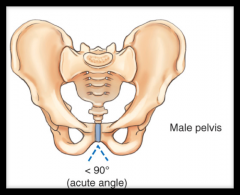
Males Pelvic Girdle: Heaver, narrow and deeper Angel at pubic sympahsis is acute (90 degree) |
|

|
Female Pelvic Girdle: Wider, Shallow and light Angle at pubic sympysis is OBTUSE |
|
|
|
|
|
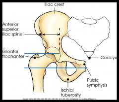
Localizing Anatomy of Hip and Pelvic Girdle: Bony Structures include: –Iliac crest–ASIS –Pubic symphysis –Greater trochanter –Ischialtuberosity –Tip of coccyx |
|
|
Projection of the Pelvis and Upper Femora: |
AP MSP centered to midline Lower limbs and feet medially rotated 15-20 degrees IR 1-1 1/2 inch above crest |
|
|
Projection of Femoral Necks: |
AP Oblique( modified Cleaves- Bilateral/Uni) IR 1 inch above public symphsis CR perpendicular to MSP @ 1 inch above pubic symphsis |
|
|
Projections of the Hip: AP |
AP Hip Lower limb and foot medially rotated 15-20 degree Femoral necks parallel to IR CR perpendicular to femoral necks |
|
|
Projections of the Hip: Lateral ( Lauenstein Method) |
Lateral(mediolaterial) Hip Rotate towards AFFECTED SIDE Affected hip to midline of grid CR enters perpendicular through hip( midway between ASIS and Pubic Symphsis) |
|
|
Projections of the Hip: Lateral ( Hickey Method) |
Lateral(mediolateral) Hip CR angled 20 degrees cephalic and enters hip joint |
|
|
Projections of the Hip: Axiolateral ( Danelius-Miller Method) |
Rotate AFFECTED limb 15-20 degrees medially IR: Vertical with upper boarder in crease above iliac crest CR: Horizontal( the tube) and perpendicular to along axis of femoral neck |
|
|
Projections of the Acetabulum: AP Oblique( Judet; modified Judet) |
AP Oblique( Judet;modified Judet) INTERNAL OBLIQUE Recumbent 45 degrees with AFFECTED side up CR: Perpendicular to IR and enters 2 inches INFERIOR to ASIS of affected side Internal oblique used to show POSTERIOR RIM of acetabulum and iliopubic |
|
|
Projections of Acetabulum: AP oblique( Cont) |
AP Oblique( Judet-Method) EXTERNAL OBLIQUE Recumbent 45 degrees with AFFECTED SIDE DOWN CR: Perpendicular to IR and enters Pubis Symphysis External oblique used to show ANTERIOR RIM of acetabulum |
|
|
Clements-Nakayama(Axiolateral) Projection |
Used for bilateral fractures when Daneilus-Miller cannot be used IR: lower than femoral necks and tilt back 15 degrees CR: Directed 15 degrees posteriorly and perpendicular to femoral necks |