Reading...
![]()
Play button
![]()
Play button
![]()
Use LEFT and RIGHT arrow keys to navigate between flashcards;
Use UP and DOWN arrow keys to flip the card;
H to show hint;
A reads text to speech;
22 Cards in this Set
- Front
- Back
|
Describe the elbow joint:
1. Articular surfaces 2.Type 3. Capsule 4. Ligaments 5. Special features 6. Movements |
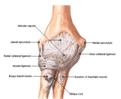
1. Articular surface: Trochlear & capitulum of humerus. Trochlear notch of ulna. Head of radius
2. Type: Hinge synovial joint 3. Capsule: Weak strengthened by collateral ligaments 4. Ligaments: Radial collateral blends with anular. Ulnar collateral 5. Special features: Synovial membrane extends into the proximal radioulnar joint. Carrying angle 165 degrees 6. Movements: Flexion & extension |
|
|
Describe the features of the radio-ulnar joint
1. Type 2. Articular surfaces 3. Ligaments 4. Movements 5. Special features |
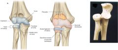
1. Type: Synovial pivot joint
2. Articular surfaces: Head of radius : radial notch of ulna 3. Ligaments: Annular Ligament 4. Movements: Permits rotation of radius about ulna = pronation 5. Special features: sacciform recess is a continuation of synovial membrane from the elbow joint to the radio-ulnar joint under the annular ligament. This helps remove the friction of movement of the radius |
|
|
Describe the features of the distal radio-ulnar joint:
1. Articular surfaces 2. Type 3. Capsule 4. Movements |
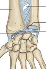
1. Articular surfaces: Head of the ulna with the ulnar notch of the radius
2. Type: Synovial pivot joint 3. Capsule: Fibrocartilagenous disc binds the ends of the bones 4. Movements: pronation and supination = radius rotates around the ulna |
|
|
Describe the features of the wrist joint:
1. Type 2. Articular surfaces 3. Capsule 4. Ligaments 5. Movements |
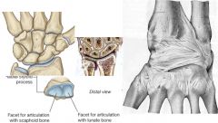
1. Condyloid synovial joint
2. Radius: scaphoid and lunate 3. Articular disc between bones and sacciform reccess of synovial continues up to form between distal radio-ulnar joint - Radio-carpal and collateral ligaments 5. Flexion, extension, adduction, abduction and circumduction |
|
|
Describe the joints of the phalanges
|
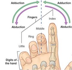
1. Carpo-metacarpal joint = plane synovial joint. Allows some gliding
2. Metacarpo-phalangeal joint = condylar synovial joint. Allows flexion, extension, abduction and adduction 3. Proximal and Distal interphalangeal joints = hinge synovial joint. Allows flexion and extension Thumb carpo-metacarpal: - Saddle synovial joint. Allows full circumduction |
|
|
What is the cubital fossa and describe its boundaries
|
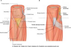
- Triangular hollow area on the anterior surface of the elbow
- Bounded: • Superiorly by line between the epicondyles of the humerus • Medially by Pronator Teres • Laterally by Brachioradialis • Floor formed by Brachialis and Supinator • Roof by Fascia and Bicipital Aponeurosis |
|
|
What are the 4 major contents of cubital fossa?
|
1. Tendon of biceps brachii
2. Median nerve 3. Brachial artery 4. Median cubital vein (on top) |
|
|
Describe the features of pronator teres:
1. Origin 2. Insertion 3. Nerve supply 4. Action |
1. Origin :
• Medial epicondyle of humerus • Coronoid process 2. Insertion : Middle of lateral surface of radius 3. Nerve Suppy : Median nerve C 6 & 7 4. Action : Flexion of the elbow joint & pronates the forearm |
|
|
Name the muscles that make up the superficial layer of the flexor compartment of the forearm
|
1. Flexor carpi radialis
2. Flexor carpi ulnaris 3. Palmaris longus - Inserts into the common flexor tendon |
|
|
Name the muscles that make up the intermediate layer of the flexor compartment of the forearm
|
Flexor digitorium superficialis
|
|
|
Name the muscles that make up the deep layer of the flexor compartment of the forearm
|
1. Flexor digitorum ptofundus
2. Flexor pollicis longus |
|
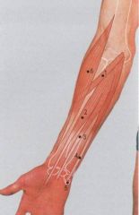
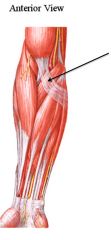
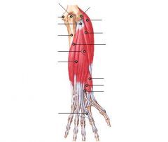
Label this diagram
|
1. Pronator Teres
2. Flexor Carpi Radialis 3. Palmaris Longus 4. Flexor Carpi Ulnaris 5. Pisiform 6. Brachioradialis |
|
|
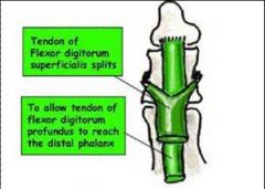
Describe how flexor digitorum superficialis and profundus insert onto the phalanges
|
|
|
|
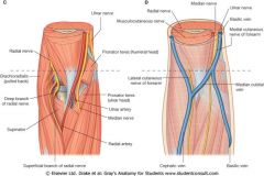
List the order of structures on the anterior surface of the distal forearm
|
Lateral to medial:
1. Superficial branch of radial nerve 2. Brachioradialis 3. Radial artery 4. Flexor carpi radialis 5. Median nerve with its palmar cutaneous branch 6. Palmaris longus 7. Flexor digitorum superficialis (4 tendons) 8. Ulnar artery 9. Ulnar nerve with its palmar cutaneous branch 10. Flexor carpi ulnaris |
|
|
What are the boundaries of the carpal tunnel?
|
- Roof = flexor retinaculum, attaching from the tubercle of scaphoid and the trapezium and inserting into the hook of hamate and the pisiform
- Floor and walls = carpus |
|
|
Describe the contents of the flexor retinaculum
|
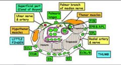
1. 4 tendons of flexor digitorum superficialis
2. 4 tendons of flexor digitorum profundus 3. Tendon of flexor pollicis longus 4. Median nerve 5. Flexor carpi radialis (within its own compartment) |
|
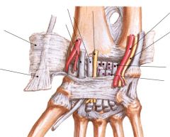
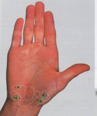
Label this diagram
|
See instant anatomy pg 212 if unsure
|
|
|
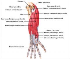
What muscles make up the superficial extensors of the wrist and digits?
|
1. Extensor carpi radialis longus
2. Extensor carpi radialis brevis 3. Extensor digitorum 4. Extensor digiti minimi 5. Extensor carpi ulnaris |
|
|
What muscles make up the deep extensors of the wrist and digits?
|
1. Abductor pollicis longus
2. Extensor pollicis brevis 3. Extensor pollicis longus 4. Extensor indicis |
|
Label this diagram
|
|
|
|
Describe the boundaries of the anatomical snuffbox and its contents
|
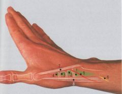
- Floor = scaphoid and trapezium
- Medially = Extensor pollicis longus - Laterally = extensor pollicis brevis and abductor pollicis longus - Roof = fascia, skin etc. - Contents: Radial artery and nerve and 1st dorsal interossei |
|
|
Why is the anatomical snuffbox clinically significant?
|
- Tenderness in the snuffbox is a good indicator of scaphoid fractures, which are extremely difficult to X-ray and diagnose
- This is particularly important, as the scaphoid receives its blood supply from distally rather than proximally, so can get ischaemic necrossi if damaged |