![]()
![]()
![]()
Use LEFT and RIGHT arrow keys to navigate between flashcards;
Use UP and DOWN arrow keys to flip the card;
H to show hint;
A reads text to speech;
17 Cards in this Set
- Front
- Back
|
what are the four main regions of the oviducts
|
- infundibulum - ampulla - isthmus |
|
|
describe the mucosa of the oviducts
|
mucosa: - endosalpinx, columnar epithelial cells submucosa: - myosalpinx, smooth muscle, connective tissue, vessels |
|
|
what type of cells does the epithelium of the oviducts contain
|
ciliated cells: - highly motile - extend into lumen - %age of ciliated cells decreases towards isthmus, max in fimbriae and infundibulum non-ciliated cells: - secretory cells - contain granules, which secrete components of lumen fluid - apical surface covered in microvilli |
|
|
what is the function of the ciliated cells in the oviduct
|
the action of their beating enables oocyte with cumulus cells (layer of cells that has to be penetrated for fertilisation to occur) to be striped from surface of collapsing follicles they beat in synergy with the oviduct contractions meaning that the oocytes are in constant rotation helping the oocyte and sperm come together |
|
|
what are the three histological layers of the uterus
|
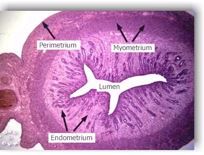
2. myometrium 3. endometrium |
|
|
describe the perimetrium layer of the uterus
|
- serous layer (tunica serosa) - supportive layer - connective tissue, smooth muscle, vessels, nerves |
|
|
describe the myometrium layer of the uterus
|
- outer thin layer of longitudinal smooth muscle - middle vascular layer - inner thick layer of circular smooth muscle - contractions in synergy with oviducts |
|
|
describe the endometrium layer of the uterus
|
epithelium: - simple, columnar (mare, bitch, queen) - simple, stratified columnar - epithelial cells relatively tall during oestrus, but become low and cuboidal a couple of days after oestrus - uterine glands - widespread except in ruminants where they are absent from the caruncular areas - varies in thickness and vascularity with stage of cycle and gestation |
|
|
describe the histological structure of the cervix
|
epithelium: - simple,columnar - mucous secreting - secretions vary throughout cycle |
|
|
what are the three histological layers of the vagina
|
2. tunica muscularis 3. tunica mucosa |
|
|
describe the tunica adventitia layer of the vagina
|
- serous layer of connective tissue and longitudinal smooth muscle |
|
|
describe the tunica muscularis layer of the vagina
|
inner, thick circular smooth muscle and outer thinner longitudinal smooth muscle |
|
|
describe the tunica mucosa layer of the vagina
|
- 2-3 layers during anoestrus/prepubertal but multiple layers during oestrus - superficial layers cornify (keratinised) but not much in ruminant and depends on stage of cycle - microridges on surface of vaginal cells aid firmness of vagina (copulation and parturition) |
|
|
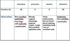
describe the different observations seen in the reproductive tract during anoestrus, proestrus, oestrus and dioestrus
|
|
|
|
what is responsible for the bloody discharge seen from the vagina during proestrus
|
high E2 levels which leads to capillary breakdown and leakage through the uterine epithelium |
|
|
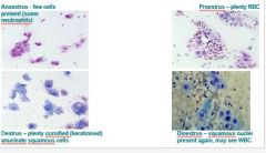
other side shows the histological appearance of the tubular tract during anoestrus, proestrus, oestrus and dioestrus
|
|
|
|
what characteristic change in epithelial cells of the reproductive tract marks dioestrus
|
decline in number of cornified (keratinised) epithelial cells |