Reading...
![]()
Play button
![]()
Play button
![]()
Use LEFT and RIGHT arrow keys to navigate between flashcards;
Use UP and DOWN arrow keys to flip the card;
H to show hint;
A reads text to speech;
17 Cards in this Set
- Front
- Back
- 3rd side (hint)
|
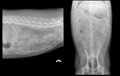
Small Bowel
|
Evaluation
Size - diameter of lumen or wall thickness; overall, hard to tell shape - countour of bowel loops margin - serosal surface definition architecture - mucosa/bowel wall smoothness; use other modalities position - location within abdominal cavity; can be mass effect of full bladder or masses opacity - bowel wall and luminal contents motility - intermittent contractions small bowel diameter: Dogs - <twice the width of a rib; <1.6 x L5 height cats - <12mm; <2 x L4 height can mask foreign bodies, don't always show foreign body, esp distal obstruction |
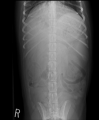
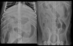
reduced detail on VD view
overlying muscles and detail in abdomen is reduced typically use lateral view |
|
|
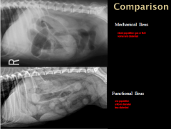
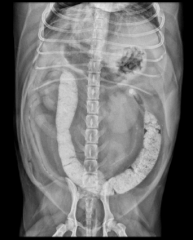
Mechanical vs Function
|
mechanically obstructed bowel is usually larger diameter
-usually fluid and gas filled -usually mixed bowel population, will see distended and normal bowel functional is predominately either gas filled or fluid filled, more uniform opacity -generalized involvement of bowel, intestinal lumen is patent |
|
|
|
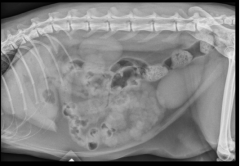
Functional Ileus
|
Viral enteritis
-parvovirus Chronic mechanical obstruction Vascular compromise -intestinal strangulation/entrapmetnt -mesenteric volvulus -segmental jejunal arterial thrombosis neuromuscular disease -spinal trauma -dysautonomia -generalized change to whole GI tract |
|
|
Enteritis
|
generally fluid filled
mildly distended enteritis and colitis |

|
|
|
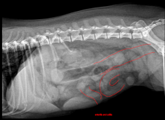
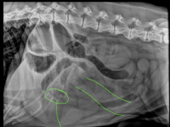
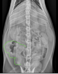
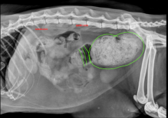
Ileus from peritoneal effusion
|
urinary bladder rupture and leakage of urine into abdomen
post cystogram images positive contrast media throughout abdomen sometimes effusion in abdomen causes paralysis/hypomotility, causes distention uniform distension - leakage of contrast from bladder rupture |
mixed population; gas or fluid; normal and distended
one population, uniform diameter, less distended |
|
|
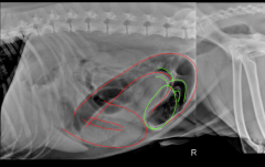
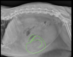
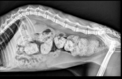
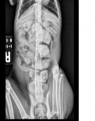
mesenteric volvulus
|
occlusion of cranial mesenteric artery
ischemic necrosis gastrointestinal toxin release shock GSD and other large breed dogs overrepresented previous surgery, anastomosis, enterotomy uncommon vomiting blood, bloody diarrhea - bad systemic illness |
gas filled predominately, severely distended, vascularly compromised, intestines going all different ways
|
|
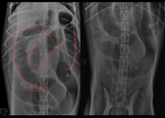
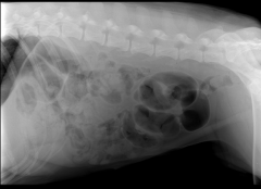
Mechanical Ileus
|
occlusion of intestinal lumen
foreign objects intussusception mural mass extrinsic lesions determine the cause radiographic signs: variable - depends on location, duration, vom complete or partial location in the GI tract duration -variable degree of bowel loop dilation oral to obstruction - some distended, some normal -stacked - gas and fluid - abdomen compartment squeezes together -gravel sign - contents of intestines become opqaue - more distal mechanical obstruction |
partial
-may only have partially dilated bowel loops -appearance is less severe -fluid and gas can pass the partial obstruction - large parts may not duodenal -variable dilation of duodenum and stomach -depends on frequency of vomiting linear -plicated bowel -only mild bowel loop distention -some gas becomes trapped in some of the plicated segments and will have a short tubular, tapered, or tear dropped shaped appearance mixed population of bowel fluid and gas; normal and distended |
|
|
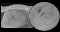
Chronic Distal Obstruction
|
Gravel Sign
ileocecal stenosis opaque ingestion - dessication |
distal ileal obstruction
|
|
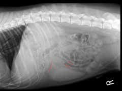
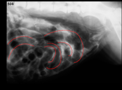
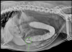
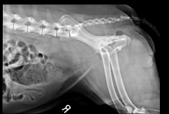
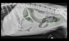
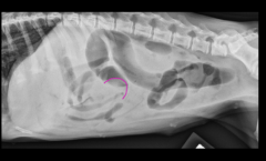
Linear Foreign Body - Dog
|
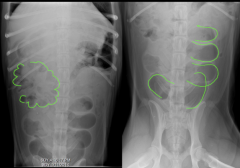
(distended gas filled bowel, crescent shaped, stacking of segments, sharp turns in cranial abdomen)
abnormal shape and contour of bowel loops abnormal gas pattern crescent shaped, comma shaped pleated or plicated appearance clumped, bunched *check under the tongue of cats! |
abnormal shape and contour
abnormal transit through the abdomen |
|
|
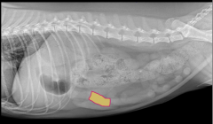
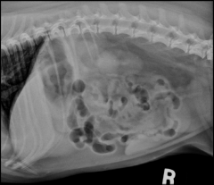
Linear Foreign Body - Cat
|
|
severe plication with scalloped margins
some gas trapped in segments tear drop, cresecent shaped |
|
|
Infiltrative Bowel Disease
|
Generalized or segmental infiltration of the bowel wall
-nonseptic inflammation -infection -neoplasia overall increase in thickness of bowel ultrasound upper GI study loss of detail in ventral abdomen can't see serosal margin of urinary bladder |
bowel associated masses
no abnormalities focal loss of serosal detail mechanical obstruction -acute or chronic etiologies -intestinal wall abscess -fungal -pythium -neoplasia -polyp |
|
|
Large Bowel
|
uniform size when distended
larger than small bowel should not exceed the length of L4 dogs - <1-1.5X L7 length cats - normal < 1.28 X L5 length megacolon >11.48 X L5 length colonic wall is thinner than small bowel at 2mm |

|
|
|
common colonic diseases
|
decreased size (stricture)
-mural masses (neoplasia, granuloma) -extramural masses (lymph nodes, prostate) -inflammation (colitis) -spasm (may or may not be associated with colitis) increased size -generalized (megacolon) -localized (proximal to colonic obstruction, mass lesions, intussception) |
|
|
Segmental
|
impaction
mechanical obstruction mural disease -stricture extramural tumor narrowed pelvic canal |

|
|
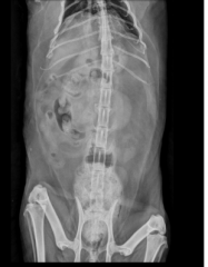
Caudal retroperitoneal mass
|

|
|
|
Megacolon
|
Constipation
-infrequent or difficult defecation associated with the retention of feces obstipation -constipation that is refractory to treatment, which occurs where there is permanent loss of function Megacolon -a persistent irreversible increase in colon diameter -this is the most advanced stage in the spectrum of chronic constipation Radiographic diagnosis of megacolon -history of chronic constipation/obstipation that that is nonresponsive to medical treatment -the presence of an enlarged distended colon filled with fecal material chronic constipation and obstipation -nutritional -metabolic -mechanical spinal anomalies -cauda equina syndrome -sacrocaudal agenesis neuromuscular disorders -dysautonomia metabolic disorders congenital anorectal anomalies |
|
|
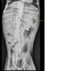
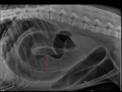
Intussusception
|
(soft tissue/gas interface)
Various types - based on location (ileocolic, cecocolic and colocolic) appearance is variable -soft tissue mass associated with colon -may appear as homogenous soft tissue opacity of the colon -meniscus sign contrast studies and ultrasound are a useful aid in diagnosis |
meniscus sign
|