![]()
![]()
![]()
Use LEFT and RIGHT arrow keys to navigate between flashcards;
Use UP and DOWN arrow keys to flip the card;
H to show hint;
A reads text to speech;
43 Cards in this Set
- Front
- Back
|
Primary Brain Vesicles (developing tube) |
Prosencephalon (forebrain) Mesencephalon (midbrain) Rhombencephalon (hindbrain) |
|
|
Prosencephalon secondary brain vesicles |
Telencephalon Diencephalon |
|
|
Telencephalon adult brain structures |
Cerebrum: Cerebral Hemispheres: - cortex - white matter - basal nuclei |
|
|
Diencephalon adult brain structures |
Diencephalon: - thalamus - hypothalamus - epithalamus
Retina |
|
|
Mesencephalon Secondary brain vesicles |
Mesencephalon (brain stem: midbrain)
|
|
|
Rhombencephalon secondary brain vesicles + adult brain structures |
Metencephalon (brain stem: pons; cerebellum) Myencephalon (brain stem: medulla oblongata)
4th ventricle is not part of the rhombencephalon itself but is in this area |
|
|
CNS Development |
Ectoderm and neural tissue (notochord) interact - forms the neural plate
Neural plate edges fold upward and inward and meet midline - forms a neural crest and tube
The neural tube 'zips up' from either side until we have closed tube with openings at either end - lumen in the middle forms the ventricles - the brain vesicles develop on either side of the neural tube |
|
|
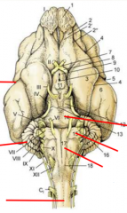
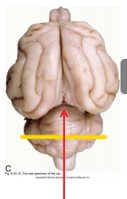
Clockwise starting on right side:
Pons Trapezoid bodies (part of the pons) Pyramidal tract (in medulla oblongata) Spinal cord Cerebellum Cerebrum |
|
|
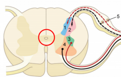
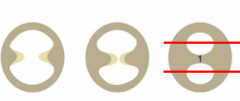
dorsal horn = afferent ventral horn = efferent
1= somatic afferent 2= visceral afferent 3 = visceral efferent 4 = somatic efferent 5 = dorsal root ganglion (afferent neurons from peripheral system)
red circle = central canal |
|
|
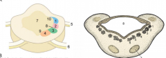
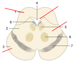
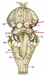
organization of the medulla oblongata/pons - we now find cranial nerves
in pons: Central canal (7) expands greatly and becomes the 4th ventricle
Tracts take on a different orientation (afferents are more lateral and efferents are more medial)
2 more tracts join - special visceral afferent - special sensory afferent
Medulla picture (left): 1 = somatic afferent column 2 = visceral afferent column 3 = visceral efferent column 4 = somatic efferent column 5 = dorsal root 6 = ventral root 7 = central canal 8 = sulcus limitans 9 = basal lamina 10 = alar lamina
Pons (on right) 1 = somatic afferent column 2 = visceral afferent column 3 = special visceral afferent column 4 = visceral efferent column 5/6 = somatic efferent column 7 = nuclie of pons 8 = fourth ventricle |
|
|
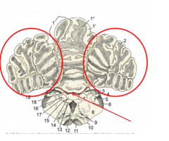
Cerebellum:
red circles = cerebellar hemispheres red arrow = 4th ventricle/choroid plexus
1 = vermis 3 = vasciculus gracilis and cuneatus 4 = gracile and cuneate nuclei 11 = pyramidal tracts |
|
|
red arrow = transverse fissure AKA tentorum cerebelli
- fold of the dura mater between the cerebrum and cerebellum
- falx cerebri (fold of dura mater) is between the two sides of the cerebrum |
|
|
red arrows clockwise:
cerebellum 4th ventricle medulla oblongata pons |
|
|
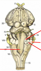
Rhombencephalon with cerebellum removed (dorsal view)
red line on left: median sulcus of 4th ventricle
Right (top to bottom): - rostral peduncle - caudal peduncle |
|
|
What are peduncles? |
Roots (pairs) that attach cerebellum to medulla beneath it - transmit various nerve structures to and from the pons and medulla
Rostral peduncle: most efferent parts of the cerebellum
Middle peduncle: receive afferent, connected to pons
Caudle peduncle: redceive afferent, connected to medulla |
|
|
Medulla Oblongata functions |
Involved in Autonomic and Visceral systems
Contains nuclei of cranial nerves involved in:
breathing (CN#10 - Vagus nerve) taste (CN#11 - Glossopharyngeal nerve) |
|
|
Pons functions |
"Bridge" between hind brain and higher centres in mesencephalon and telencephalon
Contains nuclei of cranial nerves involved in:
hearing (CN#8 - vestibulocochlear nerve) facial expressions (CN#7 - facial nerve) facial sensation (CN#5 - trigeminal nerve) |
|
|
Cerebellum functions |
Coordination of posture, balance, proprioception
*DOES NOT initiate these, but regulates and coordinates them |
|
|
4th ventricle functions |
CSF production (choroid plexus)
gateway into subarachnoid space |
|
|
Mesencephalon adult features |
Tectum - Colliculi Mesencephalic aqueduct Tegmentum - CN nuclei Crus cerebri (most ventral) |
|
|
Mesencephalon
1 = Tectum (2 colliculi) 2 = Tegmentum (CN Nuclei) 3 = Crus cerebri 4 = mesencephalic aqueduct (CSF, connects 3rd and 4th ventricles) 5 = oculomotor nucleus (CN#3) 6 = red nucleus 7 = substantia nigra 8 = locus coeruleus |
|
|
Mesencephalon with cerebellum removed
Top red arrow: rostral colliculus - related to optic pathways to and from eyes to occipital lobe
Bottom red arrow: caudal colliculus - related to auditory pathway |
|
|
Red arrow on right side: crus cerebri = "cross" * only thing we can see on outside of mesencephalon
- is a connection between telencephalon and brain stem
- origin of CN#3 (oculomotor - visceral efferent and somatic efferent nerves related to the eye) |
|
|
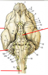
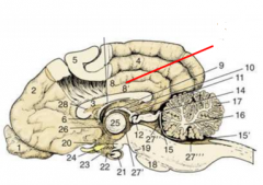
Diencephalon Red arrows clockwise, starting top right
- Hypothalamus - Mamillary bodies (connect hippocampus to other parts of the brain) - Spinal Cord - Cerebellum - Cerebrum - Infundibulum (attaches pituitary gland AKA hypophysis to the brain)
Major parts: - Thalamic areas - 3rd ventricle (not technically part of brain tissue but found in this area) - retina/CN#2(optic) |
|
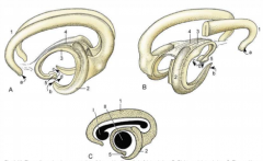
Formation of Diencephalon |
- Two portions of neural tube merge toward each other - a bridge is formed between both sides of the thalamus (= interthalamic adhesion) - this creates an upper and lower portion of the 3rd ventricle and divides the thalamus into 3 regions
Epithalamus, thalamus, hypothalamus (top to bottom) |
|
|
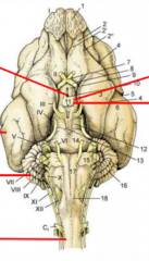
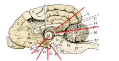
Diencephalon Red arrows clockwise starting at top:
Epithalamus Epiphysis (pineal gland) Mamillary body Hypophysis (pituitary gland) Tuber Cinereum (infundibulum) Interthalamic adhesion
*Epithalamus contains the epiphysis (pineal gland) *Hypothalamus contains the hypophysis (pituitary gland) and the mamillary body
#3 = corpus callosum (telencephalon) |
|
|
Epithalamus function |
Contains the Epiphysis (pineal gland) - Circadian rhythms |
|
|
Thalamus functions |
integrates all sensory info from body and special senses to cortex, EXCEPT from olfactory bulb *nuclei relay the info to the cortex
Lateral geniculate nuclei: optic info medial geniculate nuclei: auditory info
Also involved in sleep/wakefulness |
|
|
Hypothalamus functions |
Contains the Hypophysis (pituitary gland) = master gland of endocrine control
- secretes hormones that control parts of the endocrine system
Contains the mamillary bodies - connection between hippocampus and amygdala - involved in memory formation |
|
|
3rd ventricle functions |
makes CSF
*not part of diencephalon brain tissue but is located in this area |
|
|
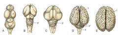
Phylogenetic brains
A = fish B = reptile C = bird D = mammal E = human
1 = telencephalon 2 = mesencephalon 3'/3" = metencephalon 3' = archicerebellum 3" = neocerebellum 4 = myelencephalon 5 = spinal cord |
|
|
Telencephalon |
The Cerebrum itself
Divided into two hemispheres by longitudinal fissure (contains falx cerebri - folds of dura mater from roof of cranium toward the floor of the cranial cavity)
pallium = cortex - covered with folds (sulci) and gyri(ridges) - (increase surface area for greater neuronal connectivity and complexity)
Corpus callosum - connects the two hemispheres
enlarged cortex is unique to mammals and supports high level cognitive functioning |
|
|
3 portions of the pallium |
Pallium = cortex
Paleopallium = olfactory bulbs and piriform bulbs Neopallium = cerebral hemispheres Archipallium = cingulate, hippocampal formation - can't really distinguish this one but it is between the two hemispheres |
|
|
Basal Nuclei |
Part of telencephalon
nucleus = collection of neuronal cell bodies basal ganglia = collection of nuclei
Function: initiation and regulation of motor control
3 basal nuclei: - Caudate nucleus - Lentiform nucleus - Amygdala |
|
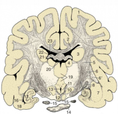
Identify the basal nuclei |
Caudate nucleus = #3 Lentiform Nucleus = #6, 6' Amygdala = #7
#2 = corpus callosum |
|
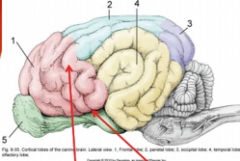
Neopallium/Paleopallium |
Neopallium = cortex Part of telencephalon
1 = frontal lobe - executive functions and motor function 2 = parietal lobe - spatial and sensory function 3 = occipital lobe - visual functions 4 = temporal lobe - association cortex, auditory functions
Paleopallium 5 = olfactory bulb/piriform lobe |
|
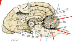
Archipallium |
Areas of the telencephalon - Arches around various other structures
A = lateral view B = right caudolateral view C = positions of corpus callosum (I) and thalamus (II)
1 = supracallosal and cingulate gyri 2 = hippocampus 3 = fornix 4 = commissure of fornix 5 = hypothalamus with mamillary body a = input from medial olfactory tract b = input from piriform lobe c = output to mamillothalamic tract d = output to brain stem |
|
Identify parts of the archipallium |
8 = cingulate gyri 8' = supracollasal gyri 7 = fornix (cut off at the top) |
|
|
Arterial supply to the brain |
Done by the cerebral arterial ring - receives main sources from lower down in body (left and right internal carotids, come up through the head from the neck) - single basilar artery runs up spinal column
*they anastamose and form the ring |
|
|
Important branches of the Cerebral arterial ring |
Rostral, middle and caudal cerebral arteries *rostral meet in middle to form rostral part of the ring *middle provide to the temporal lobe and other structures in brain *caudal goes deep into the brain
More caudal branches are rostral and caudal cerebellar arteries
Receives important contribution from vertebral artery |
|
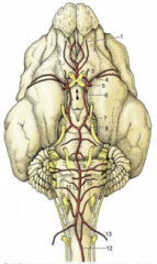
Name the important arteries of the Cerebral arterial ring |
#5 - left and right internal carotids #11 - single basilar artery #2 - rostral cerebral artery #4 - middle cerebral artery #7 - caudal cerebral artery #8 - rostral cerebellar artery #10 - caudal cerebellar artery #13 - vertebral artery
Not part of ring but important: #12 - ventral spinal artery |
|
|
Venous drainage of brain |
Drainage is done via sinuses
1) Dorsal sagittal sinus (drains CSF via villi) 2) Transverse sinus (meets with dorsal sagittal, runs along back of tentorum cerebelli) 3) Straight sinus (unpaired, goes deep into brain and drains parts like the midbrain) 4) Cavernous sinus (rostral end of skull, contributions from ophthalmic veins) 5) Basilar (petrosal) sinus (unpaired)
all of these collect and drain into internal jugular vein - exit jugular foramen in the skull |
|
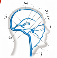
Name these! |
1 - opthalmic veins 2 - cavernous sinus 3 - basilar (petrosal) sinus 4 - dorsal sagittal sinus 5 - straight sinus 6 - transverse sinus 7 - internal jugular vein |