![]()
![]()
![]()
Use LEFT and RIGHT arrow keys to navigate between flashcards;
Use UP and DOWN arrow keys to flip the card;
H to show hint;
A reads text to speech;
173 Cards in this Set
- Front
- Back
|
Cnidarian |
Phlyum of carnivorous animals that sting. Radially symmetrical. Diploblastic. Sesile polyp or mobile medusa. Hydrostatic skeleton for digestion |
|
|
Bilateria |
Phylum that diverged from cnidarians. Tripoblastic and bilateral symmetry. Diverges into Protostomes and Deuterostomes |
|
|
Coelom |
Acts as a fluid filled body cavity. Functions: circulate/store metabolites, provides a cushioned space for organs, increases size without more cells, hydrostatic skeleton. |
|
|
Coelomate |
Body cavity fully lined by mesoderm e.g humans, segmented worm |
|
|
Pseudocoelomate |
Body cavity where the outer wall is lined by mesoderm. E.g roundworms |
|
|
Acoelomate |
Body cavity containing a non-split coelom. E.g flatworms |
|
|
Deuterostomes |
Diverged from cnidaria so are tripoblastic and have bilateral symmetry. Have radial cleavage. Anus develops first in the blastopore. pockets of mesoderm form. |
|
|
Protostomes |
Derived from bilateria/deuterostomes. They are bilaterally symmetrical and are tripoblastic(develop mesoderm and coelom). Spiral cleavage. Anus is formed second in the blastopore(mouth is formed first). |
|
|
Echinodermata |
Marine animals that are sessile and slow moving (looks like an echidna). They have secondary bilateral symmetry(bilateral in embryo which resembles humans) |
|
|
Chordata |
All animals after echinoderm. 4 characteristics: notochord, dorsal hollow nerve, pharyngeal slits, post-anal tail. |
|
|
Craniates |
Phylum with a carnium/skull/eye structures/neural crest. e.g Myxini(hagfishes) |
|
|
Vertebrates |
Have a backbone and paired appendages. Can be either an exo or endo skeleton. But lampreys lack the paired apdgs. e.g Petromyzontida(lampreys) |
|
|
Gnathostomes |
Jawed vertebrates. Derived from Pharyngeal slits. e.g sharks rays, chondrichthyes |
|
|
Chondrocythes |
Cartilagenous fishes. Skeleton predominantly cartilage. e.g shark, rays, chimaeras |
|
|
Osteichthyans |
Development of lungs/lung derivatives. Fins are supported by dermal rays and not muscle. e.g Actinopterygii (ray-finned fishes) |
|
|
Lobe fins |
Have rod shaped bones. Muscles. Can now walk underwater. This is the first sign of limb development. e.g actinistia, lung fishes |
|
|
Tetrapods |
Limbs! And a stronger vertebral column. Ribs neck and supporting girdles. e.g amphibia |
|
|
Amniotes |
produce an amniotic egg. have thicker skin(keratin). e.g reptiles and birds. Diverged into reptillia and mammalia. |
|
|
Amniotic egg |
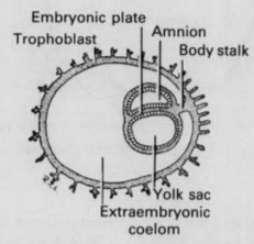
Inthe blastocyst, an inner cell mass is formed. Within the inner cell mass, twofurther cavities appear (the amnion and yolk sac) separate by the bilaminardisc. Reptiles and birds are known to have a yolk sac, but human embryos havethem as well. The yolk sac attaches to the outside of the developing embryo,which acts as a preliminary circulatory system delivering nutrients and iseventually absorbed into the gut of the embryo. the aminotic cavity is lined by epiblast. hypoblast lines the yolf sac. |
|
|
Mammals |
Defined by the existence of mammary glands producing milk and hair. Development of a larger brain and increased metabolism. |
|
|
Two migration hypothesis theories |
Multiregional: When Homo erectus left Africa 2 mya and dispersed into other portions of the Old World, regional populations slowly evolved into modern humans. Continuity with neanderthals. Replacement/Out of Africa: 2 migrations out of africa. No continuity with neanderthals |
|
|
Components of blood |
Buffy coat(smallest layer, platelets and leukocytes), plasma(rises to the top), erythrocytes(RBC's, lowest layer) |
|
|
Erythrocyte formation |
RBC's are produced in the bone marrow. When they form they eject their nucleus and so cannot replicate because of their lack of DNA. |
|
|
Blood vessel tunica |
3 layered wall: tunica externa, tunica medica, tunica intima. |
|
|
Pericardium |
A complex sac of multiple layers that provides a space for the heart to move and contract . Consists of the fibrous pericardium(outer layer) and the serous pericardium(inner layer which secretes fluid for lubrication for movement by the heart). |
|
|
Hepatic Portal Circuit |
Blood circulation through the liver. The function is to detoxify and metabolise. Takes oxygen poor nutrient rich blood from the GI tract to the liver via the portal vein. |
|
|
The lymphatic system |
Single celled wall lumen parallel to blood vessels which contains drained interstitial fluid. This checks on overall organism health(which may lead to an activated immune response). |
|
|
Spleen and it's 2 pulps |
This is the largest lymphatic organ located in the 2nd quadrant. Contains 2 pulps: white pulp(lymphocyte storage). Red pulp(reservoir for RBC's and platelets. |
|
|
Upper Respiratory Tract |
The Nasal Cavity and Pharynx. |
|
|
Nasal cavity |
Part of the URT. Consists of pseudo-stratified columnar epithelium, the nasal septum, external nares, conchae. |
|
|
Nasal septum |
central wall of bone and cartilage that divides the nasal cavity |
|
|
External nares |
Nostrils, bilateral opening of the paranasal sinuses. When this is inflamed it blocks the passageway which can cause a headache |
|
|
Conchae |
Bony plates and soft tissue found inside the nose. It increases surface area of mucous membranes for humidifying air and increasing temperature. Also important for directing the air to the olfactory bulb. |
|
|
Pharynx |
Funnel shaped common space used by both the respiratory and digestive tracts. Directs air to the larynx/bronchi and ingested food to the esophagus. Pharynx walls are lined with mucosa (stratified squamous epithelium) and skeletal muscle. There is the nasopharynx, oropharynx, laryngopharynx. |
|
|
Lower Respiratory Tract |
conducting airways: larynx, trachea, bronchi etc. |
|
|
Larynx |
Cartilaginous structure that connects the pharynx to the trachea. |
|
|
Epiglottis |
Cartilage which deflects food into the esophagus |
|
|
Thyroid cartilage |
Partial ring of cartilage: not complete so that esophagus doesn't rub against from the movement of swallowing. |
|
|
Cricoid cartilage |
Complete ring of cartilage |
|
|
Trachea |
A flexible and slightly rigid tube with 15-20 cricoid cartilage rings. Trachea splits into 2 primary bronchi. |
|
|
Bronchioles |
Cartilage is being replaced by smooth muscle leading to one cell thick alveoli |
|
|
Hilus |
region where blood vessels enter/leave |
|
|
Visceral Pleura |
Lines the surface of each lung |
|
|
Parietal Pleura |
Lines the internal abdominal walls: thoracic walls, mediastinum, diaphragm |
|
|
Pleural cavity |
The space between the visceral and parietal pleura which contains pleura which is serous and secretes fluid. The fluid creates hydrostatic tension which supports the lungs. |
|
|
Nervous control of Respiration |
Receives regular rhythmic signals from the ANS |
|
|
Accessory digestive organs |
Liver, gallbladder, pancreas |
|
|
Assisting organs |
tongue, salivary glands, teeth |
|
|
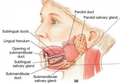
Salivary Glands |
Consists of 99% water and 1% enzymes(amylases and lysozymes). 1-1.5L is secreted daily. 3 glands: submandibular, parotid, sublingual. Duct tubes transport the saliva to the mouth. |
|
|
Parotid glands |
Largest salivary gland, which overlays the bottom teeth. *accidentally biting on your cheek bit, para(tid)aalysed. produce saliva (20%-30%) |
|
|
Submandibular Glands |
Produce most of the saliva(60-70%) and are located inferior to the body of the mandible but the ducts open on either side of the tongue. It has many lymph nodes associated with it. |
|
|
Tongue |
Assessory digestive organ made of skeletal muscle and lightly keratinised stratified squamous epithelium. Contains papillae and lingual tonsils. |
|
|
Esophagus |
made up of skeletal muscle(superior) and smooth muscle(inferior) and connects the pharynx to the stomach. undergoes peristalsis and segmentation. |
|
|
Histology of the GI tract |
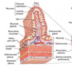
Lumen consists of 4 connective layes/tunics: mucosa(epithelium and connective tissue), submucosa(connective tissue and blood vessels), muscularis(layers of smooth muscle), serosa(outer connective tissue covering) |
|
|
Nerve plexuses |
Complex interconnections of neurons between the tunics. 2 types: Meissner's submucosal, Auerbach's(myentric) |
|
|
Meissner's(submucosal) nerve plexus |
Located in the submucosa layer of the GI tract, and regulates SECRETION |
|
|
Auerbach's(myentric) nerve plexus |
Located in the muscularis externus tunic layer of the GI tract and regulates MOTILITY |
|
|
Parietal Peritoneum |
Lines the abdominal(body) cavity. Secretes peritoneal fluid and stores fat. |
|
|
Visceral peritoneum |
Lines the outer layer of the wall of the gut components |
|
|
Mesentries |
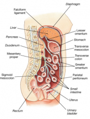
Double layers of peritoneum that attaches viscera to posterior abdominal wall; contains vessels, nerves and lympatics. Omenta is a double layer of mesentry |
|
|
Intraperitoneal organs |
Organs that are suspended from the body wall by a mesentry. e.g stomach, transverse colon, Small intestine |
|
|
Retroperitoneal |

Organs that do not have a mesentry and are held against the posterior body wall. e.g kidneys, baldder, ascending and descending colons. (organs outside the peritoneum. |
|
|
Stomach |
Located inferior to the diaphragm. divided into the cardia, fundus, body and pylorus. It has large circular folds called rugae and 3 layers of smooth muscle. |
|
|
Small intestine |
Duodenum, Jejunum, Ileum. Absorbs most of the nutrients |
|
|
Large intestine |
1.5m long. absorbs most of the water, electrolyes and only some remaining nutrients. |
|
|
Liver |
Accessory digestive organ |
|
|
Mesonephros |
Multiple Glomeruli merge all of their ducts together to form the mesonephric duct which drains into a region called the cloaca(receives urinary and digestive secretions) and becomes the gonads. |
|
|
Metanephros |
3rd type of kidney that has evolved over time. the urinary and reproductive tract are separate and then connect. |
|
|
Kidney development |
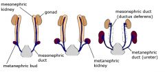
As the pronephros and mesonephros start degrading, the kidney changes position and walks its way up from the pelvis and to the abdominals. It does this by making arterial connections and breaking them(cos it needs to stay functional using a blood supply while it's migrating. The mesonephros turns into the gonads
|
|
|
Bladder |
An expandable skeletal reservoir for urine made of 4 tunics. The ureter enters the bladder at a steep angle at the bottom, so it fills the bladder from the bottom up. A triangle shaped area at the bottom of the bladder called the trigone ensures that urine will flow down the urethra even when the bladder wall contracts, to ensure there is no back flow in the ureters. |
|
|
Urethra |
Tube connecting the urinary bladder to the exterior containing a mucous lining secreted by urethral glands. Smooth muscle fibers surround the mucosa and help propel urine to the outside of the body. |
|
|
Female urethra |
only 3-5cm long because it only transmits urine from the bladder to vestibules |
|
|
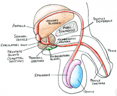
Male urethra |
18-20cm long common passageway for both urine and semen. Prostatic urethra(3cm) --> membranous urethra(1cm)--> spongey urethra(15cm) |
|
|
Tripoblast |
Gives rise to the 3 germ layers (endoderm, mesoderm, ectoderm). Germ(sex) cells differentiate separately. |
|
|
Urogenital system germ layer derivation |
Derived from intermediate mesoderm which later develops into the urogenital system. |
|
|
Sex determination |
The common gonad starts off as a bipotential gonad and then a transcription factor SRY found on the Y chromosome witches on a cascade of genes responsible for male gonad development. Testis development also requires DHT and AMH. For ovary development there must be the absence of the SRY gene but also must have 2X chromosomes and other factors. |
|
|
Bipotential reproductive system |
Twin set of ducts and tubes formed within the first 10 weeks of development. Contains both The Mullerian(female) ducts and Wolffian ducts(male) |
|
|
Mullerian Ducts |
Duct in the bipotential reproductive system. In a female embryo it develops into the fallopian tubes, uterus and vagina. The wollfian ducts degenerate |
|
|
Wollfian ducts |
Duct in the bipotential reproductive system. In a male embryo its develops into the vas deferens and prostate. |
|
|
Labia majora |
In females it stays the way it is. In males it develops as the scrotum, into which the testes will descend at birth. |
|
|
Pathway of spermatozoa |
Spermatozoa produced in the testes --> mature in the epididymis --> pass through the ductus deferen tubes --> ejaculated out. Sperm needs to pass through all these places to make sure they are as viable as possible for fertilisation. |
|
|
Testes structure |
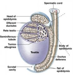
Consists of seminiferous tubules(site of spermatogenesis). The rete testis collects sperm from these tubules via efferent ducts. |
|
|
Testes location |
The testes stay outside the body to stay cooler since the optimal temperature of some enzymes in spermatogenesis is lower than body temp. 34 deg. |
|
|
Epididymis |
A coiled tube (4-5m) along the side of the testis which acts a site of sperm storage and maturation(development of a tail). |
|
|
Testes Muscles |
Cremaster and Dartos |
|
|
Cremaster muscle |
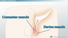
Testes muscle that raises testes during sex or a fear stimulus (keeps it tucked away and not swinging side to side when trying to escape) |
|
|
Dartos muscle |

Wrinkled appearance(like the labia majora in females), and it's role is to aid in temperature regulation. The wrinkles increase SA required for heat loss. important in spermatogenesis. |
|
|
Testosterone |
Before birth, it drives the development of the external genitalia, where the testes descend out from the frontal pelvis and into the scrotum. After birth, testosterone secretion ceases until puberty, where thereis a surge of testosterone. It eventually decreases but they produce it at high levels for most of their lives. This results in enlargement of testes, penisand scrotum, facial hair, pubic hair etc. Spermatogenesis begins at this pubescent stage. |
|
|
Spermatic cord |
Thick-walled tube consisting of muscle and fascial layers formed by the vas deferens, surrounding tissue, veins and arteries that runs from the deep inguinal ring in the abdominal wall down to each testicle. |
|
|
Seminal vesicles
|
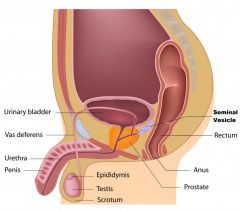
An accessory gland sperm must pass through. Located in the posterior surface of the bladder thete secrete a viscous fluid of fructose and prostaglandins. Prostaglandins dilate the cervix so that it can get inside the uterus and up to fertilise the egg. |
|
|
Prostate gland |
Accessory gland which is walnut shaped and inferior to the bladder which secretes citric acid(nourishment), seminal plasmin, PSA that liquefies semen after ejaculation.
|
|
|
Bulbourethral (cowper's gland) |
Inferior to the prostate gland. Secretes a clear vicous mucin which protects the urethra and serves as a lubricant during sex. |
|
|
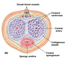
Penis |
|
|
|
3 Sexual Functions of the Penis |
Erection: Parasympathetic innervation - dilation Emission: Movement of secretions from accessory glands which provide sperm with nourishment. |
|
|
The Primordial germ cell(PGC) journey |
(After gastrulation but before Gonad formation) Specification: Around 6 cells in the embryo express Blimp1 , which suppresses all genes that would've turned them into somatic type cells. Commitment: After several rounds of proliferation, a subset of the Blimp1 positive cells express Stella, and commit to the germline. The rest of the cells go off and become other tissues. Migration: PGCs undergo both passive and active migration to the gonads. Extensive proliferation occurs during this migration Colonisation: PGCs reach and colonise the gonad(week 6-8 of gestation), and undergo sex determination |
|
|
Gonocytes |
Germ cells that enter the testis. They undergo mitosis-arrest-mitosis, and don't enter meiosis/spermatogenesis until puberty |
|
|
Oogonia |
Germ cells that enter the ovary. Entermeiosis but arrest at prophase I of meiosis until puberty (fertilisation bysperm) |
|
|
Disrupted development in the male germline |
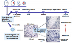
Gonocytesarrest in their potential to differentiate and become completely resistant to the surrounding tissues that tell them to start spermatogenesis. There is progression into malignancy and tumours will start to grow. can result in seminoma or non-seminoma. |
|
|
Seminoma |
Malignant tumour where thetestis will seem very disorganised |
|
|
non-seminosa |
testicular cancer |
|
|
Meiosis in females |
1 oogonium --> 1 ovary + 3 polar bodies. There is only 1 because there is not enough material to produce 4 fully supplied ovaries. 1 ovary requires a large supply of nutrients for its first stages of development. The polar bodies to help direct early embryonic axes. The anterior posterior axis of the embryo is set up at the first cleavage division of the embryo. |
|
|
Meiosis in males |
1spermatogonium --> 4 spermatozoa(& |
|
|
Advantages of meiosis |
Crossing over of alleles, independent assortment, random fertilisation |
|
|
oogenesis |
primordial cell -> primary cell --> secondary cell (puberty) |
|
|
FSH |
Hormone needed for the survival of follicles and their continued maturation |
|
|
Menstruation |
Up to 50 oocytes will mature but only one will dominate (defined around 7 days prior to ovulation). Once the dominant follicle enlarges, it becomes FSH independent, and secretes high levels of inhibin. The inhibin suppresses pituitary FSH production, causing the remaining 49 semi-matured follicles to degenerate and die. |
|
|
Spermatogenesis |
spermatogonium start in the coiled seminiferous tubules --> 2 rounds of meoisis --> spermatozoa in the inner layer. Supported by leydig, sertoli and immune cells. |
|
|
Leydig cells |
Testosterone producing cells between the tubules in the testes. |
|
|
Sertoli cells |
'Nurse cells' that extend from the basement membrane to the lumen which express the SRY gene and keep the germ cells that start the process healthy and nourished. |
|
|
The syncytium |
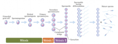
Cyotplasmic connections between male germ cells that ensures there are an even number ofsperm with X and with Y. *for a 1:1 girl boy ratio to exist. |
|
|
Behavioural reproductive war |
Males: social structures, seduction or dominance, mate guarding. |
|
|
Human skeleton: Number of bones |
Composed of 206 bones: |
|
|
Molecular structure of bone |
Connective tissues containing specialised cells and mineralised matrix of crystalline calcium phosphate and calcium carbonate. Consists of an organic(collagen -elastic) and inorganic(mineral crystal Hydroxyapatite - contribute to rigidity) component. The proportion/ratio of these components vary, i.e between antlers(high collagen), auditory ossicles(high mineral) and long bones(iso). Without mineralisation, the bones will be very brittle, dry and easy |
|
|
Bone cells |
Osteoblast: immature bone cell which secretes organic components of matrix Osteocyte: mature bone cell(from osteoblasts) that maintains bone matrix Osteoprogenitor cell: stem cell whose division produce osteoblasts Osteoclast: secretes acis and enzymes to dissolve bone matrix |
|
|
Bone structure |
Compact bone, spongy bone, bone marrow |
|
|
Bone marrow |
A tissue that fills the internal cavity in bones, it is dominated by blood cell or adipose tissue. |
|
|
Spongey bone |
Inner layer of the bone which is a composed of a network of body struts(trabeculae). there is many empty cavities therefore no need to have a canal system. The nutrients are passed through by diffusion. Thetrabeculae of spongey bone are aligned with the direction of stress. Theychange shape to give resistance to stress and strains on the bone |
|
|
Compact bone |
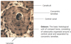
Solid dense bone on the outer layer which contains parallel osteons. Osteonsare cylindrical in shape and consist of osteocytes organised around a centralcanal(blood vessels, nerves, lymphatic vessels) and are separated by concentriclamellae. |
|
|
Periosteum |
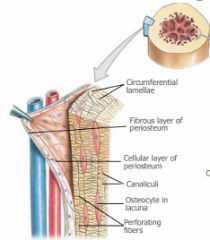
A very dense, fibrous and tough outerlayer of the bone. |
|
|
Endosteum |
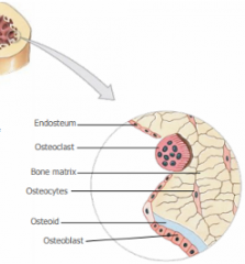
The single cellular layer composed of loose connective tissue lining the trebaculae |
|
|
Long bone anatomy |
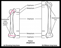
Epiphysis: The head of the long bone Diaphysis: The shaft(middle bit)of a long bone Metaphysis: between the epiphysis and diaphysis. It corresponds to the location of the epiphyseal cartilage of the developing bone. |
|
|
Cartilage |
Dense connective tissues composed of chondrocytes with a more gelatinous, less calcified matrix(glycoprotein material-chondroitin) than bones and has an abundance of matrix. It is an avascular tissue that provides support and allowsfor some flexibility of movement. |
|
|
Cartilage anatomy |
Perichondrium, chondrocytes, lucunae |
|
|
Types of cartilage |
Hyaline, Elastic, Fibrocartilage |
|
|
Bone development 2 processes |
Intramembranous 'ossification in membrane', endochondral 'ossification in cartilage' |
|
|
Intramembranous 'ossification in membrane' |
no cartilage precursor, but rather directly from connective tissue where osteoblasts lay down bone in fibrous tissue from mesenchymal cells. |
|
|
Endochondral 'ossification in cartilage' |
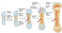
Preexisting hylaine cartilage is gradually destroyed and replaced and occurs at the epiphyseal plate. Osteoblasts die from lack of nutrients, and then blood vessels form |
|
|
Bone remodelling |
The rate of osteoclasts destroying bone and osteoblast forming bone tissue are roughly equal. old age: formation < degredation. Bones become weaker. |
|
|
Bone Fracture and Repair |
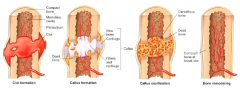
If there is a bone fracture where blood vessels are broken, blood clots and coagulates. The periosteum forms a callus to hold everything together which is soft initially. New born cells causes it to become a hard callus. Through the activity of osteoclasts, the callus is removed. After several months it is completely repaired |
|
|
Classification of Joints: Function |
Synarthrosis(no movement): bony edges interlock like a jigsaw e.g skull Amphiarthrosis (little movement): stronger than a freely moveable joint. connected by collagen fibers or cartilage. Diarthrosis(free movement): a form of joint articulation/synovial joint that permits movement. |
|
|
Classification of joints: Structure |
Cartilaginous: held by a thin layer of hyaline and/or fibro-cartilage e.g epiphyseal plate Between vertebrae Fibrous: are those joints where the articular surface of the bones are attached to each other through fibrous connective tissues. e.g joined bones of the skull e.g attachment of the fibula nd tibia to the ligament Synovial joints:(most common)are freely movable joints covered with a layer of articular cartilage within a joint capsule that contains synovial fluid. The fluid is lined with synovial membrane and reinforced by a fibrous capsule and ligaments |
|
|
Muscle organisation |
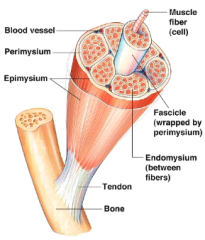
Epimysium: surrounds the entire muscle and separates it from surroudning tissue and organs. Perimysium: divides the skeletal muscles into a series of compartments. Surrounds the fascicl Endomysium: surrounds the individual skeletal muscle cells(muscle fibers) |
|
|
Fast muscle fibers |
Large in diameter, low number of capillaries, fast contraction, rapid fatigue. |
|
|
Slow muscle fibers |
Smaller in diameter than fast fibers, take three times as long to contract. are fatigue resitance, contain a large amount of myoglobin, have a more extensive network of capillaries. |
|
|
Intermediate muscle fibers |
Have properties between fast and slow muscle fibers. |
|
|
Lateral mesoderm |
The flatter end of mesoderm at which limb development occurs |
|
|
Appearance of limb buds and bud growth |
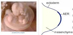
They start to appear as out-pocketings from the body wall. At the tip is a thickening of the limb bud called the apical ectodermal ridge (AER) which is important for the outgrowth and extension of the limb. This forms all the way around the developing limb. |
|
|
Apical ectodermal ridge (AER) |
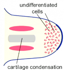
Located on the tip of the thickened limb bud outgrowth of the body wall. The AER keeps cells proliferating and undifferentiated. But as the limbs grow out the inner cells(farther from the AER) lose influence of the AER and so they stop proliferating and then start differentiating. The cells at the tip, closest to the AER, are still undifferentiated. |
|
|
Forming the appendicular skeleton |
Mesenchyme causes condensations when losing influence from the AER and produces Cartilage Templates(CT's) which ossify and become bone. Cartilages are laid down in a proximo-distal sequence/patterning. This is done by HOX genes |
|
|
Hox genes |
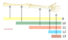
Gradual sequencing. Proximal distal patterning |
|
|
Anterior-posterior patterning (sonic hedgehog patterning) |
Regulated by the zone of polarising activity (ZPA) which initiates sonic hedgehog expression(shh). Shh expression dictates digit location. Whe a shh bead is placed in another spot, it causes mirror image duplication. |
|
|
Creating a cartilage template |
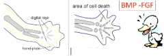
The distal limb flattens to become a hand-plate. The condensed digital rays form and become cartilage. Finger webbing is removed by cell death/apoptosis. - (BMP - FGF signalling family) |
|
|
Developing Limb muscles |
Limb muscles originate in the somites and myoblasts migrate into the limb bud and arrange on opposite sides of the cartilage template. The limb muscle cells arrange into masses and cleave into individual muscle cells |
|
|
Phocomelia |

Absence of most of the limb, reduced limb features at proximal regions. failure of the limb bud to grow. |
|
|
Syndactyly |

Fusion of digits or joining of digits with soft tissue (still retains the finger webbing). Possibly due to apoptosis/ proliferation problems. This is a normal feature in kangaroos however. |
|
|
Basic patterning of limb skeleton |
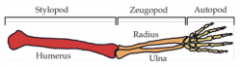
As a result of divergent evolution, most vertebrates have the similar structure of stylopod(humerus), zeugod(ulna) and autopod(hand) |
|
|
Somites |
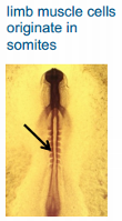
Anything from the neck down during development. |
|
|
Hyoid bone |
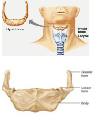
Supports the larynx with its greater horns and is the attachment site for muscles of the larynx, pharynx and tongue. It is inferor to the manidble and acts as a moveable base for the tongue. It is the only bone in the body that does not move with another bone. It's position is in the body can determine vocals produced. |
|
|
Vertebral column |
Function is to protect spinal cord and spinalnerves, support body weight, posture and locomotion, flexibility. Consists of 33 vertebrae arranged in 5 regions: cervical, thoracic, lumbar, sacral, coccygeal. |
|
|
Spine curvature development |
During foetal development the spine is C shaped (primary curve - thoracic and sacral). The lumbar and cervical S-shaped curvatures(secondary curves) develop during childhood in association with lifting the head and assuming upright setting. Curves are important because they increases column strength, help maintain balance, absorbs shock during walking. |
|
|
Abnormal curvatures |
Kyphosis - excessive posterior thoracic - hunchback Lordosis- excessive anterior curvature - bum sticks out Scoliosis - excessive lateral curvature |
|
|
Evolution of upright posture (CoG and vertebrae size)
|
Centre of gravity moves from the thorax(gorilla) to the pelvis(human). Vertebrae size increases across evolution because humans have to carry a larger load due to gravity. |
|
|
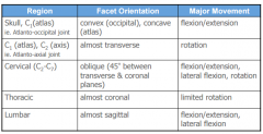
Cervical vertebrae
|
C1- C7. start at the top of the skull. C1(atlas), C2(axis). Bodyis small and wide laterally, to distribute the weight of the head across alarger surface-provides more support. Vertebral foramen is prominent, largeand triangular because spinal cord is larger at the top. Transverse foramina - passage forblood vessels |
|
|
Lumbar vertebrae |
L1-L12. Extends to accommodate for pregnancy. |
|
|
Intervertebral dics |
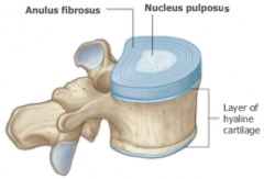
Cushion-like pads between vertebrae that act as shock absorbers. they are composed of: nucleus pulposus and the anulus fibrosus. |
|
|
Anulus fibrosus |
Outercollar made of ligaments and fibrocartilage. Its function is to bind vertebraetogether, resist tension on the spine, and absorb compressive forces. |
|
|
Nucleus pulposus |
Gelatinous inner sphere of the intervertebral disc. Enables spine to absorb compressive stresses |
|
|
Intervertebral articulations |
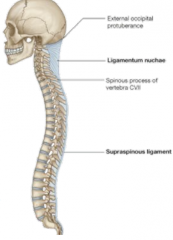
Ligaments: Ligamentum nuchae: supraspinous ligament that extends from the base of the skull to C7. Interspinous ligament: Fills space between the spinous processes. Supraspinous ligament: interconnects the tips of the spinous processes of each vertebrae from C7 to the sacrum |
|
|
Vertebral movements |
|
|
|
Ribs/The thoracic cage |
The costal cartilage start to fuse together before it attaches to the sternum. |
|
|
Sternum |
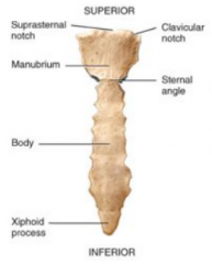
A flat bone in the middle of the chest/thoracic wall, Consists of 3 parts: Manubrium, Sternal body of the pectoral girdle, The xiphod process
|
|
|
True Ribs and Falser ibs
|
True Ribs 1-7: Connected to the sternum s.cn False Ribs 8-12: Do not attach directly to the sternum. Ribs 11-18: floating ribs and are only attached to back muscle |
|
|
Tendons |
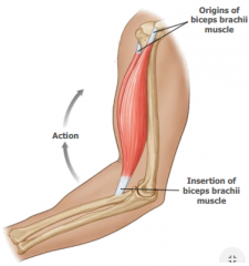
Connect bones to muscle. Origin - place where the fixed end of the muscle attaches to a bone/cartilage and where the it moves the least, usually proximal. Insertion- site where the movable end of the muscle attaches to another structure. |
|
|
Erector spinae muscles |
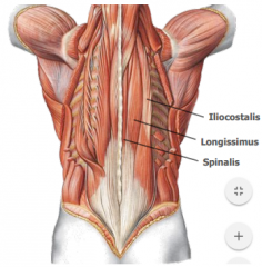
Lies in the groove of each side of the vertebral column, and is the major extensor of the vertebral column. |
|
|
Transversospinalis muscle group |
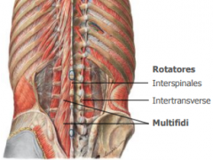
Consists of three groups of muscles found in the groove between the transverse and spinous processes |
|
|
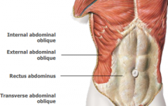
Thoracic and abdominal wall muscles |
|
|
|
Phylogeny |
The evolutionary development and diversification of a species or group of organisms, or of a particular feature of an organism. |
|
|
Ontogeny |
The development of an individual organism or anatomical or behavioural feature from the earliest stage to maturity. |
|
|
Difference between CT scanning and MRI
|
CT scans use emission of rotating X rays with multiple detectors. MRI measures hydrogen atom emission of a radio frequency in a strong magnetic field and constructs imagery from the detected frequencies picked up by a coil around the object. |
|
|
Female external genitalia |
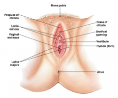
collectively called the vulva:composed of the mons pubic, labia majora and minora, clitoris, vestibule(urethral opening and vaginal orifice), prepuce(fold of labia minora covering the clitoris), urethra. |
|
|
Internal female genitalia |
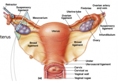
Consists of the fallopian tubes, uterus, vagina |
|
|
Uterus |
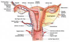
A pear shaped muscular organ which leads to the cervix and then the vagina. It undergoes changes every month and consists of 3 layers: (peritmetrium and underlying connective tissue). Myometrium(thick layer of smooth muscle-high proliferative capacity for belly expansion during pregnancy). Endomentrium, simple columnar epithelium with underlying connective tissue. |
|
|
Vagina |
elastic tube of stratified squamous epithelium located between the urinary bladder(anterior) and rectum(posterior).open sinto the vestibule and leads up through the cervix and into the uterus. |