Reading...
![]()
Play button
![]()
Play button
![]()
Use LEFT and RIGHT arrow keys to navigate between flashcards;
Use UP and DOWN arrow keys to flip the card;
H to show hint;
A reads text to speech;
121 Cards in this Set
- Front
- Back
|
Chest Pain
|
most important symptom of cardiac disease
may also indicate intestinal, gallbladder, musculoskeletal, or pulmonary disorders exam: serious to minor causes cardiac: freq. described as crushing substernal pain that does not disappear with rest |
|
|
Rib Fractures (p.54)
|
1st rib rarely fractured b/c of protected position
frac. leads to risk of brachial plexus and subclavian vessel damage frac. result from blows or crushing injuries; weakest part is anterior to angle The middle ribs are the ones most commonly fractured (7th and 10th) when several ribs are broken in several places a flail chest results danger of damage to lung or spleen from broken end diaphragmatic hernia is tear in diaphragm can be caused by rib fracture painful b/c broken parts move during respiration |
|
|
Flail Chest (p.54)
|
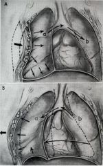
multiple rib fractures allow segment of t. wall to move freely
moves paradoxically to respiration (inward and outward on expiration) diagram: A. Inspiratory phase. Chest wall collapses inward (a), forcing air out of bronchus of involved lung into trachea and bronchus to uninvolved lung and causing shift of mediastinum to uninvolved side (b). B. Expiratory phase. Chest wall balloons outward (a) so that air expelled from lung on uninvolved side (b) enters lung on involved side and mediastinum shifts toward involved side (a). This is a very inefficient form of respiration, and the patient will die of hypoxia and exhaustion if it occurs in an extreme phase and is not relieved. extremely painful and impairs vent. fixed by hooks and wires so it cannot move |
|
|
Thoracotomy, Intercostal Space Incisions, and Rib Excision (p.55)
|
surgical creation of an opening through thoracic wall to access pleural cavity
anterior (H-shape) at the anterior chest wall - wide segment of rib removed allows entry through periosteal sheath, spares intercostal muscles can be used for pneumonectomy. also a critical maneuver in the management of traumatic cardiac arrest (wikipedia) bone may be used as a graft in reconstruction procedures - will regenerate in periosteal sheath posterior - 5th to 7th intercostal but can go up to 4th. lateral appproach gives best access- pt. lying contralateral, and upper limb fully abducted to change position of scapula for access to the 4th intercostal space. is considered the approach of choice for pulmonary resection (pneumonectomy and lobectomy) complications: pneumothorax, infection, bleeding, air leaks, and resp. failure |
|
|
Supernumerary Ribs (p.54)
|
usually have 12 ribs on each size
can have too many or failure of 12th pair to form can confuse identification or numbering of levels on radiographs |
|
|
Protective Function and Aging of Costal Cartilages (p.54)
|
costal c. contribute to elasticity for thorax and provide resilience to t. cage; prevent blows from fracturing sternum/ribs
chest compression can cause injury without fracture in elderly, costal c. lose elasticiy from calcification and become brittle = radiopaque and less resilient |
|
|
Ossified Xiphoid Process
|
normal in mid 40s to discover a hard lump in "pit of stomach"
patients may fear it's a tumor such as stomach cancer; just calcification at epigastric fossa |
|
|
Sternal Fractures (p.54)
|
not common
can occur from crush trauma (ex. forced into steering column in crash) usually "comminuted fracture" most common site is at sternal angle - results in dislocation of the manubriosternal joint. concern of possible heart damage with mortality ~25-45% chance ALL patients with sternal injury should be evaluated for visceral trauma |
|
|
Median Sternotomy (p.54)
|
to gain access to mediastinum by dividing sternum in median plane and retract.
(ex. for CABG surg., or removal of superior lobe tumors) halves of sternum are reunited with wire sutures |
|
|
Sternal Biopsy (p.54)
|
sternal body often used for bone marrow needle biopsy b/c of its breadth and subcutaneous position
obtain specimens for transplant or for detection of metastatic cancer and blood dyscrasias |
|
|
Sternal Anomalies (p. 54)
|
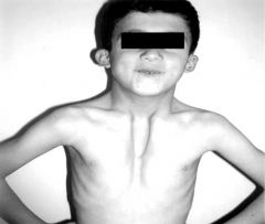
sternal halves may not fuse in fetus = Sternal Cleft (above pic)
repair in infancy w/ direct apposition and fixation of cartilaginous sternal halves sternal foramen due to incomplete fusion of plates (not clinically significant, except for misinterpretation on medical image as a bullet wound) |
|
|
Dislocation of Ribs (p.55)
|
slipping rib syndrome or dislocation of a sternocostal join
displacement of a costal cartilage from the sternum injury produces lump at dislocation site common in body contact sports can damage nearby nerves, vessels and muscles |
|
|
Thoracic Outlet Syndrome (p.55)
|
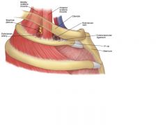
involving compression at the superior thoracic outlet
Superior TA: Anatomically is called t."inlet", and Clinically is called t."outlet" Arteries/Viens, and brachial plexus nerves emerge trough aperture to enter lower neck and upper limbs TOS (t. outlet syndrome) = implies thoracic location, obstruction is in root of the neck, manifestations of syndrome variably involve upper limb. (eg.: costoclavicular syndrome = diminished radial pulse resulting from compression of the subclavian artery b/w clavicle and first rib) |
|
|
Separation of Ribs (p.55)
|
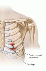
refers to dislocation of a costochonral junction between rib and its costal cartilage
if occurs in 3 - 10 = usually tears perichondrium and periosteum causing ribs to move superiorly, overriding rib above it and causes pain |
|
|
Paralysis of the Diaphragm (p.54)
|
Innervated by cervical motor neurons C3-C5 via the phrenic nerves
hemidiaphragm paralysis as result of injury to its motor supply from phrenic nerve (each dome has separate nerve supply) will move paradoxically when paralyzed the affected dome descends during expiration as it is pushed down by the positive preassure in the lungs |
|
|
Dyspnea: Difficulty Breathing (p.64)
|
when people with resp. problems (eg. asthma or CHF) struggle to breath, they use accessory respiratory muscles to assist expansion of t. cavity
leaning on chair or knees can "fix" pectoral girdle (clavicle and scapula) so these accessory muscles are better able to act on rib attachments and expand the thorax. |
|
|
(Extrapleural Intrathoracic Surgical Access)
|
endothoracic fascia is a natural cleavage plane in surgery
separate costal parietal pleura from the thoracic wall = access to lymph nodes can place instruments without risking opening of potential space of pleural cavity surrounding lungs |
|
|
Herpes Zoster Infection of the Spinal Ganglia (p. 64)
|
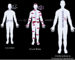
shingles = viral disease of spinal ganglia
dermatomally distributed skin lesion pathogenesis; 1)Entry through upper respiratory tract 10–21 day incubation 2)Travels to regional lymph nodes, liver and spleen (primary viraemia) 3) Travels to skin/mucous membranes: vesicular rash (secondary viraemia) 4) Replication in epidermal cells, entry into nerve endings and transport to dorsal root ganglia (DRG) where it establishes latency in sensory neurones 5)Reactivation in DRG → infection of nerves and dermatome → herpes zoster the herpes virus invades a spinal ganglion and is transported along the axon to the skin. causes red, vesicular rash confined to a dermatome; sharp burning pain in affected region after a few days, the skin of the dermatome becomes red and vesicular eruptions appear VZV (varicella-zoster virus) for shingles and small pox |
|
|
Intercostal Nerve Block (p.64)
|
injecting a local anesthetic near intercostal nerves between paravertebral line and area of desired anesthesia
each area of skin gets innervation from two adjacent nerves so overlapping dermatomes occurs. therefore complete loss of sensation usually does not occur unless several intercostal nerves in adjacent intercostal spaces are anesthetized. Herpes zoster or shingles pain in the chest is commonly treated with intercostal blocks http://www.youtube.com/watch?v=BOLdHJC6_50 |
|
|
Changes in the Breasts (p.58)
|
branching of the lactiferous ducts may occur during menstruation or pregnancy
Colostrum = creamy white premilk; starts in last trimester; rich in protien, immune agents, growth factors breasts increase in size with multiple pregnancies (multiparous) breasts decrease in size wuth age and loss of fat and atrophy of glands |
|
|
Breast Quadrants (p.58)
|
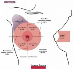
radial coordinate system to refer to location of tumors or cysts in breasts
divide into 4 equal quadrants, nipple as center reference point |
|
|
Carcinoma of the Breast (p.58)
|
lymph drainage important (lymphogenic metastasis - metastasis of cancer by means of lymph); usually pass 2 or 3 sets of nodes before reaching venous drainage
Symptoms: lymphedema, peau d'orange sign, dimples, breast elevation with pectoral contraction (if invading muscle) most common site of metastasis is axillary lymph nodes since most of the lymphatic drainage of breast goes there. usually adenocarcinomas (from epithelial cells of lactiferous ducts) subareolar breast cancer - may cause inversion of the nipple spread of cancer to cranium and brain through azygos/hemiazygos system of viens (where the posterior intercostal viens drain) p. 59 cancer can also spread by contiguity (invasion of adjacent tissue). see clinical sign of advanced cancer p. 59 |
|
|
lymphedema (p.58)
|
edema or excess fluid backup in breast upon blockage of lymph drainage by cancerous mass
|
|
|
peau d'oragne sign (p.58)
|
prominent / puffy skin between dimples pores
results from lymph backup in cancerous breast |
|
|
Mammography (p.59)
|
radiographic breast examination
carcinoma appears as large jagged density in the mammogram guide when removing breast tumors, cysts, and abscesses |
|
|
Supernumerary Breast and Nipples: Polymastia, Polythelia, and Amastia (p.58)
|
supernumerary breasts (polymastia), or accessory nipples (polythelia) = occur superior or inferior to normal on mammary ridge
usually rudimentary = easily mistaken for mole until they change color during pregnancy |
|
|
Breast Cancer in Men
|
1 in 100 breast cancers affect men and only about 10 in 1 million develop breast cancer (webMD)
usually metastasizes to lymph nodes (also to bone, pleura, lung, liver, and skin) presents with visible or palpable subareolar mas or nipple secretion and tends to invade pectoral fascia |
|
|
Gynecomastia
|
male breast hypertrophy after puberty - quite rare
may be age or drug related; or imbalance between estrogenic and androgenic hormones treat as a symptom, check suprarenal and testicular cancer and cirrhosis 40% Klinefelter's (XXY) have gynecomastia |
|
|
Injuries to the Cervical Pleura and Apex of Lung (p.70 - 71)
|
apex of lung (covered by cervical pleura) project through opening of the superior thoracic aperture into the base of the neck, just posterior to inferior attachment of sternocleidomastoid
risk of pneumothorax with penetrating wounds at base of neck children are especially vulnerable |
|
|
Injury to Pleurae and Pleural Pain (p.78)
|
visceral pleura - insensitive to pain
parietal pleura - sensitive to pain (esp. costal pleura = somatic intercostal and phrenic n.) pain referred to dermatomes of same posterior root ganglia costal / peripheral diaphragmatic pleura thoracic and abdominal walls mediastinal / central diaphragmatic pleura = C3-C5 dermatomes |
|
|
Pulmonary Collapse (p.77)
|
with penetrating wound, air will be sucked into pleural cavity and breaks surface tension adhearing visceral to parietal pleura, and the lung collapses because of its inherent elasticity (elastic recoil)
it's possible that only one lung can collapse because of the separation of pleural sacs pressure in pleural cavities in subatmospheric (-2mmHg, during inspiration: -8mmHg) eg. in open-chest surgery, maintain inflation with positive-pressure pump leads to pneumothorax |
|
|
Pneumothorax
|
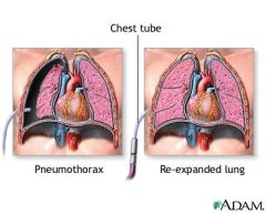
if air enters the pleural space, the lung will collapse= pneumothorax.
If the chest wall is penetrated, which may occur as a result of an injury, air can enter the pleural space from the outside. Air can also enter from the inside, from the lung itself, if the lung is torn or ruptured. One of the most common causes of spontaneous non-traumatic pneumothorax is a pulmonary bleb. This is a weakness and out-pouching of the lung tissue, which can rupture. This introduces air into the pleural space. The immediate treatment for pneumothorax is tube thoracostomy, or the insertion of a chest tube Pulmonary blebs are a common cause of spontaneous pneumothorax in young children and adults. They can be resected to prevent future pneumothorax |
|
|
Pneumothorax, Hydrothorax, Hemothorax, and Chylothorax (p.77)
|
Air, Water, or Blood in the pleural cavity
pneumo = air from penetrating wound hydro = fluid from from pleural effusion hemo = blood injury to a major intercostal/internal thoracic vessel chylo= chyle (pale white/yellow lymph fluid) from a torn thoracic duct |
|
|
Thoracentesis (p.78)
|
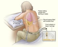
insertion of hypodermic needle through intercostal space into pleural cavity to sample fluid or remove blood or pus
needle should be just superior to rib to avoid neurovascular bundle pleural effusions accum. (when sitting) in costodiaphragmatic recess- space located between costal pleura and diaphragmatic pleura. used for thoracocentesis because of less risk of puncturing the lungs insert needle at 9th intercostal space to avoid lung (& point up to avoid liver); also posterior superior to the rib, high enough to avoid the collateral branches. (posterior because the interspaces are wider. superior because neurovascular bundle is closer to the inferior margin of the rib posteriorly) |
|
|
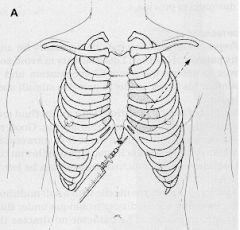
Insertion of a Chest Tube (p.78)
|
placed for removal of large amounts of air, blood, fluid, or pus
incision in 5th/6th intercostal space in midaxillary line direct superior - remove air direct inferior - drain fluid connect outside end of tube with underwater drainage system with controlled suction (to prevent backflow) |
|
|
Thoracoscopy (p.78)
|
diagnostic/therapeutic procedure for examination of pleural cavity for cancer
incisions in intercostal space allow placement of scope (take photos, biopsies; disrupt adhesions or remove plaques) |
|
|
Pleuritis (Pleurisy) (p.77)
|
normally respiration/expiration produces no detectable noise from pleurae (by asucultaion)
but with inflammation, this makes the lung surfaces rough = pleural rub and can be heard with stethoscope acute pleuritis = sharp, stabbing pain friction = "pleural rub" this is audible with stethoscope |
|
|
Lung Resections (p.78)
|
pneumonectomy = whole lung
lobectomy = a lobe segmentectomy = one or more bronchopulmonary segments for tx. for lung cancer/tumor, or abscesses (collection of pus) understand bronchopulmonary segments and their relationship to the bronchial tree for plan of drainage and clearance techniques (especially for pts. with pneumonia or cystic fibrosis) tumors often localize in a bronchopulmonary segment, when resecting a bp. segment, surgeons follow the interlobar veins to pass between the segments. |
|
|
Variations in the Lobes of the Lung (p.77)
|
can have extra fissure or one may be absent
azygos lobe = most common accessory lobe, appears in RL 1% of pop. azygos vein arches over apex, not hilum, creating new (azygos) lobe in medial part of apex |
|
|
Inhalation of Carbon Particles (p.78-79)
|
lungs are light pink in children, non-smokers in clean environment
foreign bodies most likely enter the right main bronchus commonly dark / mottled in adults in urban / agricultural areas as a result of inhaled particles (smoker's cough) lymph carries "phagocytes" that phogocytose carbon particles. These particles color the surface of the lungs gray to black. |
|
|
Auscultation of the Lungs and Percussion of the Thorax (p.78)
|
listening with steth; tapping to detect sounds
A: asses airflow in lungs. also used to detect heart murmurs. use steth. P: percussion of the lungs - tapping the chest over the lungs with the finger = establishes state of underlying tissue = air filled (resonant), fluid (dull), or solid (flat) some places should be flat b/c of bone structure. Always include root of the neck (apicies); base of lung refers to inferoposterior part, listen at inferoposterior aspect of thoracic wall = T10 |
|
|
Lung Cancer and Mediastinal Nerves
|
involvement of phrenic nerve may paralyze diaphragm
apical cancers may involve recurrent laryngeal nerve (branch of vagus); presents as hoarseness due to paralysis of vocal cored |
|
|
Aspiration of Foreign Bodies (p.78)
|
R bronchus is wider, shorter, and runs more vertically than L
foreign bodies tend to lodge in the R main bronchus or one of its branches dentists may insert a rubber dam in oral cavity to prevent aspiration during procedures |
|
|
Bronchoscopy (p.79)
|
uses bronchoscope
carina (keel) = ridge seen between orifices of main bronchi; lays in sagital plane and has definite edge carina will be distorted with enlarged lymph nodes in angle - morphologic changes important for diagnosis from a bronchogenic carcinoma mucus membrane covering carina associated with cough reflex |
|
|
Segmental Atelectasis
|
blockage of segmental bronchus prevents air from reaching supplied segment
air will be gradually absorbed and segment will collapse (does not require compensating space in pleural space; adjacent segments expand) |
|
|
Pulmonary Embolism (p.79)
|
embolus forms when blood clot, fat globule, or air bubble travels to lung from a leg vein
embolus travels through the R heart to pulmonary artery and to the lung. may block artery or one of it's branches: Pulmonary embolism blocks a pulmonary artery = leads to partial or complete obstruction of blood flow = acute respiratory distress right side of heart may become dilated (inc. venous return collateral circulation can compensate for PE in younger people pulmonary infarct = medium size embolus blocks an artery supplying bronchopulmonary segment, and results in an area of necrotic lung tissue = tx. resection. |
|
|
Lymphatic Drainage after Pleural Adhesion
|
lymphatic vessels in lung and visceral pleura drain to axillary lymph nodes
presence of C particles is evidence of pleural adhesion |
|
|
Bronchogenic Carcinoma
|
usually squamous or small-cell carcinoma arising from bronchus (now refers to any lung cancer)
main cause: cigarette smoking most arise in mucosa of large bronchi; present w/ persisten, prouctive cough metastises to blood, bone, lymph, adrenal |
|
|
Mediastinoscopy and Mediastinal Biopsies
|
incision approximately 1 cm above the suprasternal notch of the sternum, or breast bone
used to conduct minor surgical procedures or biopsy of lymph nodes to test for bronchiogenic carcinoma |
|
|
Widening of Mediastinum
|
often observed after trauma from lacerated great vessels/ serious hemorrahge
also indicates: malignant lymphoma enlargement of heart (hypertrophy) with CHF |
|
|
Surgical Significance of Transverse Pericardial Sinus (p.82)
|
after the pericardial sac has been opened anteriorly, a finger can be passed through the TPS posterior to the aorta and pulmonary trunk.
must ligate these vessels during cardiac surgery (eg. CABG) |
|
|
Pericarditis and Pericardial Effusion (p.83)
|
inflammation of pericardium = chest pain
no detectable sound during auscultation but causes pericardial friction rub (pericarditis makes surface rough which results in friction) = sounds like the rustle of silk with steth. results in compressed heart - unable to expand and fill fully and ineffectual |
|
|
Pericardial Friction Rub p.83
|
inflammation causes friction along roughened surfaces
sounds like rustle of silk, hairs rubbing together |
|
|
Pericardial Effusion p.83
|
("fluid around the heart") is an abnormal accumulation of fluid in the pericardial cavity
leaky pericardial capillaries or pus accumulation during inflammation also occurs during CHF without inflammation negative effect to heart = cardiac tamponade! |
|
|
Cardiac Tamponade p.84
|
heart compression caused by tough and inelastic pericardium. consequently, heart volume is compromised by the fluid outside the heart.
occurs when the pericardial space fills up with fluid faster than the pericardial sac can stretch caused by a large or uncontrolled pericardial effusion, (eg. the buildup of fluid inside the pericardium) slow increase in size of hear = cardiomegaly hemopericardium = blood enters the pericardial cavity, caused by trauma (eg. stab wounds) pericardiocentesis = drainage of serous fluid from pericardial cavity |
|
|
Pneumopericardium
|
air from pneumothorax dissecs percardial sac
|
|
|
Pericardiocentesis
|
drainage of fluid from pericardial cavity
insert needle in left 5th or 6th intercostal space near sternum ("bare area of pericardium") |
|
|
Positional Abnormalities of the Heart
|
dextrocardia : apex directed right instead of left
mirror image positioning of great vessels |
|
|
Percussion of the Heart p.91
|
]defines the density and size of the heart.
percuss at 3rd, 4th, and 5th intercostal spaces from left anterior axillary line to right anterior axillary line. change from resonance to dullness indicates edge of heart |
|
|
Atrial Septal Defects (ASDs) p. 91
|
congenital anomalies of interatrial septum- usually related to incomplete closure of the ovale foramen after birth
small-size ASDs have no clinical significance large-size allow O2 rich blood from lungs to be shunted from the LA thorugh the defect into the RA, causing enlargement of the RA and ventricle and dilation of the pulmonary trunk |
|
|
Ventricular Septal Defects (VSDs) p.91
|
commonly on membranous part of IV septum
25% of all forms of congenital heart disease size of defect 1 to 25mm causes LV to RV shunt of blood through the defect. increases pulmonary blood flow (hypertension) and may cause cardiac failure much less common for the muscular part of the septum to remain open (usually closes in childhood) |
|
|
Thrombi / Stroke / CVA p.91
|
"cerebral vascular accident" aka stroke
thrombi (clot) detach and pass into systemic circulation, occluding peripheral arteries occlusion of an artery supplying brain = CVA affects vision, cognition, sensory and/or motor fxn. previously controlled by the now damaged area of the brain |
|
|
Valvular Heart Disease p.91
|
VHD produces either stenosis or insufficiency
stenosis = valve cannot fully open slowing blood flow from a chamber insufficiency / regurgitation = valve cannot fully close. cusps do not meet or align, allowing blood to flow back both result in increased workload, produce audible turbulence (murmurs or thrills) turbulance sets up eddies (small whirlpools) that produce vibrations that are audible as murmurs thrills = superficial vibratory sensations, that may be felt on the skin over an area of turbulance vulvuloplasty = surgery can replace valves with artificial valve prostheses (synthetic or xenografted from other species) |
|
|
Mitral Valve Insufficiency / Prolapse p.91
|
valves are "floppy" - where one or both leaflets are enlarged/redundant and extending back into the LA during systole.
blood regurgitates back into LA when the LV contracts, produces a characteristic murmur freq. present w/out clinical significance |
|
|
Pulmonary Valve Stenosis p.92
|
narowed central opening - the valve cusps are fused
infundibular pulmonary stenosis : conus arteriosus is underdeveloped, producing a restriction of RV outflow during diastole |
|
|
Aortic Valve Stenosis p.92
|
most frequent valve abnormality
often results in LV hypertrophy freq. degenerative calcification, appears after 60 y/o |
|
|
Aortic Valve Insufficiency
|
produces a heart murmur and collapsing pulse (a forcible impulse that rapidly diminishes)
|
|
|
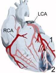
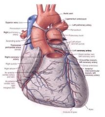
Coronary Artery Disease (CAD) p.100
|
leading cause of death
occlusion of major artery resulting in reduced blood supply to myocardial tissue (MI or coronary atherosclerosis) m. infarction - sudden occlusion of major artery by embolus = vessel bloodless (infarcted) leads to necrosis - pathological tissue death common sites: Anterior IV LAD branch of the LCA, RCA, and circumflex branch LCA |
|
|
Myocardial Infarction (MI) p. 100
|
area of myocardium has undergone necrosis due to sudden occlusion of major artery
common locations: LCA (left main coronary artery) RCA (right main coronary artery) Circumflex branch 1) LAD branch (left anterior descending) |
|
|
Coronary Atherosclerosis p. 100
|
the most common cause of ischemic heart disease
atherosclerotic process = lipid accumulations in the intima (lining layer), or on internal walls of arteries eventually results in stenosis collateral channels may compensate, but may not be able to provide enough O2 insufficiency of blood supply to heart leads to myocardial ischemia/infarction |
|
|
Angina Pectoris
|
transient, but moderately severe pain / tightness in thorax
caused by ischemia of myocardium results from narrowed/hardened coronary arteries anaerobic metabolism of myocytes produces lactic acid -pain receptors sensitive to drop in pH treat w/ sublingual nitroglycerin to dilate coronary arrteries |
|
|
Coronary Bypass Graft (CABG) p. 101
|
segment of artery or vein connects proximal part of artery to ascending aorta, distal to blockage/stenosis
great saphenous vein is often used b/c 1) has the same or greater than diameter than of the coronary arteries. 2)it can be easily dissected from the lower limb. 3) it offers relatively lengthy portions with a minimum occurrence of valves or branching. can also use radial artery revascularization of the myocardium may also be achieved by surgically anastomosing an internal thoracic artery with a coronary artery. |
|
|
Coronary Angioplasty p. 101
|
percutaneous transluminal coronary angioplasty = catheter with small inflatable balloon flattens plaque against vessel wall to increase lumen size to improve blood flow
in other cases thrombokinase (enzyme) is injected trhough the catheter, to dissolve blood clot intravascular stent - introduced to maintain dilation |
|
|
Coronary Occlusion and Heart Conduction Effects
|
LAD supplies AV bundle
RCA supplies SA and AV nodes heart block may occur upon occlusion of these vessels |
|
|
Artificial Cardiac Pacemaker
|
implanted to induce heart contraction at a predetermined rate
electrode passed through SVC into RA, through tricuspid to RV and firmly fixed to traveculae carnae in ventricular wall |
|
|
Restarting the Heart
|
CPR -- firm pressure over thorax forces blood into systemic circulation, release of pressure allows for refilling
|
|
|
Fibrillation of Heart
|
multiple, rapid, circuitous contractions or twitching of muscle fibers
atrial fibrillation : rapid, irregular uncontrolled twitchings of atrial walls ventricular fibrulation : twitchings do not pump / maintain circulation. most disorganized of all dysrhythmias; fatal if persistant |
|
|
Defibrillation of Heart
|
electric shock causes cessation of all cardiac movements to induce normal beating
|
|
|
Cardiac Referred Pain p. 102
|
actual heart insensitive to touch, pain, or temp.
ischemia and metabolic products stimulate pain endings in myocardium, through T1-T5 axons (left side) noxious stimuli in heart perceived as pain on superficial part of body (i.e. left upper limb) visceral pain = transmitted by visceral afferent fibers accompanying sympathetic fibers anginal pain = radiates from substernal and left pectoral regions to left shoulder and medial aspect of upper limb |
|
|
Age changes in Thymus
|
thymus replaced by adipose tissue in adults, but continues to be important
number one source of T cells in body (that's why immune defense of older people go down) |
|
|
Aneurysm of Ascending Aorta p. 112
|
aneurysm = localized dilation
evident on a chest film (radiograph) or a magnetic resonance angiogram results from force of LV contraction and a localized dilation symp: chest pain that radiates to back aneurysm of arch of aorta may exert pressure on the trachea, esophagus, and reccurent laryngeal nerve, and results in difficulty breathing and swallowing |
|
|
Variations in the Great Arteries p. 112
|
most superior part of the aorta = ~2.5 cm inferior to the superior border of the manubrium.
different branch patterns of aortic arch (in 35% of people) R. arch or the aorta = over R lung double arch or retroesophageal right subclavian artery = vascular ring around the esophagus and trachea. -coarctation of aorta : abnormal narrowing, producing obstruction to blood flow *most commonly near ligamentum arteriosum; collateralization provides many years of life |
|
|
Injury to Recurrent Laryngeal Nerves p.112
|
supply all intrinsic muscles of larynx, except one
affected by bronchial/esophageal carcinoma, enlargement of mediastinal lymph nodes, or an aneurysm of aortic arch (the nerve may be stretched by the dilated arch of the aorta) |
|
|
Laceration of Thoracic Duct p.112
|
vulnerable to inadvertent injury during investigative / surgical procedures because it's thin walled and colorless
results in lymph escaping into thoracic cavity at very high rates, and pleural cavity causing chylothorax fluid may be removed by thoracentesis and ligation of duct may be necessary |
|
|
Alternate Venous Routes to the Heart
|
azygos, hemiazygos, and accessory hemiazygos all offer alternate means of venous drainage to heart if there is obstruction of the IVC
|
|
|
Subarealoar breast cancer (p. 58)
|
may cause inversion of the nipple by similar mechanis involving the lactiferous ducts
|
|
|
surgical incision of breast (p.59)
|
placed in the inferior quadrants when possible since they are less vascular
|
|
|
Mastectomy (p.59)
|
breast excision
not as common as it once was simple mastectomy = breast is removed down to the retromammary space. radical mastectomy = more extensive that involves removal of the breast, pect. muscles, fat, fascia, and many lymph nodes lumpectomy or quadrantectomy aka breast-conserving surg. = often only remove the tumor and surrounding tissues - followed by radiation therapy |
|
|
Role of costal cartilages p. 50 & 54
|
contribute to the elasticity of the t. wall
in elderly people, the costal cartilages undergo calcification, making them radiopaque and less resilient |
|
|
Dyspnea p.64
|
shortness of breath (SOB), or air hunger
may be caused by asthma or emphysema or with heart failure fix pectoral girdles (clavicle and scapula) to expand thorax. |
|
|
Muscles of thoracic wall (link)
|
http://home.comcast.net/~wnor/thoraxmuscles.htm
|
|
|
Bones of the thorax (link)
|
http://home.comcast.net/~wnor/thoraxbones.htm
|
|
|
Arterial Blood and Nerves supply to the thorax (link)
|
http://home.comcast.net/~wnor/thoraxves&nerofwall.htm
|
|
|
Lung Dissection (link)
|
http://home.comcast.net/~wnor/thoraxlesson2.htm
|
|
|
Pleura (link)
|
http://home.comcast.net/~wnor/thoraxlesson3.htm
|
|
|
The Heart (link)
|
http://home.comcast.net/~wnor/thoraxlesson4.htm
|
|
|
Posterior Mediastinum (link)
|
http://home.comcast.net/~wnor/thoraxlesson5.htm
|
|
|
Superior Mediastinum (link)
|
http://home.comcast.net/~wnor/thoraxlesson6.htm
|
|
|
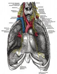
The Mediastinum p. 80
|
Superior Mediastinum: suprior thoracic aperture to the sternal angle (T4-T5)
Inferior: anterior, middle, and posterior mediastinum |
|
|
Inferior mediastinum p. 80
|
Anterior contains: thymus, lymph noes, fat, and connective tissue
Middle contains: pericardium, heart, roots of the great vessels, arch of the azygos vein, main bonchi Posterior contains the : esophagus, thoracic aorta, azygos and hemiazygos veins, thoracic duct, vagus nerves, SVC, sympathetic trunks, and splanchnic nerves |
|
|
Nerve supply of the pericardium p. 82
|
Phrenic nerves (C3-C5) - primary source of sensory fibers; pain sensations - skin of top of the shoulder of the same side
Vagus (CNX) - fxn uncertain. Sympathetic trunks - vasomotor |
|
|
apex beat p. 92
|
impulse that results from the apex being forced against the anterior thoracic wall when the left ventricle contracts
the location of the apex beat (mitral area) varies in position; it may be located in the 4th or 5th intercostal spaces. |
|
|
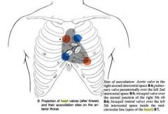
Areas of auscultation p. 94
|
Aortic valve; second intercostal space to the right of the sternal border
Pulmonary valve; second intercostal space to the left of sternal border Tricuspid valve; near left sternal border in fifth or sixth intercostal space Mitral valve; apex of the heart in fifth intercostal space in midclavicular line SEE p. 93 |
|
|
The cardiac cycle p.98 (link)
|
http://www.youtube.com/watch?v=jLTdgrhpDCg
|
|
|
Conducting system of the heart p. 97 (link)
|
http://www.youtube.com/watch?v=te_SY3MeWys&feature=related
|
|
|
Dissection (link)
|
http://www.youtube.com/watch?v=87iMg1dMXiA&feature=related
|
|
|
Coarctation of Aorta p. 113
|
the arch of the aorta or descending aorta has a abnormal narrowing (stenosis) that decreases the size of its lumen.
most common site is near the ligamentum arteriosum. |
|
|
septomarginal trabecula p. 89
|
moderator band
significance: it carries part of the right bundle branches of the AV bundle of the conducting system of the heart and the anterior papillary muscles this facilitates conduction time, allowing coordinated contraction of the anterior papillary muscle. |
|
|
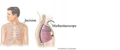
Mediastinoscopy
|
a procedure that enables visualization of the contents of the mediastinum, usually for the purpose of obtaining a biopsy.
often used for staging of lymph nodes of lung cancer or for diagnosing other conditions affecting structures in the mediastinum such as sarcoidosis or lymphoma. involves making an incision approximately 1 cm above the suprasternal notch of the sternum, or breast bone A scope (mediastinoscope) is then advanced into the created tunnel which provides a view of the mediastinum |
|
|
Pulmonary valve stenosis p.92
|
valve cusps are fused, forming a dome with a narrow central opening.
|
|
|
Vagus Nerve Pathways aka Pneumogastic nerve/ Cranial Nerve X
|

The vagus nerve supplies motor parasympathetic fibers to all the organs except the suprarenal (adrenal) glands, from the neck down to the second segment of the transverse colon. The vagus also controls a few skeletal muscles
The vagus nerve is responsible for such varied tasks as heart rate, gastrointestinal peristalsis, sweating, and quite a few muscle movements in the mouth, including speech (via the recurrent laryngeal nerve) and keeping the larynx open for breathing (via action of the posterior cricoarytenoid muscle, the only abductor of the vocal folds). Parasympathetic innervation of the heart is controlled by the vagus nerve. To be specific, the vagus nerve acts to lower the heart rate. Effects of Vagus nerve lesions:The patient complains of hoarse voice, difficulty in swallowing (dysphagia), and choking when drinking fluid. There is also loss of gag reflex. Uvula deviates away from the side of lesion, and there is failure of palate elevation |
|
|
Phrenic Nerve
|
originates mainly from the 4th cervical nerve, but also receives contributions from the 5th and 3rd cervical nerves (C3-C5)
contain motor, sensory, and sympathetic nerve fibers provide the only motor supply to the diaphragm as well as sensation to the central tendon. In the thorax, each phrenic nerve supplies the mediastinal pleura and pericardium Path : Found in the middle mediastinum, both phrenic nerves run from C3, C4 and C5 along the anterior scalene muscle deep to the carotid sheath. - The right phrenic nerve passes over the brachiocephalic artery, posterior to the subclavian vein, and then crosses the root of the right lung anteriorly and then leaves the thorax by passing through the vena cava hiatus opening in the diaphragm at the level of T8. The right phrenic nerve passes over the right atrium. - The left phrenic nerve passes over the pericardium of the left ventricle and pierces the diaphragm separately. phrenectomy will paralyze half of the diaphragm |
|
|
Azygos venous system
|
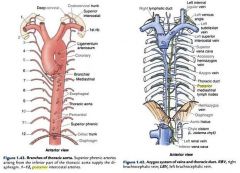
Azygos vein= runs to R of T4-T12, arches on root of R lung, and empties into SVC. Serves to drain most of the posterior intercostal veins on the right side of the body,
hemiazygos vein and the accessory hemiazygos vein drain most of the posterior intercostal veins on the left side of the body Accessory hemiazygos = drains the fifth through eighth intercostal spaces on the left side of the body. Joins hemiazygos v. or runs sup to join brachiocephalic v. Hemiazygos v. = runs superiorly in the lower thoracic region, just to the left side of the vertebral column. |
|
|
Heart valves
|
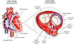
The four valves are known as:
The tricuspid valve The pulmonic or pulmonary valve The mitral valve The aortic valve |
|
|
Heart Vascular supply (ant.)
|
|
|
|
Heart vascular supply (post)
|
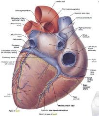
|
|
|
Sympathetic chain
|
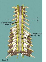
The sympathetic trunk is a fundamental part of the sympathetic division of the autonomic nervous system. It allows nerve fibers to travel to spinal nerves that are superior and inferior to the one in which they originated. Also, a number of nerves, such as most of the splanchnic nerves, arise directly from the trunks
|
|
|
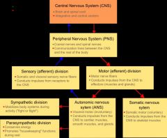
the nervous system
|
|
|
|
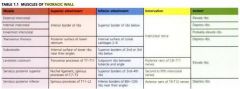
Muscles of the thoracic wall
|
|
|
|
Posterior Thorax dissection (link)
|
part1 : http://www.youtube.com/watch?v=YathjWGgmEc&feature=related
part 2 : http://www.youtube.com/watch?v=4J-fgyb4spk |
|
|
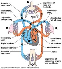
Circulatory Sys.
|
|