![]()
![]()
![]()
Use LEFT and RIGHT arrow keys to navigate between flashcards;
Use UP and DOWN arrow keys to flip the card;
H to show hint;
A reads text to speech;
26 Cards in this Set
- Front
- Back
|
The skeleton |
Consists of Bones Joints Cartilage Ligaments composed of 206 named bones Axial skeleton: 80 bones Appendicular skeleton : 126 bones |
|
|
Axial skeleton |
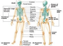
Skull , vertebral column, bony thorax |
|
|
Skull |
Body most complex bony structure Skull: formed by cranial and facial bones 8 cranial bones : ethmoid , frontal , occipital , sphenoid, parietal 2 , temporal 2 14 facial bones : mandible , vomer , inferior nasal conchae 2 , lacrimal 2 , maxilla, 2 , nasal 2 , palantine 2 . zygomatic 2 |
|
|
Skull |
Facial bones : Form framework of face form cavities of sense organs of sight , taste , and smell Provide opening for passage of air and food Hold teeth Anchor facial muscles Cranium bones: Encloses and protects brain Provides attachment for head and neck muscle |
|
|
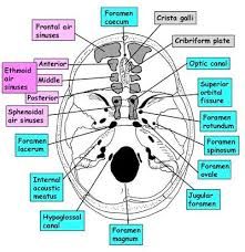
Cranial Fossae |
- Internally prominent bony ridges divide skull into disticnt fossae - Anterior Cranial Fossa : frontal lobe of cerebum - Middle Cranial Fossa : temporal lobe of cerebum Posterior Cranial Fossa : cerebellum |
|
|
Small cavities of skull |
1) middle and inner ear cavities 2) Nasal cavity 3) orbits 4) Air filled sinuses |
|
|
Skull has 85 opening |
Foramina , canals and fissures - provide opening for important structures spinal cord , blood vessels , cranial nerves |
|
|
Cranial bones |
Formed from eight bones - paired bones include Temporal , Parietal Unpaired bones include : frontal , occipital , sphenoid , ethmoid |
|
|
Parietal bones and assoicated sutures |
Parietal bones form superior and lateral parts of skull Coronal suture - runs in the coronal plane and is located where parietal bones meet frontal bone Squamous suture - occurs where each parietal bone meets a temporal bone inferiorly Lambdoid suture - occurs where parietal bones meet the occupital bone posteriorly Saggital suture : occurs where right and left parietal bones meet superiorly Sutural bones : small bones that occur within sutures , irregulat in shape , size and location. Not all people have sutural bones |
|
|
Frontal bones |
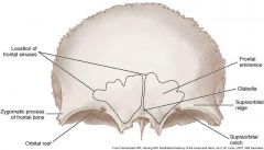
Forms the forehead and roofs of orbits Supraorbital margin - superior margin of orbits Glabella - smooth part of frontal bone between supercilliary (eyebrow) arches Frontal sinuses- within frontal bone Contributes to anterior cranial fossa |
|
|
Occipital bone |
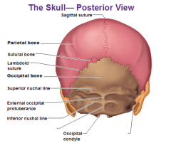
articulates with the temporal bones and parietal bones Forms the posterior cranial fossa Superior and inferior nuchal lines occipital condyles Hypoglossal canal through which CN XII runs Foramen magnum located at its base |
|
|
Temporal bone |
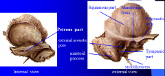
Lie inferior to parietal bones Contributes to the middle and posterior cranial fossae Form the inferolateral portion of the skull Squamous region- flat area of bone which contains bar like zygomatic process; zygomatic process projects anteriorly to meet zygomatic bone of face and contribution of these two bones to makeup the zygomatic arch Tympanic region- surronds the external acoustical meatus Styloid process- extends down from inferior temporal bone and is muscle attachment site Mastoid region- site for neck muscle attachment contain air sinuses Petrous region - projects medially , contributes to cranial base - appears as a boney wedge between occipetal bone posteriorly and sphenoid bone anteriorly - house cavities of middle and internal ear |
|
|
Foramina of Temporal bone |
Carotid canal Jugular Foramen ( at boundary with occipital bone ) Foramen lacerum ( at boundary with sphenoid bone and occipital bone) Internal and external acoustic meatus |
|
|
The sphenoid bone |
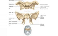
Spans the width of the cranial floor Resembles a butterfly or bat The superior part of the body bears a saddle shaped prominence called sella turcica The seat of this saddle contains the hypophyseal fossa which holds the pituitary gland Greater wings Lesser wings Pterygoid processes |
|
|
Sphenoid bone opening |
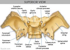
Superior orbital fissure : long slit between greater and lesser wings Optic canal: lies just anterior to sella Tursica Foramen Rotundum: in medial part of greater wing Foramen Oval : Posteriolateral to foramen Rotundum Foramen Spinosum : Posteriolateral to foramen ovale Foramen Lacerum : at boundary with temporal bone and occipital bone |
|
|
The ethmoid bone |
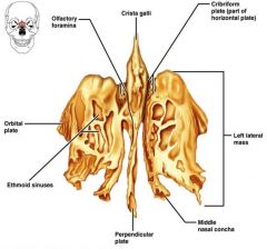
Lies between nasal and sphenoid bones Forms most of the medial bony region between the nasal cavity and orbits Crista Galli- attachment for falx cerebi, the large vertical sheet of connective tissue which lies between cerebral hemispheres Cribriform plate - superior surface of the ethmoid bone; contain olfactory foramina Perpendicular plate - forms superior part of nasal septum Lateral masses- contain air cells Superior and middle nasal conchae - extend medially from lateral masses |
|
|
Left lateral wall of nasal cavity |

|
|
|
The facial bones |
Form framework of the face Form cavities for the sense organs of sight taste smell Provide opening for the passage of air and food Hold teeth in place Anchor muscles of the face Unpaired bones Mandible Vomer Paired bones Maxillae Zygomatic Nasal Lacrimal Inferior nasal conchae Palatine |
|
|
Mandible |
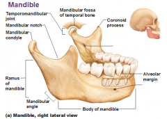
The Lower jawbone is the largest and strongest facial bone Composed of two main parts Horizontal body Two upright rami Mandibular condyle Temporomandibular joint- interface of mandibular condyle and mandibular fossa of temporal bone Mandibular notch Coronoid process Ramus of mandible Mandibular angle Body of mandible Alveolar margin Mental foramen Mandibular foramen |
|
|
Maxillary bone |
Articulate with all other facial bones except mandible are the keystone bones of the face Contain maxillary sinuses- largest paranasal sinuses Forms part of the inferior orbital fissure Alveolar margin Inferiomedial surface of orbit Infraorbital Foramen Forms part of hard palate |
|
|
Paired bones of the face |
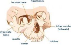
Maxilla bone Zygomatic bones - form lateral wall of orbits Lacrimal bones- located in the medial orbital walls Nasal bones- form bridge of nose Inferior nasal conchae - thin curbed bones that project medially and form the lateral walls of the nasal cavity Palatine bones - Complete the posterior part of the hard palate |
|
|
Other bone |
Vomer : forms the inferior part of the nasal septum and is an upaired bone |
|
|
Nasal septum |
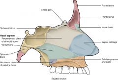
Perpendicular plate of ethmoid bone Vomer bone Septal Cartilage |
|
|
Paranasal sinuses |
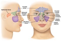
Air filled sinuses are located within Frontal bone Ethmoid bone Sphenoid bone Maxillary bone Lined with mucous membrane |
|
|
Orbit walls and wall opening |
Roof Lateral wall Medial wall Floor Superior orbital fissure Interior orbital fissures Optic canal |
|
|
Orbit walls are formed by 7 bones |
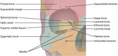
Frontal Sphenoid Zygomatic Maxillary Palatine Lacrimal Ethmoid |