![]()
![]()
![]()
Use LEFT and RIGHT arrow keys to navigate between flashcards;
Use UP and DOWN arrow keys to flip the card;
H to show hint;
A reads text to speech;
128 Cards in this Set
- Front
- Back
|
Located in the vertebral foramen. It begins at the foramen magnum and ends at the corpus medullaris in the lumbar region. It conducts sensory impulses to the brain and motor impulses from the brain to the body. |
Sinal Cord |
|
|
Looking for Spinal Cord |
|
|
The largest part of the brain in mammals. It is composed of the frontal, parietal, and temporal lobes. |
Cerebrum |
|
|
They have motor functions, but also deal with aggression, mood, foresight, motivation, and social judgements.These structures lie between the frontal bone and the central sulcus, and above the eye orbit. |
Frontal Lobes |
|
|
Integration of sensory information with the exceptions of vision, hearing, and smell.Between the parietal bones and the central sulcus. |
Parietal Lobes |
|
|
Receives and interprets visual signals. Between occipital bone and the patio-occipital sulcus. |
Occipital Lobe |
|
|
Functions in memory, vision, learning, hearing, and emotional behavior. between Lateral sulcus and temporal bone. |
Temporal Lobes |
|
|
Thick folds in the surface of the cerebrum. |
Gyri (Gyrus) |
|
|
Shallow groves in the surface of the cerebrum. |
Sulk (Sulcus) |
|
|
A deep groove separating the cerebrum, into right and left halves. |
Longitudinal fissure |
|
|
Divided by the Longitudinal Fissure into right and left portions called: |
cerebral hemisphere |
|
|
Second larges part of the brain in mammals largest part in birds. Involved in the regulation of posture and balance, fine motor control of skeletal muscles, and repetitive movements. |
Cerebellum |
|
|
An endocrine gland directly attached to the hypothalamus. It is divided into anterior and posterior portions. Anterior portion produces hormones which regulate other endocrine glands, and directly affects large cells. Posterior portion functions to store and release hormones produced by the hypothalamus. |
Pituitary Gland |
|
|
Functions to integrate all sensory information (with the exception of smell) from the body, and channels it into proper processing regions in the cerebrum. |
Thalamus |
|
|
Major integration system between various organ system and the nervous system. It coordinates activities of both the nervous and endocrine systems, and between voluntary and autonomic activities. It is attached directly to the pituitary gland. |
Hypothalamus |
|
|
Processes olfactory information and contains centers for reflex movements involved in eating, such as chewing, licking, and swallowing. |
Mammillary Body |
|
|
The region that regulates the day/night cycle. Secretes the hormone melatonin, which effects sleepiness. |
Pineal Body |
|
|
Contains the nerve tracts and physically joins the two cerebral hemispheres. |
Corpus Callosum |
|
|
Located above the pons and is the smallest part of the brain stem. The oculomotor, trochlear, and trigeminal cranial nerves originate in this area. |
Midbrain |
|
|
Located above the medulla, on the brain stem. It works with the medulla to control respiration and helps regulate sleep. It is the origin for the trigeminal, abducens, facial , and vestibulocochlear cranial nerves. |
Pons |
|
|
Base of the brain stem. It contains nerve centers for the regulation of the heart rate, blood vessel diameter, respiration, swallowing, vomiting, coughing, sneezing, and hiccoughing. |
Medulla Oblongata |
|
|
Bottom-center of the brain where two optic nerves cross. |
Optic Chiasma |
|
|
Located just below the frontal lobes. They function in the sense of smell. |
Olfactory Bulbs |
|
|
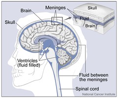
A vascular layer of connective tissue which function to protect the brain and spinal cord |
Arachnoid Layer of meninges |
|
|
Space between the arachnoid and pia mater containing cerebrospinal fluid |
Subarachnoid space |
|
|
Thin, transparent layer of connective tissue on the surface of the brain and spinal cord. Contains blood vessels that nourish the spinal cord. |
Pia Matter |
|
|
Surround the gray matter. Composed of both myelinated and unmyelinated axons. It has three regions, anterior, lateral, and posterior columns. |
White matter of spinal cord |
|
|
Shaped like a butterfly or the capital letter "H" in cross-section. It is composed of neuron cell bodies, neuroglia, dendrites, and unmyelinated axons. It has 3 regions: anterior, lateral, and posterior horns. The central canal is in the center of the gray matter. |
Gray matter of spinal cord |
|
|
These structures contain the axons of the autonomic sensory neurons. |
Dorsal Root of spinal nerve |
|
|
contains the cell bodies of somatic and autonomic sensory neurons. |
Dorsal root ganglion |
|
|
contains the cell bodies of the somatic motor neurons that innervate the skeletal muscles. |
Ventral root of spinal nerve |
|
|
nerve tracks that connect the spinal cord with various regions of the body. |
Spinal nerve |
|
|
Most superficial layer of connective tissue surrounding the brain and spinal cord. It functions to protect the brain and spinal cord. |
Dura mater |
|
|
Originates in the olfactory epithelium of the nasal cavity and terminates in the olfactory lobe. |
Olfactory Nerve |
|
|
Originates in the retina and goes to he optic chiasma. The right nerve goes to the left hemisphere and the left nerve goes to the right hemisphere. |
Optic nerve |
|
|
A mixed nerve originating in the brain and termination at the eye. |
Oculomotor |
|
|
A mixed nerve which originated in the brain and terminates in the eye |
Trochlear nerve |
|
|
Sensory nerve functioning in the sense of smell. |
Olfactory nerve |
|
|
Sensory nerve involved in vision. |
Optic nerve |
|
|
Sensory function is to provide information on the position of the eye. Motor function include eye and eye lid movement, controlling pupil diameter, and focusing |
Oculomotor |
|
|
Sensory function is to provide information on the position of the eye. Motor functions are lateral and inferior movement of the eye. |
Trochlear nerve |
|
|
A mixed nerve dividing into three branches. The branches originate in the face, jaws, mouth, tongue, and scalp and terminate in the pons. |
Trigeminal nerve |
|
|
A mixed nerve originating in the pons and innervating the later rectus eye muscles. |
Abducens nerve |
|
|
A mixed nerve originating in the pons. It innervates the muscles of the face, scalp, neck, and salivary glands. |
Facial nerve |
|
|
A mixed nerve originating in the inner ear and terminating in the thalamus |
Vestibulocochlear |
|
|
Mixed nerve with motor fires originating in the medulla, and traveling to the pharyngeal region. Sensory fibers originate in the pharyngeal region, middle and external ear, rear of the tongue, and the carotid arteries. |
Glossopharyngeal Nerve |
|
|
Sensory function is to transport information from various touch receptors on the face. Motor function is chewing. |
Trigeminal nerve |
|
|
Sensory function is to provide information on the position of the eye. Motor function is the lateral movement of the eye. |
Abducens Nerve |
|
|
Sensory functions are reception of taste stimuli from the anterior 2/3 of the tongue and the position of the face and scalp muscles. Motor functions include controlling facial expressions and secretion from the salivary glands. |
Facial Nerve |
|
|
Sensory nerve functioning in hearing and equilibrium. Motor functions include a response by the head and neck to changes in equilibrium. |
Vestibulocochlear |
|
|
Sensory functions are taste and touch by the tongue, the gag reflex, regulation of blood pressure, and reparation. Motor functions include the control of pharyngeal muscles in swallowing, speech, salivation, and the gag reflex. |
Glossopharyngeal Nerve |
|
|
A mixed nerve with motor fibers originating in the medulla and terminating in the pharyngeal region, digestive, reparatory, and cardiovascular systems. The sensory fibers originate in the thoracic and abdominal cavities, the pharyngeal region, and external ear. |
Vagus |
|
|
A mixed nerve originating in the medulla and the upper cervical portions of the spinal cord. |
Accessory nerve |
|
|
Primarily a motor nerve originating in the upper cervical portions of the spinal cord and terminating in the muscles of the tongue. |
Hypoglossal Nerve |
|
|
Sensory functions involve sensations from the respiratory tract, pharyngeal region, external ear canal, and of hunger and fullness. Motor functions include controlling swallowing, coughing, speech and smooth muscles of the respiratory and digestive systems. |
Vagus |
|
|
Sensory function is to provide information on the position of the muscles of the head, neck, and shoulders. Motor functions include controlling those muscles and voluntary swallowing. |
Accessory nerve |
|
|
Sensory function is to provide information on the tongue movement. Motor function is to control muscles involved in food maculation, swallowing, and speech. |
Hypoglossal Nerve |
|
|
Innate responses designed to protect the body. posture maintenance. balance, -Achilles reflex -Patellar reflex |
Somatic Reflexes |
|
|
Reflexes controlled by the autonomic nervous system. Involuntary responses.Involve reactions such as smooth muscle, cardiac muscles, and glands |
Autonomic Reflexes |
|
|
Somatic Reflex Quadriceps Femoris Kicked foward |
Patellar Reflex |
|
|
Somatic Reflex Gastrocnemius Plantar Flexion |
Achilles Reflex |
|
|
Somatic Reflex Afferent- Trigeminal Nerve Efferent- Facial Nerve Blinking |
Corneal Reflex |
|
|
Autonomic Reflex Afferent- Optic nerve Efferent- Oculomotor Nerve Constriction of Pupil |
Pupillary Reflex |
|
|
Autonomic Pupil dilation |
Ciliospinal Reflex |
|
|
Widespread, superficial, found in hairy and hairless skin. Light touch, temperature, and pain. |
Free nerve endings |
|
|
A dendrite wrapped around a hair follicle. Light touch when the hair is displaced. |
Hair follicle receptors |
|
|
Found in the dermal papillae of hairless skin. Light touch and low- frequency vibration. |
Meissner's corpuscles |
|
|
Found in the stratum basal of hairy and hairless skin. pressure touch |
Merkel's nerve complex |
|
|
In the dermis, joint capsules, some viscera, genitals, and breasts. High frequency vibration. pressure, stretch, and tickling. |
Lamellate or Pacinian Corpuscles |
|
|
In the dermis and joint capsules. Pressure touch. |
Ruffini's Cylinders |
|
|
Recording the minimum distance between the jaws of the caliper at which you can feel two points. |
Two-point touch threshold |
|
|
What are the 5 different tastes? |
Salty Sweet Bitter Sour Umami |
|
|
Form capsules surrounding the taste receptor cell. These cells support and protect the receptor cells. |
Supporting Cells |
|
|
Has a microvilli, called a "taste hair", protruding through a pore on the apical surface of the taste bud. These hairs are the receptor surface for taste stimuli. |
taste receptor cells |
|
|
Found peripherally on the base of the taste bud. They develop into supporting cells and then into receptor cells which live about 10 days. |
Basal Cells |
|
|
Small, spike-like projections found all over the tongue. They are the most abundant papillae, but lack taste buds. They roughen the tongue and aid in food manipulation. |
Filiform Papillae |
|
|
These Papillae form parallel bands on the sides of the posterior 2/3 of the tongue. They have few taste buds. |
Foliate Papillae |
|
|
These papillae, as the name implies, are mushroom-shaped projections found all over the tongue, although they tend to be concentrated on the tip and sides. Each papillae has about 5 taste buds. |
Fungiform Papillae |
|
|
These are large circular papillae with a depression in the middle. There are about 12 of them arranged in a V-shaped row on the back of the tongue. They contain from 100 to 300 taste buds. |
Vallate Papillae |
|
|
Comprises the two olfactory organs in the nasal cavity. |
Olfactory Epithelium |
|
|
The connective tissue beneath the olfactory epithelium. |
Lamina Propria |
|
|
Columnar epithelia cells found in the olfactory epithelium between the olfactory receptor cells. |
Supporting cells |
|
|
Bipolar neuron found in the olfactory epithelium. The dendrite is enlarged into a bulb-shaped "olfactory vesicle" on the surface of the olfactory epithelium. Odors cause depolarization on these hairs |
Olfactory Receptor Cells |
|
|
Replace lost or damaged olfactory receptor cells at the base of the olfactory epithelium. |
Basal Cells |
|
|
Muscle glands found in the lamina propria that moisten olfactory epithelium and dissolve odor molecules |
Bowman's Glands |
|
|
The fleshy cartilaginous external ear flap located on the side of the heat. it functions to collect sound waves and direct them into the external auditory canal. |
Auricle or pinna |
|
|
The passageway that directs sound waves from the auricle to the tympanic membrane. |
External auditory canal |
|
|
Commonly call the "ear drum". It separates the outer ear and middle ear. vibrates when struck by sound wave - mechanically transfers sound to the middle ear. |
Tympanic Membrane |
|
|
secrete cerumen, or earwax, into the external auditory canal. located at the base of the hairs that line the canal. Helps prevent foreign substances from reaching the delicate tympanic membrane |
Ceruminous Glands |
|
|
Attached to inside surface of the tympanic membrane. Articulates with the incus and transmits vibrations from the tympanic membrane to the incus. Hammer |
Malleus |
|
|
Articulates with the malleus and stapes and transmits vibrations from the malleus to the stapes. Anvil
|
Incus |
|
|
Articulates with the incus and the oval window and transmits vibrations from the incus to the oval window. |
Stapes |
|
|
Opening between middle and inner ear. Transfers vibrations to fluid in the inner ear. |
Oval window |
|
|
opening directly below the oval window. covered by a secondary tympanic membrane. bulges out into the middle ear to dissipate the pressure waves within the cochlea, after they have been detected by the inner ear. |
Round window |
|
|
tube connecting the inner ear and nasopharynx. equalizes air pressure in middle ear |
Eustachian Tube |
|
|
Small skeletal muscle which protects the oval window by dampening the vibration of the stapes in response to loud noises. |
Stapedius |
|
|
limits movement of ossicles and increases tension of the tympanic membrane to prevent damage in response to loud, prolonged noises. |
Tensor Tympani |
|
|
Series of interconnected passageways in the temporal bone. |
bony labyrinth |
|
|
series of interconnected fluid-filled tubes found within the bony labyrinth |
membranes labyrinth |
|
|
a part of the bony labyrinth resembling a snail shell. It contains the cochlear duct. |
Cochlea |
|
|
A part of the membranous labyrinth found within the cochlea. It contains the hearing receptor cells. |
Cochlear duct |
|
|
a part of the bony labyrinth containing the saccule and utricle. |
Vestibule |
|
|
A pair of membranous sacs found within the vestibule that contain the receptor cells for gravity and linear acceleration. |
Saccule and utricle |
|
|
A part if the bony labyrinth containing the semicircular ducts. |
semicircular canals |
|
|
series of 3 fluid-filled ducts found within the semicircular canals. they are oriented at the right angles to each other on 3 planes. the receptors in the ducts provide information on the position of the head and body in space, acceleration, and deceleration. |
Semicircular ducts |
|
|
located superior to the eye. partially shade eyes. protect from sweat. |
eyebrows |
|
|
over the eye. blink to moisten the eye and sweep foreign substances from the eye's surface. |
eyelids |
|
|
margin of the eyelids. prevent foreign substances from entering into the eye. |
eyelashes |
|
|
lacrimal gland and lacrimal ducts. secretion or tear from the gland moisten the eye and washes away foreign substances. |
lacrimal apparatus |
|
|
epithelial covering on the inside of the eyelid and the anterior surface of the eye. helps keep the cornea moist and clean. |
Conjunctiva |
|
|
superior- rotates the eye downward and medially inferior- rotates the eye upward and medially |
Oblique eye muscles |
|
|
4 muscles are the superior, inferior, medial, and lateral rectus muscles. these muscles move the eye up, down, medially, and laterally. |
Rectus eye muscles |
|
|
jelly-like fluids that fit the interior of the eye. |
Aqueous and vitreous humours |
|
|
tough, tense-like layer continuos with the dura mater of the brain around the optic nerve. shares they eye. insertion point for the 6 muscles which control eye movements. Eye White |
Sclera |
|
|
Most anterior layer of the eye the is continuos with the sclera. transparent. supplied with nerve endings for pain, reflex blinking, and to stimulate lacrimal secretions. It also lacks blood vessels. |
Cornea |
|
|
separates fibrous and sensory tunics. dens capillary bed - oxygen and nourishment for eye. many melanocytes - gives the choroid its dark appearance. |
Choroid |
|
|
beneath the cornea. eye color. 2 layers of pupillary muscles that control diameter of the pupil, and thus the amount of light entering the eye. |
Iris |
|
|
round central opening in the center of the eye. |
Pupil |
|
|
Thick ring of tissue attached to and just beneath the iris. anchored by choroid. holds lens in place. cilia muscle. changes the lens shape to focus light onto the retina. |
Ciliary Body |
|
|
Suspending ligaments extending from the ciliary body hold it in place. transparent and convex in outer and inner surfaces. focuses the image on the retina by changing shape under the influence of the ciliary muscles. |
Lens |
|
|
innermost layer of the eye. neural layer contains photoreceptors and neuron that react to light and transmit and integrate visual signals. pigmented later - absorbs light pass through neural layer to prevent the light from bouncing back and causing "visual echoes". |
Retina |
|
|
Photoreceptor cells very sensitive to light. enables visibility of shades of gray at dim light. |
Rods |
|
|
Photoreceptor cells responsible for high acuity color vision. only operate in bright light. 3 types: sensitive to red, green, blue. |
cones |
|
|
Synapse with dendrites of the rods and cones. transmit nerve impulses to the ganglion cells. |
bipolar neurons |
|
|
synapse with axons of the bipolar neurons. the axons combine to form the optic nerve, which sends nerve impulses to the brain. |
Ganglion Cells |
|
|
Where the optic nerve leaves the eye. Not covered by the retina. A blind spot in the eye. |
Optic disc |
|
|
Transmits visual information from the eye to the brain. |
Optic nerve |
|
|
responsible for sharp central vision. detailed vision including driving. |
Fovea Centralis |