Reading...
![]()
Play button
![]()
Play button
![]()
Use LEFT and RIGHT arrow keys to navigate between flashcards;
Use UP and DOWN arrow keys to flip the card;
H to show hint;
A reads text to speech;
55 Cards in this Set
- Front
- Back
|
The central area of the thorax, located btwn the two pleural cavities is known as the what?
|
mediastinum
|
|
|
What are the borders of the mediastinum?
|
-superior to inferior: superior thoracic aperture to diaphram
-anterior to posterior: sternum to vertebral bodies -laterally: two pleural cavities |
|
|
The mediastiunum is divided into superior and inferior mediastinum, above and below the _______________
|
sternal angle
|
|
|
The inferior mediastinum is subdivided into what 3 mediastinums?
|
anterior
middle posterior |
|
|
The pericardium serves as the border for the (anterior/middle/posterior) mediastinum.
What does this mediastinum contains? |
middle mediastinum
contains: pericardial sac heart origins of great vessels phrenic nerves (C3,4,5) pericardiacophrenic vessels |
|
|
The pericardium consists of two layers,what are they?
|
Fibrous (outer) & serous (inner)
|
|
|
The serous pericardium is further divided into the parietal and visceral layer, differentiate btwn the two
What is the potential space btwn the two layers called? |
parietal layer- lines inner surface of fibrous pericardium
visceral layer- tightly adherent to myocardium (epicardium) pericardial cavity |
|
|
The arterial supply to the pericardium is mainly from what arteries?
Branches from what other 2 arteries may also contribute? |
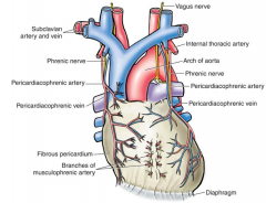
mainly pericardiacophrenic arteries
(pericardiacophrenic from internal thoracic from subclavian*) musculophrenic arteries & thoracic aorta may also contribute |
|
|
Innervation of pericardium
|
Vagus nerve
Symphathetic trunks Phrenic nerve |
|
|
Pain sensation from the pericardium is carried by what nerve?
Thus referred pericardium pain would likely be present where? |
phrenic nerve
neck/top of shoulder |
|
|
The serous pericardium forms 2 reflections where?
|
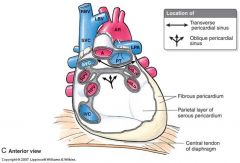
1. around the aorta & pulmonary trunk
2. around the pulmonary veins & superior & inferior vena cava |
|
|
The reflection onto the pulmonary veins form the _____________________
|
oblique pericardial sinus
|
|
|
The two reflections form a passage behind the aorta & pulmonary trunk called the _________________________
|
transverse pericardial sinus
*important surgical landmark, can put finger through sinus to separate and clamp ascending aorta & pulmonary trunk |
|
|
During development, the primordial arterial and venous ends are brought together to from __________________
|
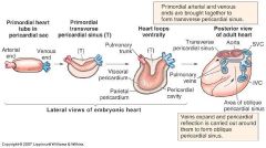
transverse pericardial sinus
|
|
|
During developement, as the _____________ expand, the pericardial reflection is carried out around them to form the oblique pericardial sinus
|
pulmonary veins
|
|
|
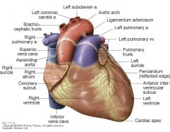
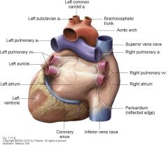
Recognize structures on the anterior heart
|
|
|
|
Recognize structures on the posterior heart
|
notice vena cava runs straight vertically
|
|
|
The base of the heart is directed posteriorly & consits of the ________________, a portion of the right atrium, and the proximal parts of the _____ veins
|

left atrium
great veins |
|
|
The apex of the heart is directed anteriorly and to the left and formed by the inferolateral part of the ___________
|

left ventricle
|
|
|
The heart has 4 surfaces and 4 chambers, what are they?
|
surfaces: anterior, diaphragmatic, right and left pulmonary
chambers: right and left atriums, right and left ventricles |
|
|
Differentiate btwn the inferior and obtuse margins of the heart
|
inferior margin- sharp edge btwn anterior & diaphragmatic surfaces
obtuse margin- separates the anterior and left pulmonary surfaces |
|
|
Identify the features w/i the Right Atrium
-Crista terminalis (sulcus terminalis) -Sinus venarum (smooth posterior wall of artium) -Fossa ovails & limbus -pectinate muscule -opening of superior vena cava & valve -opening of inferior vena cava & valve -right A-V orifice |
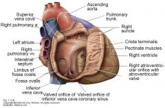
*sinus venarum is from sinus venosus
*fossa ovalis from foramen ovale (probepatency = flap of tissue from foramen ovale that is not sealed, help shut by blood, present in 25% of people) |
|
|
Identify features w/i the Right Ventricle
-trabeculae carneae -tricuspid valve (anterior, posterior, & septal cusps) -chordae tendineae -papillary muscle -septomarginal trabecula (moderator band) -conus arteriosus -pulmonary valve (anterior, right, left cusps, nodule & lunule) |
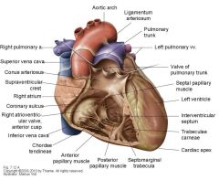
cusps are named for their embryological position*
|
|
|
Identify features w/i left atrium:
-pectinate muscle -openings for pulmonary veins -interatrial septum (valve of foramen ovale) -left A-V opening |

*valve of foramen ovale less pronounced
|
|
|
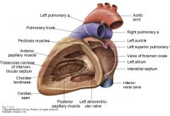
Features w/i left ventricle:
-trabeculae carnae -left A-V valve/ bicuspid valve, mitral valve (anterior and posterior cusps) -chordae tendineae -anterior and posterior papillary muscle -aortic vestibule -aortic valve (posterior, right, & left cusps, nodule & lunule) |
|
|
|
The ventricle wall of the (left/right) ventricle is MUCH thicker than the other.
|
Left ventricle thicker
|
|
|
Blood flow through the heart:
right side left side |
right side -
body--> sup/inf VC--> right atrium-->tricuspid valve-->right ventricle-->pulmonary valve-->pulmonary trunk/aa-->lungs left side- lungs-->pulmonary veins-->L atrium--> bicuspid(mitral) valve--> L ventricle--> aortic valve--> body |
|
|
When atria contract, blood is forced into ventricles and AV valves are (closed/open)
when ventricles contract blood is forced to exit via aorta & pulmonary trunk, AV valves are (closed/open) |
atria contract= open
ventricle contracts= closed |
|
|
The _______________act to prevent valve prolapse.
|
chordae tendinae (tendinious cords)
|
|
|
Closure of the A-V valves produces the (first/second) heart sound
Closure of the aortica and pulmonary valves produces the (first/second) heart sound |
first (lubb) sound
second (dubb) sound |
|
|
The pulmonary & aortic valves are open during (aortic/ventricular) contraction, allowing blood to escape
|
ventricular contraction
|
|
|
After ventricular contraction, the recoil of blood fills the ________ & __________ sinuses, forcing the valves closed (& preventing backflow into ventricles)
|
aortic & pulmonary sinuses
|
|
|
The proper examination of the heart includes:
|
visual inspection
palpation percussion ausculation ****know where you are!! |
|
|
Where are the heart borders in relation to surface anatomy?
|
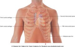
Upper limit:
3rd costal cartilage (R) 2nd intercostal space (L) Right margin: 3rd to 6th costal cartilage Left marigin: 2nd(near sternum) to 5th intercostal (near midclavicularline) space Lower Limit: 6th costal cartilage at sternum (R) to 5th intercosta space near midclavicular line (L) |
|
|
Where do you listen to heart sounds on chest and w/i heart?
(hint: A PeT Monkey) |
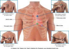
Aortic valve
-medial end of R 2nd intercostal space Pulmonary valve -medial end of L 2nd intercostal space Tricuspid valve -slightly L of sternum near 5th intercostal space Mitral valve -L 5ht intercostal space at mid-clavicular line |
|
|
T/F
heart sound are best heard upstream from the flow of blood through the valve |
FALSE!
best heard DOWNSTREAM from flow! |
|
|
calcific aortic stenosis is primarily age related but will usually occur earlier & more aggressively when?
|
in someone w/ a congenital valve malformation
|
|
|
What may calcific aortic stenosis cause?
|
systolic murmur, L ventricle hypertrophy, angina, syncope, heart failure, arrhythmia
|
|
|
What is mitral stenosis?
What are 99% of cases secondary to? |
narrowing of the orific of the mitral valve
rheumatic fever |
|
|
In Mitral stenosis, blood flow from the L atrium to L ventricle is restricted, leading to what?
what may develop as a result of increased workload? |
blood pressure increaese in L atrium & pulmonary arteries
R ventricular hypertrophy |
|
|
The myocardium is supplied by what arteries?
|
coronary arteries (R & L)
|
|
|
What are the branches off the left (left main stem) and right coronary arteries?
|
Left-
Anterior interventricular branch (LAD) circumflex branch left marginal branch Right- SA nodal branch (60% of time) right marginal branch posterior interventricular branch (PDA)(80% of time) |
|
|
How can you tell if a heart is right or left dominant/dominant coronary artery?
|
Right dominant-
posterior interventricular branch arises from the RIGHT coronary artery (most people) Left dominant- posterior interventricular branch arises from an enlarged circumflex branch of the LEFT coronary artery |
|
|
Blood from the myocardium returns to the right atrium via the __________
|
cardiac veins
|
|
|
Cardiac veins:
Anterior, small, middle, & great All drain into the coronary sinus except _______ |
anterior cardiac vein does not
|
|
|
What can happen when a coronary artery is blocked?
|
angina pectoris= intermittent chest pain cuased by REVERSIBLE cardiac ischemia
or myocardial infarction= localized area of myocardial necrosis induced by IRREVERSIBLE local ischemia (1.4 million per year in US) |
|
|
What is the most common cause of death in the US?
1/2 million or more deaths per year |
Coronary artery disease (CAD)
**50% of ppl die w/o receiving treatment |
|
|
Where are the 3 most common sites of coronary artery occlusion?
|
1. anterior interventricular branch of LCA (40-50%)
2. RCA (30-40%) 3. circumflex branch of LCA (15-20%) |
|
|
2 most common treatments for occlusion?
|
angioplasty
-use catheter to compact arterial walls, expanding lumen coronary artery bypass surgery -use another vessel to bypass occlusion |
|
|
T/F
Atherosclerosis leads to the formation of plaque along the walls of blood vessels |
FALSE!!!!!
plaque forms WITHIN the vessel wall |
|
|
What is the cardiac skeleton and what is its function?
|
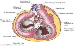
dense, fibrous CT, sits btwn atria & ventricles & forms a rings around each of the 4 heart valves
Functions: -maintain structural integrity of openings -provide attachment point for valve cusps -electrically isolates atria from ventricles |
|
|
T/F
The heart consist of neural tissue |
FALSE
consists of specialized myocardial cells NOT neural tissue |
|
|
The heart is it's own conducting system & does not need input from the nervous system to beat rhythmically, HOWEVER the rate or force of contraction may be influenced by outside input via _____________
|
cardiac plexus
|
|
|
The cardiac plexus gets its main contributions from what?
|
vagus nerve and cardiac branches from sympathetic trunk (T1-T4 cord levels)
-has deep and superficial parts |
|
|
The heart's conducting system consists of what?
|
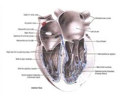
sinoatrial node (SA node)
atrioventricular node (AV node) AV bundle R & L bundle branches Purkinje fibers |