![]()
![]()
![]()
Use LEFT and RIGHT arrow keys to navigate between flashcards;
Use UP and DOWN arrow keys to flip the card;
H to show hint;
A reads text to speech;
74 Cards in this Set
- Front
- Back
- 3rd side (hint)
|
What kind of skin covers the palpebrae externally? How is this type of skin characterized?
|
Thin skin - relatively thin stratesfied squameous epithelium, dermis, fine hairs, sweat and sepaceous glands
|
|
|
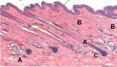
ID these structures in a section of skin
|

A - Sweat gland
B - Secaceous gland C - hair follicle |
|
|
|
T or F:
Bulbar conjunctiva is continuous with the corneal epithelium |
True!
|
|
|
|
T or F:
Palpebral conjunctiva can be stratefied columnar. |
True!
|
|
|
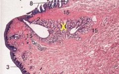
ID the structure indicated by the yellow X.
Where does this structure open? |

Tarsal gland (meibomian gland)
Opens on the cunjunctival part of the eyelid margin. |
|
|
|
What are the three layers of a tear film and what is the composition of each?
|
Superficial - lipids
Middle - aqueous Inner - mucoid |
|
|
|
Which region secretes the superficial tear film layer and what is the function of this layer?
|
Tarsal glands and ciliary glands.
Limits evaporation & provides binding effect for tear film |
|
|
|
Which region secretes the middle tear film layer and what is the function of this layer?
|
Lacrimal and superficial gland of 3rd eyelid.
Flushes foreign material and lubricates surfaces. |
|
|
|
Which region secretes the inner mucoid tear film layer and what is the function of this layer?
|
Conjunctival cells.
Binds film to cornea |
|
|
|
What are the three tunics of the eye?
|
Fibrous
Vascular Nervous |
|
|
|
What structures comprise the outermost layer of the eye?
|
Fibrous layer - cornea and sclera
|
|
|
|
What structures comprise the middle layer of the eye?
|
Vascular layer - Choroid, iris, tapetum lucidum, and ciliary body
|
|
|
|
What structures comprise the innermost layer of the eye?
|
Retina and optic disc
|
|
|
|
T or F:
The cornea is avascular, anervous, and receives its dissolved O2 from the tear film. |
False! It is avascular and does receive its O2 from the tear film but it is HIGHLY innervated
|
|
|
|
What are the layers of the cornea?
|
Anterior corneal epithelium
Anterior limiting membrane Corneal stroma Posterior limiting membrane Posterior corneal epithelium |
Hint - there are 5
|
|
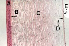
ID these corneal layers
What is the eponymous name for B and D? |

A - Anterior corneal epithelium
B - Anterior limiting membrane (Bowman's membrane) C - Corneal stroma D - Posterior limiting membrane (Descemet's membrane) E - Posterior corneal epithelium |
|
|
|
T or F:
The anterior limiting membrane of the cornea is only present in certain species. |
True!
Present in birds, humans, giraffes, and some cetaceans. |
|
|
|
What is the histology of the Corneal stroma? What substance makes up this material?
|
Dense regular CT of collagen fibers.
|
|
|
|
Which corneal layer is the strongest barrier between the anterior chamber and the outside? What substance tests for a breach of this layer?
|
Descemet's membrane
Flourescin dye testing |
|
|
|
Which corneal membrane maintains corneal transparency? How does it do this?
|
Posterior corneal epithelium pumps water out and provides nutrients to substantia propria
|
|
|
|
When is the cornea vascularized?
|
During repair processes
|
|
|
|
Of these four animals, which is the most sensitive to eye injury? The least sensitive to eye injury?
Horse, Cow, Cat, Dog |
Horse most sensitive
Cow least sensitive |
|
|
|
T or F:
The dog develops more neovascularization than the cat upon corneal injury. |
True!
|
|
|
|
What is the transition zone between the cornea and the sclera called?
|
Limbus
|
|
|
|
T or F:
The limbus is avascular like the cornea. |
False! The limbus is highly vascular.
|
|
|
|
What are the three layers of the sclera? What is the bony part of the sclera called in owls and hawks?
|
Lasmina fusca
Sclera proper Episclera Scleral ossicles |
|
|
|
Which scleral layer provides nutrition to the sclera?
|
Episclera (and vascular tunic)
|
|
|
|
What is the scleral area where the optic nerve exits called?
|
Area cribosa
|
|
|
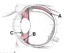
ID these three regions of the eye. Which tunic of the eye do these comprise?
|

A - Choroid
B - Ciliary Body C - Iris A + B + C = Uvea |
|
|
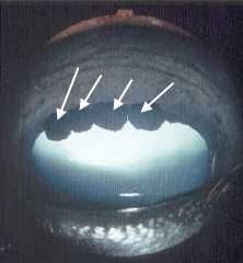
Que son estas punctas nigras en este ojo de caballo?
|

Iridic granules (corpora nigra)
|
|
|
|
T or F:
The epithelium of the iris is vascular and nervous. |
False!
While the iris does have a vascular and nervous part, it has NO TRUE EPITHELIUM |
|
|
|
What is the pupillary shape in a dog? Pig? Cat? Herbivore?
|
Dog and pig - round
Cat - vertical Herbivore - horizontally ovoid |
|
|
|
What are the two tunics of the iris?
|
Vascular tunic (anterior stroma)
Nervous tunic (pars iridica retinae) |
|
|
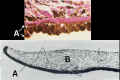
ID these regions of the Iris. Which region contains the pupillary muscles?
|

A - Pars iridica retinae
B - Anterior stroma (contains muscles) |
|
|
|
What structure allows for the change in shape of the lens?
|
Ciliary body and processes
|
|
|
|
Which structures form the blood/aqueous barrier?
|
Ciliary body and pars ciliaris retinae (ciliary processes)
|
|
|
|
What innervates the ciliary muscle? What does contraction of this muscle do? What type of stimulation causes this?
|
CN III innervates
Contraction RELEASES tension on suspensory ligaments and lens becomes thicker. Parasympathetic stimulation |
|
|
|
What are the layers of the choroid? Which contains the tapetum lucidum?
|
Suprachoroidea
Large vessel layer Medium sized vessel layer - contains tapetum lucidum Choriocapillaris |
Hint - there are 4
|
|
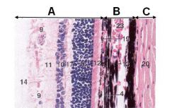
ID these layers of the ojo
|

A - Retina
B - Choroid C - Sclera |
|
|
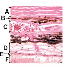
YO IDENTIFY THIS EYEBALL SHIZ MUTHAEFFA!!!
|

A - Sclera
B - Suprachoroidea C - Large vessel layer D - Tapetum lucidum (part of medium sized vessel layer) E - Choriocapillaris F - Retina |
|
|
|
T or F:
The large vessel layer has numerous melanocytes irregardless of the presence of the tapetum lucidum. |
FALSE!
Irregardeless isn't a word you dolt! Other than this word, the statement is actually TRUE!!! :) |
|
|
|
What is the name of the membrane between the choriocapillaris and the retinal pigment epithelium?
|
Bruch's membrane
|
|
|
|
What are the two types of tapetum lucidum? Which species is each found in?
|
Fibrous - herbivores
Cellular - carnivores |
|
|
|
How are squirrel and llama eyes the same?
|
Neither has a tepetum lucidum
|
|
|
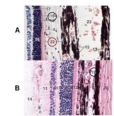
Which of these has a tapetum lucidum present? What number is it?
|

A #22
|
|
|
|
What holds the lens in place? Where do these structures insert?
|
suspensory ligaments insert on lens capsule
|
|
|
|
T or F:
The lens has no pigment or blood within it. |
True!
|
|
|
|
What is the lens epithelium morphology?
|
Squamous to cuboidal centrally; columnar near equator
|
|
|
|
What is the protein in the lens called?
|
Crystimine
|
|
|
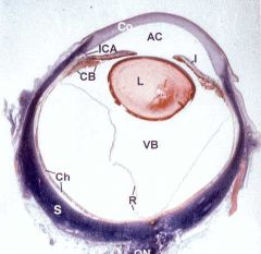
See the little "Y" in the lens? What is this called?
|

Suture line
|
|
|
|
T or F:
Birds have more flexible lenses than whales. |
Um. Yeah.
|
|
|
|
How many layers in the visual retina? Of these layers, how many are neural retinal layers? Which is the odd man out?
|
10 layers, 9 are neural retinal; Retinal pigmented epithelium is odd man out
|
|
|
|
What is the blood/retina layer formed by?
|
Retinal pigmented epithelium
|
|
|
|
T or F:
The retinal pigmented epithelium is always pigmented. |
False, actually! It DOES NOT contain pigment where it is adjacent to tapetum lucidum
|
|
|

ID 19 and 22 in these slides
Is 19 pigmented in both cases? |

19 - retinal pigmented epithelium (unpigmented on the right)
22 - Tapetum lucidum |
|
|
|
What are the functions of the retinal pigmented epithelium?
|
Nutrient transport from choroid to outer retina.
Insulate photoreceptors and increase individual sensitivity. Phagocytose outer segments of photoreceptor cells. |
|
|

What are A and B?
|

A - Retinal pigmented epithelium
B - Photoreceptive layer |
|
|
|
T or F:
The nuclei for rod and cone cells are located in the photoreceptive layer. |
False. Only the dendritic processes are here. Cell bodies are in the outer nuclear layer
|
|
|
|
What is the light sensitive pigment in a rod? In a cone?
|
Rod - rhodopsin
Cone - iodopsin |
|
|
|
Which photoreceptive cell perceives color? Detects motion?
|
Color - cone
Motion - rod |
|
|
|
T or F:
Rods are more sensitive to light than cones. |
True! This is why rods are better in dim light
|
|
|
|
T or F:
Rods are inactivated by constant bright light. |
True again! Cones are for bright light vision.
|
|
|
|
What is the area of the retina at the center of the vision field? What photoreceptor is most dense here?
|
Macula (or area centralis) has density of cones
|
|
|
|
What is so special about the macula in birds and primates that it gets its own name (and what is the name)?
|
Fovea - area of retina with cones ONLY
|
|
|
|
What are the sustentacular fibers of the retina?
|
Muller's fibers
|
|
|
|
What is found in the outer nuclear layer? In the outer plexiform layer?
|
Nuclear - rod/cone nuclei
Plexiform - rod/cone axon terminals (synapse on interneurons) |
|
|
|
What is found in the inner nuclear layer and inner plexiform layer?
|
Nuclear - nuclei of interneurons for interneurons and muller's cells
Plexiform - synapses between bipolar interneurons and amacrine cells and dendrites of ganglion cells |
|
|
|
What is found in the ganglionic cell layer? In the optic nerve fiber layer?
|
Ganglion - ganglia, neuroglia, BVs
Optic - axons of ganglion cells coursing to optic disc |
|
|
|
What is attached to the inner limiting membrane of the eye?
|
Vitreous humor
|
|
|
|
Which cell(s) interconnects photoreceptor cells?
|
Horizontal cells
|
|
|
|
Which cell(s) modifies visual stimuli from other retinal cells?
|
Horizontal and amacrine cells
|
|
|
|
Which cells serve as interneurons between photoreceptor cells and ganglia of the optic tract?
|
Bipolar cells
|
|
|
|
What are the layers of the non-visual retina? How many cell layers in each? Which is more pigmented?
|
Pars ciliaris retinae and pars iridica retinae
2 cell layers in each iridica is more pigmented |
|
|
|
T or F:
Dogs are pretty much red/green colorblind. |
Yup.
|
|

