Reading...
![]()
Play button
![]()
Play button
![]()
Use LEFT and RIGHT arrow keys to navigate between flashcards;
Use UP and DOWN arrow keys to flip the card;
H to show hint;
A reads text to speech;
54 Cards in this Set
- Front
- Back
|
What lesion is part of a group of diseases known as histiocytosis X, or Langerhans cell histiocytosis?
a. lymphoma b. leukemia c. eosinophilic granuloma d. osseus metastasis e. multiple myeloma |
c. EG
|
|
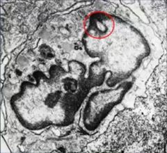
What is the circled phenomenon in the EG Langerhans cell?
|
Birbeck's granule
|
|
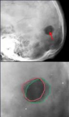
What feature is shown on the bottom image that is unique to eosinophilic granuloma in the skull?
|
hole within a hole
|
|
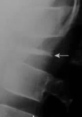
The photo above is an example of fracture in anterior body wall (hard to see). What lesion causes this, and what are the 3 names for the vertebrae at the arrow?
|
1. Eosinophilic granuloma
2. Vertebra plana, silver-dollar vertebra, coin-on-edge vertebra |
|

What lesion is shown in the photo above?
|
Eosinophilic granuloma (within the mandible)
|
|
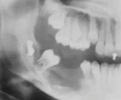
What is the term used to describe the phenomenon in EG above?
|
floating teeth sign
|
|
|
In EG, what general area of the spine is most commonly involved? Least involved?
|
Thoracic most common, cervicals least common
|
|
|
What is the term for EG in a skull's feature showing centrally located residual bone?
|
button sequestrum
|
|
|
Can EG ever degenerate or morph into a malignancy of bone?
|
No - it is not a true neoplasm
|
|
|
What are some differential diagnoses for EG?
|
Ewing's
Osteosarcoma Osteomyelitis Leukemia/Lymphoma |
|
|
What is the age range for EG? What is the specific age of most common involvement?
|
0-30 years old (rarely ever over 30)
Most commonly between 5-10 years of age. |
|
|
Approximately 40% of cases are associated with skin lesions such as: papules, erosions, ulcerations, and purpura.
a. Lymphoma b. Leukemia c. Osteomyelitis d. EG e. Osseus Mets |
d. EG
|
|
|
True/False?
EG often presents with systemic pain and swelling clinically. |
False - localized pain and swelling
|
|
|
In EG (especially in the vertebral body), what is the preferred method of treatment?
|
Don't touch - spontaneous resolution
|
|
|
How often does secondary malignant bone tumors occur compared to primary malignant bone tumors? (ratio)
|
25 to 1
|
|
|
Regarding metastasis to bone, what does BPLKT stand for?
|
B-Breast, P-Prostate, L-Lung, K-Kidney, T-Thyroid
80% of secondary bone mets originated from here, in descending order: BPLKT |
|
|
In a child, where is the most likely location to find secondary mets?
a. long bones b. axial skeleton |
a. long bones (more red marrow)
|
|
|
In an adult, where is the most likely location to find secondary mets?
a. long bones b. axial skeleton |
b. axial skeleton
|
|
|
With secondary bone mets coming from the prostate, what vertebral segments are most likely to be involved?
a. cervical b. thoracics c. lumbar d. sacral |
d. sacral (this is an exception to the rule)
|
|
|
In adults, what vertebral segment is most likely to get secondary bone mets?
a. cervical b. thoracics c. lumbar d. sacral |
c. lumbars (except when from prostate cancer)
|
|
|
__________ refers to the mechanism by which neoplastic cells separate from the primary tumor and gain access to the systemic circulation.
|
Intravasation
|
|
|
Regarding successful secondary bone mets spread, is it more likely to spread in arteries or venous systems?
|
venous systems - arterial channels seem to possess immunity
|
|
|
Most secondary bone mets tumors are __________ (osteolytic/osteoblastic) primarily?
|
osteolytic
|
|
|
What type of primary tumor causes osteoBLASTIC effects in secondary bone metastasis? (Where does the tumor cells originate from?)
|
Prostate
|
|
|
Which of the following are common secondary bone mets lab findings? MACA
a. hydroxyprolinemia b. elevated alkaline phosphatase c. hydroxyprolinuria d. decreased PTH e. hypercalcemia |
a, b, c, e
|
|
|
Which of the following can secondary bone mets look like on plain film?
a. geographic b. moth-eaten c. permeative d. all of the above e. none of the above, it's usually blastic |
d. all of the above
|
|

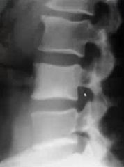
If you knew this was secondary bone mets, what is the location of the primary tumor based on the characteristics of the vertebral bodies?
|
Prostate - because almost all secondary bone mets is lytic except when from the prostate.. It's then blastic, aka solid white!
|
|
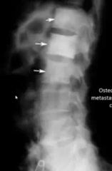
What is the radiographic name for the extremely white/blastic vertebral body shown here? (Secondary bone mets from prostat)e.
|
Ivory vertebra apperarance
|
|

Explain why the middle blastic vertebrae is not an example of Paget's disease (assuming the age of patient matched up).
|
Pagets causes bone to appear blastic, but it also causes bone to expand. Since these VB's are symmetrical, Pagets is ruled out. Diff Dx: Bone Mets & Lymphoma
|
|
|
True/False?
Secondary bone metastasis often has a periosteal reaction. |
False - usually doesn't
|
|
|
True/False?
Secondary bone metastasis never has a periosteal reaction. |
False - although it USUALLY doesn't.. It can.
|
|
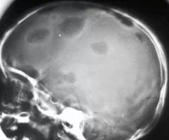
Explain why these lesions on the skull are more likely to be secondary lytic mets instead of Multiple Myeloma.
|
MM = raindrop skull - SYMMETRIC lesions in size. This image has varied size thus it's more likely to be lytic bone mets.
|
|
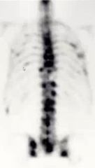
Does this image have high sensitivity or high specificity?
|
High sensitivity, low specificity (note: it is secondary bone mets)
|
|
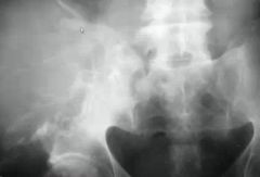
What is the imaging feature displayed here in the right hemi-pelvis? ____-___ metastasis
|
Blow-out metastasis
|
|
|
What are the 4 common primary locations of tumors that will display "blow-out metastasis" in bone?
|
RATS
Renal, Adrenal, Thyroid, Skin |
|
|
When secondary bone mets presents distal to the knee or the elbow, where is the tumor most likely originating from?
(Hint: 2 places) |
GI tract
bronchogenic carcinoma |
|
|
When secondary bone mets presents distal to the knee or elbow, what is this called? _____ metastasis
|
Acral
|
|
|
Non-Hodgkin's lymphoma occurs at a __:__ rate compared to Hodgkin's lymphoma.
|
3:1
|
|
|
What is the age range for N-HL (non hodgkin's)? Gender predeliction?
|
20-50 YOA
Males (we always get hosed) |
|
|
True/False?
NonHodgkin's lymphoma usually has a soft tissue mass associated with it. |
True
|
|
|
What is the pattern of destruction seen in NonHodgkin's Lymphoma?
|
poorly-defined, aggressive, lytic, medullary based but spreading to cortex, soft tissue mass, occasional periosteal reaction
|
|
|
If you suspect NH Lymphoma, and the patient is over 40 years of age.. What are your two leading differentials?
|
Secondary Mets
Multiple Myeloma |
|
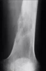
Name this lesion
|
Non Hodgkin's Lymphoma
|
|
|
Hodgkin's Lymphoma histologically are identified by having what large cells, often with bi-lobed nuclei giving an "owl's eyes" appearance?
|
Reed-Sternberg cells
|
|
|
Where does Hodgkin's lymphoma often appear at in the body?
a. distal femur b. proximal tibia c. radioulnar joint d. vertebrae e. clavicle |
d. vertebrae
|
|
|
What kind of pattern does Hodgkin's Lymphoma usually show on plain film?
|
Lytic, mixed and blastic (basically anything is possible)
|
|
|
If Hodgkin's lymphoma is sclerotic in the spine, what is the term for this view in plain film x-rays?
|
ivory vertebrae
|
|
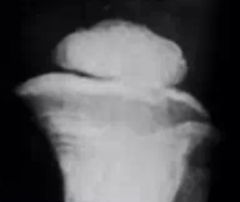
What's the term for this vertebrae's sclerotic appearance? Name 3 differentials for it.
|
Ivory vertebrae
1. lymphoma 2. osseus metastasis 3. Paget's (although since it's not expansile, HIGHLY UNLIKELY) |
|
|
T/F?
Non Hodgkin's Lymphoma and Leukemia form solid cancerous masses in the body. |
False. NH Lymphoma does, but leukemia never does.
|
|
|
With Leukemia, is an older patient more likely to develop the acute or chronic type?
|
chronic
|
|
|
What is the most common childhood malignancy?
a. Leukemia b. Non Hodgkin's Lymphoma c. Secondary mets d. Ewing's Sarcoma e. Paget's |
a. Leukemia
|
|
|
Non-Hodgkin's lymphoma is most likely to occur in the upper or lower extremities?
|
lower
|
|
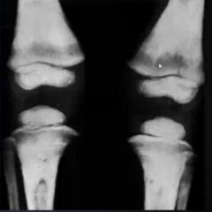
The black bands in the distal femur are indicative of what disease?
|
Acute Leukemia
|
|
The lines to the left of the image, just inferior to the growth plate.. What are they called in a patient with Leukemia?
|
Harris growth arrest lines
|