Reading...
![]()
Play button
![]()
Play button
![]()
Use LEFT and RIGHT arrow keys to navigate between flashcards;
Use UP and DOWN arrow keys to flip the card;
H to show hint;
A reads text to speech;
66 Cards in this Set
- Front
- Back
|
What is the demarcation between the false and true pelvis?
|
The pelvic brim, following along the superior margin of the pubic symphysis, pectin of the pubis, arcuate line of the ilium, ala of the sacrum and sacral promontory.
|
|
|
What are the three bones that make up the hip bone?
|
Ilium, ischium, and pubis.
|
|
|
What is the structure where the two hip bones unite anteriorly?
|
Pubic symphysis.
|
|
|
What part of the sacrum unites with the hip bones posteriorly?
|
The ala of the sacrum articulates with the ilium and are held in place by strong sacroiliac ligaments. There is actually a joint which is reasonably nonmotile, the sacroiliac joint.
|
|
|
What ligaments and bony prominences are involved in the formation of the greater and lesser sciatic foramina?
|
The sacrotuberous and sacrospinous ligaments that connect the ischial tuberosity and ischial spine to the sacrum. The space between the sacrotuberous and sacrospinous ligaments is the lesser sciatic foramen.
|
|
|
In the anatomic position what two bony features of the pelvis lie on the same vertical plane?
|
The anterior part of the pubic symphysis and anterior superior iliac spine.
|
|
|
Concerning differences in male and female bony pelvises:
Which has the widest subpubic angle? Which has the smallest pelvic inlet and outlet? Which has the shallowest true pelvis? |
Female.
Male. Female. |
|
|
What is the pouch of Douglas and what is its clinical significance?
|
The rectouterine space or peritoneal space between the uterus and anterior rectum. Clinically, it is an important area adjacent to the posterior fornix of the vagina. The thin vaginal wall is all that separates the peritoneal cavity from the outer environment. It can easily be punctured, thus transmitting infection to the peritoneal cavity.
|
|
|
What is the sacral plexus?
|
The sacral plexus is composed of ventral primary rami of segments S1 to S5 and the lumbosacral trunk from L4-5. These nerves combine to form the major nerves that leave the pelvis to supply the lower limb.
|
|
|
What are the major nerves derived from the sacral plexus? (6)
|
Sciatic, superior and inferior gluteal, pudendal, posterior femoral cutaneous, and pelvic splanchnics. (Don't worry about which segments supply each nerve.)
|
|
|
What are the two sources for sympathetic nerves in the pelvis?
|
Sacral sympathetic trunk (or chain) and the hypogastric plexus.
|
|
|
What is the origin of the parasympathetic innervation to the pelvic viscera?
|
Pelvis splanchnics from S2-S4.
|
|
|
What are the chief arteries off the internal iliac artery? (7)
|
Superior and inferior gluteal arteries, umbilical supplying superior vesicular branches, obturator, middle rectals, internal pudendals, uterine and vaginal branches.
|
|
|
What arteries in the pelvis are unique to females? (3) and where do they originate from?
|
Uterine and vaginal branches off the internal iliac and ovarian from the lumbar aorta.
|
|
|
What is the origin for the common variation of the obturator artery?
|
It may arise off the inferior epigastric branch from the external iliac and pass over the pelvic brim to the obturator canal (20% occurrence).
|
|
|
On a rectal exam, what can be palpated anteriorly in the female?
|
Cervix of uterus.
|
|
|
What is the muscular sheet found at the pelvic outlet?
What are its components? |
Pelvic diaphragm.
Levator ani and coccygeus muscles. |
|
|
What is the name of the medial part of the levator ani that keeps the "kink" in the anus?
|
Puborectal sling.
|
|
|
What is the path for the ureters?
What are the three constrictions or narrowed areas in the ureters? |
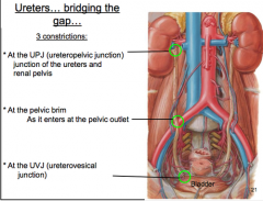
The pass down the posterior abdominal wall, over the pelvic brim, along the posterior wall of the pelvis and then anterior to the posterior wall of the bladder.
Three constrictions are 1) At the renal pelvis 2) Where they cross the external iliac artery and pass over the pelvic brim 3) As they pass through the bladder wall. These areas are where large calculi, "kidney stones", often are stopped and then cause increased pain. Small stones may pass with little or no pain. |
|
|
As the ureters approach the uterus, what structure crosses immediately over them?
|
Uterine artery.
|
|
|
What is the smooth area in the bladder where the ureters pierce the bladder wall?
|
Trigone, outlined by the inlets of the ureters and inferiorly by the outlet of the bladder.
|
|
|
What is the name of the smooth muscle found in the urinary bladder wall?
|
Detrusor muscle.
|
|
|
What is the ring of smooth muscle called at the neck of the urinary bladder?
|
Internal sphincter.
|
|
|
What is the innervation to the internal sphincter?
|
It is closed tonically by the sympathetics and stimulated to open by the parasympathetics (by inhibition of sympathetics).
|
|
|
On rectal exam what can be palpated anteriorly in the male.
|
Prostate and seminal vesicles.
|
|
|
Where is the membranous urethra located in both sexes?
|
Within the urogenital diaphragm.
|
|
|
What is the pathway for sperm after passing through the superficial inguinal ring?
|
In the vas deferens the sperm pass through the inguinal canal, then through the deep inguinal ring, over the pelvic brim, along the lateral aspect of the bladder, join the duct of the seminal vesicle to form the ejaculatory duct, passes through the prostate gland, and enter the prostatic urethra on the posterior aspect along the colliculus seminalis.
|
|
|
Into what do the ejaculatory ducts empty?
|
Prostatic urethra, along the side of the colliculus seminalis.
|
|
|
What nerve group functions during ejaculation?
|
Parasympathetics, cause contraction of urethral muscles. Sympathetics, cause movement of sperm down the vas and secretion of glands (emission).
"Parasympathetics=Point and sympathetics=Shoot" |
|
|
How does the prostate empty into the urethra compared to the ejaculatory ducts?
|
Numerous small ducts open into the prostatic urethra.
|
|
|
What is the name for the longest part of the urethra in males?
|
Spongy urethra (since it enclosed by the corpus spongiosum).
|
|
|
The female urethra opens into what space?
|
Vestibule.
|
|
|
What arteries supply the rectum? (3)
|
-Superior rectal from inferior mesenteric
-middle rectal from internal iliac - inferior rectal from internal pudendal artery. |
|
|
What are the boundaries of the perineum?
|
It is a diamond formed by the pubic symphysis, pubic rami, ischial rami, ischial tuberosities, sacrotuberous ligaments, and coccyx.
|
|
|
What are its two subdivisions, and in general what is found in each?
|
Anal triangle; anal canal, external anal sphincter, and ischiorectal fossa. Urogenital triangle; UG diaphragm, scrotum/labia, penis/clitoris, vestibule.
|
|
|
What are the boundaries of the ischiorectal fossae?
With what is it normally filled? |
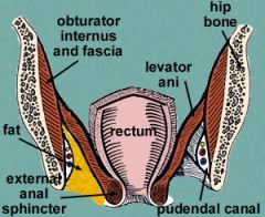
Perineal body anteriorly, laterally obturator internus muscle, levator ani.
They are filled with fat. |
|
|
What is the UG diaphragm and how does it differ from the pelvic diaphragm?
|
It stretches between the inferior pubic rami. It lies superficial to the pelvic diaphragm and contains the external sphincter of the urethra.
|
|
|
What pierces the UG diaphragm in the female?
The male? |
Female - urethra and vagina.
Male - urethra. |
|
|
What is found in the deep perineal space?
|
The urogenital diaphragm.
|
|
|
What is the superficial perineal space?
And what is found there? |
It is the area superficial to the UG diaphragm. It contains the structures associated with the penis and scrotum, or structures of the vestibule, clitoris, and labia, plus all the associated nerves and vessels.
|
|
|
What is the difference between the external and internal urethral sphincter?
|
The external sphincter is under voluntary control while the internal sphincter is controlled by autonomic nerves.
|
|
|
What is the chief nerve supply to the perineum?
|
Pudendal nerve and its branches.
|
|
|
What is the origin of the inferior rectal nerves and vessels?
|
Pudendal nerve and internal pudendal artery and veins.
|
|
|
What are the names of the nerves and arteries on the dorsum of the body of the penis? What is the function of the nerves found here?
|
Deep dorsal vein, dorsal arteries, and dorsal nerves.
Sensory to the surface. |
|
|
The crura and bulb of the penis make up what basic part of the penis?
|
Root of the penis.
|
|
|
What is the muscle layer that lies over the bulb of the penis?
What is its function? |
Bulbospongiosus, compresses the bulb and assists erection.
|
|
|
How does the path of the urethra differ between the sexes?
|
The male urethra consists of prostatic and spongy components that are not found in the female. The membranous component is common in both.
|
|
|
The bulb of the penis is continuous with what structure in the shaft of the penis?
|
Corpora spongiosum.
|
|
|
What is the mechanism of erection?
|
Upon stimulation the smooth muscle in the trabeculae and arteries of the corpora relax because of parasympathetic stimulation (via pelvic splanchnics). The arteries straighten and enlarge, allowing increased blood flow into the cavernous spaces. The bulbospongiosus and ischiocavernosus muscles compress the venous drainage at the periphery of the corpora and impede outflow of blood. As a result, the corpora becomes enlarged and rigid and erection occurs.
|
|
|
How do the drainage patterns for the superficial veins of the perineum differ from that of the deep dorsal vein?
|
The perineum is drained via the internal pudendal veins back toward the lesser sciatic foramen while the deep dorsal vein drains through a defect in the UG diaphragm directly into a plexus of veins surrounding the prostate gland.
|
|
|
What is the name of the end of the uterine tube that drapes over the ovary?
|
Fimbrae, which sweep over the surface of the ovary and pick up ovulated oocytes.
-"think Kung-fu hands"= fimbre |
|
|
What is the origin of the ovarian arteries?
|
They pass into the pelvis over the pelvic brim. They originate as paired arteries off the aorta.
|
|
|
What is the peritoneal sheet that attaches the uterus to the lateral pelvic wall?
|
Broad ligament of the uterus.
|
|
|
What is the ligamentous structure within broad ligament that contains the ovarian vessels?
|
Suspensory ligament of the ovary.
|
|
|
What ligament supports the cervix/uterus within the broad ligament?
|
Transverse cervical or "cardinal" ligament.
|
|
|
What is the usual orientation of the uterus in relationship to the bladder?
|
Anteflexed anteriorly over the bladder.
|
|
|
At what uterine anatomic landmark does the uterine artery approach the uterus?
|
It approaches at the uterine cervix from both sides and then its branches go superiorly and inferiorly to supply the uterus.
|
|
|
What are the vaginal spaces around the end of the cervix?
|
Anterior, lateral, and posterior fornices (fornix is singular).
|
|
|
The crura of the clitoris makes up structure in the body?
|
Corpora cavernosa.
|
|
|
What is the skeletal muscle that lays over the crura of the clitoris?
What is its function? |
Ischiocavernosus muscle.
Its function is to help maintain erection in the clitoris. |
|
|
What is the nerve supply to the female perineum?
|
Perineal branches of the pudendal nerve.
|
|
|
What is the surgical procedure called that prevents the UG diaphragm from ripping during childbirth?
|
Episiotomy. It is done to enlarge the vaginal orifice and prevent a jagged tear in the perineum. This heals more quickly and can prevent damage to important structures such as the perineal body that have consequences later in life in support of pelvic viscera.
|
|
|
In the pudendal nerve block, what bony feature does the clinician use as a guide to anesthetize the pudendal nerve?
|
Ischial spine.
|
|
|
To where do the lymphatics for the cervix drain?
|
They pass along the uterine vessels to the internal iliac nodes and then superior along the aorta.
|
|
|
To where do the lymphatics for the scrotum/labia drain initially?
|
They drain to the superficial inguinal nodes.
|
|
|
To where do the lymphatics of the ovary/testes drain?
|
The lymphatics follow the ovarian and testicular blood vessels and ascend to the lumbar lymph nodes (para-aortic lymph nodes)
|