Reading...
![]()
Play button
![]()
Play button
![]()
Use LEFT and RIGHT arrow keys to navigate between flashcards;
Use UP and DOWN arrow keys to flip the card;
H to show hint;
A reads text to speech;
28 Cards in this Set
- Front
- Back
|
Describe gut tube formation
|
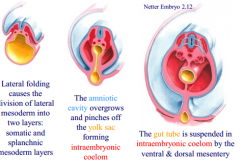
-delineated from yolk sac during process of embryonic folding (ventral bending of cranial and caudal ends, lateral bending of body).
-gut tube closed at two ends by oropharyngeal membrane cranially and cloacal membrane caudally |
|
|
Three subdivisions of developing gut tube
|
- foregut, midgut, hindgut
|
|
|
Four main events for developing gut tube
|
1. rotation of foregut 90 degrees in clockwise direction
2. development of the greater omentum and lesser peritoneal sac from the posterior (dorsal) mesentery 3. rotation of the midgut 270 degrees around the superior mesenteric artery 4. tremendous growth of the midgut loop |
|
|
Early differentiation of GI tract occurs in the __ week
|
5th
|
|
|
Foregut developmental (5 weeks)
|
-short, extends from pulmonary outgrowth to liver outgrowth/hepatic diverticulum
-posterior to septum transversum |
|
|
Midgut development (5 weeks)
|
-no ventral mesentery, communicates with yolk sac through vitello-intestinal duct (at umbilicus)
-continuous caudally with short hindgut. no distinct boundary between two parts |
|
|
Hindgut development (5 weeks)
|
-joined caudally by allantois, forming common endodermal chamber or cloaca, which receives the Wolffian ducts
|
|
|
What is the distinctive feature demarcating the two zones of the midgut and the hindgut
|
There is none
|
|
|
9-10 week differentiation of GI tract- what organs involved, starting to develop
|
-stomach forming by fusiform dilatation of foregut dorsal to septum transversum
-increase in length of trachea, esophagus -narrowing of hepatic diverticulum, forming tubular common bile duct from which gallbladder arises distally -short diverticulum of bile duct proximal to gallbladder is rudiment of ventral pancreas -dorsal to foregut at more cranial level is rudiment of dorsal pancreas |
|
|
What is the septum transversum ventral to in weeks 9-10?
|
-terminal segment of esophagus
-stomach -proximal part of duodenum as far caudally as bile duct |
|
|
foregut development weeks 9-10
|
-suspended by septum transversum ventrally, dorsal mesentery dorsally
-will give rise to pharynx, thyroid gland, trachea, lungs, esophagus in thorax, terminal esophagus, stomach, 1 and 2 of duodenum, pancreas, liver, gallbladder in abd |
|
|
midgut development 9-10 weeks
|
-begins caudally to septum transversum, no ventral mesentery
-comprises 3rd, 4th (distal) part of duodenum and midgut loop -dorsal mesentery helps suspend it -vitello intestinal duct arises from apex of loop -caudal limb -localized dilitation is primordium of cecum, appendix -elongation of midgut loop in later stages--> extra embryonic coelom, forming umbilical hernia |
|
|
Esophagus
|
-muscular tube conveying food from pharynx to stomach
-develops from foregut caudal to primordial pharynx -partitioning of common endodermal tube by tracheoesophageal septum -4 histological layers -T10 from thorax into abdomen -LARP- vagal trunks |
|
|
Function of stomach
|
-expanded part of digestive system bet esophagus and small intestine
-blender and reservoir as it performs enzymatic digestion, gastric juice coverts food into chime |
|
|
Stomach development, rotation
|
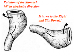
4th week-->dilatation of foregut, enlargement of caudal part of foregut-->ventrodosral enlargement,-->posterior grows, greater curvature-->rotates 90 degrees clockwise around craniocaudal axis. dorsal convex faces left, ventral concave faces right--> minor tippiing of caudal end of stomach in cranial direciton
|
|
|
Stomach is considered intraperitoneal (T/F)
|
true
|
|
|
Right surface of stomach becomes anterior surface, left surface of stomach becomes posterior surface (T/F)
|
false, opposite
|
|
|
What is omenta?
|
-double layered fold of peritoneum that extends from stomach and proximal part of duodenum to an adjacent organ in either the anterior or posterior direction
|
|
|
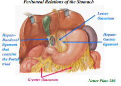
Greater omentum
|
-left side, attach stomach to posterior abdominal wall
-3 portions: gastrophrenic ligament, gastrosplenic ligament, gastrocolic ligament |
|
|
Lesser omentum
|
-right side, extends bet stomach/duodenum and liver
-two parts: hepatogastric ligament bet lesser curvature, hepatoduodenal ligament bet upper duodenum and liver |
|
|
What is the pancreas?
|
-elongated, accessory digestive gland that has exocrine and endocrine functions
|
|
|
Where is the pancreas located?
|
-behind or posterior to stomach, is covered anteriorly by transverse mesocolon, is secondary retroperitoneal
|
|
|
Pancreatic development
|
begin as dorsal and ventral primordia/buds in mesenteries--> dorsal bud becomes larger-->duodenum rotates to right, carries ventral bud and bile duct behind it and into dorsal mesentery,-->ventral pancreas fuses with dorsal, superior mesenteric vein becomes trapped bet primoordia-->ventral bud gives rise to lower head of pancreas, uncinate process. dorsal bud gives rise to remaining part of adult pancreas
|
|
|
Annular pancreas
|
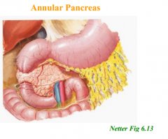
-failure of full rotation. descending portion of duodenum surrounded by pancreatic tissue, can become obstructed.
|
|
|
Ductal rearrangements of pancreas
|
-both buds possess large duct. main duct of ventral pancreas forms anastomotic connection with duct of dorsal bud
-duct of dorsal bud- tail- runs through substance of gland to head, turns into bile duct and joins it -duct of ventral bud- drains uncinate process, inferior part of head of pancreas -regression of portion of dorsal pancreatic duct bet anastomotic connection and duodenum- forming main pancreatic duct of wirsung. if no regression, then accessory duct of antorini |
|
|
Development of omental bursa (lesser sac)
|
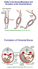
-develops within dorsal mesentery as divericulum
-dorsal mesentery bulgs to left -stomach rotated in clockwise direction -posterior wall of bursa? |
|
|
Boundaries of epiploic foramen
|
i. Posterior: hepatocaval mesentery (IVC)
ii. Roof: caudate lobe iii. Floor: first part of the duodenum iv. Anterior: hepatoduodenal ligament (portal triad) |
|
|
Spleen development
|
-proliferation of mesoderm in left wall of omental bursa- intraperiotneal
0inflitrated with lymphoid cells, by 4th month- vascular structure of red pulp takes place. 3rd-5th month important site of embryonic hematopoiesis =suspended by splenorenal ligament, part of spleen to stomach becomes gastrosplenic ligament |