![]()
![]()
![]()
Use LEFT and RIGHT arrow keys to navigate between flashcards;
Use UP and DOWN arrow keys to flip the card;
H to show hint;
A reads text to speech;
52 Cards in this Set
- Front
- Back
|
Smudge cell
Chronic Lymphocytic Leukemia |
thumbprint appearance; nuclear remnants of lymphocyte
|
|
|
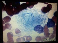
Basket cell
chronic Lymphocytic Leukemia |
net-like appearance
|
|
|
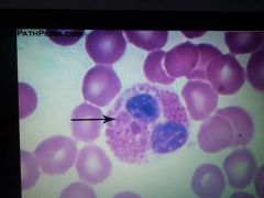
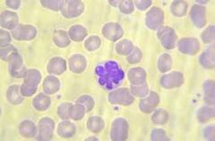
Hypersegmented neutrophils ( macropolycytes)
Pernicious Anemia |
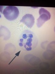
nucleus with 5-10 lobes |
|
|
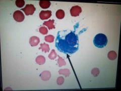
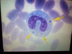
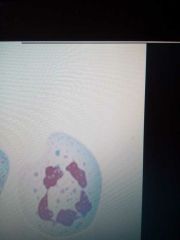
Vacuolated cells
chemical poisoning, severe infections and leukemia |
with holes or vacuoles in the cytoplasm of monocytes
|
|
|
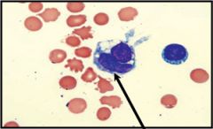
Lupus erythematosus cell
disseminated LE but not specific to SLE |
phagocytic wbc; ingested nuclear material
|
|
|
Tart cells
|
maybe seen in drug sensitivity
|
|
|
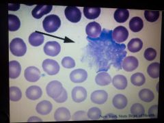
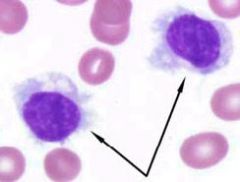
Hairy cells
|

small lymphocytes with cytoplasmic projections
|
|
|
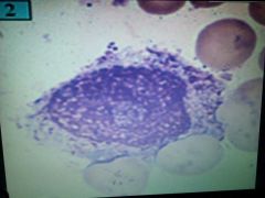
Ferrata cell
SBE- Sub bacterial endocarditis |
|
|
|
Reider cells
CLL - Chronic Lymphocytic Leukemia |
clover leaf
|
|
|
Pelger-Huet Anomaly
congenital true PHA, acquired pseudo PHA |
Pince-nez
|
|
|
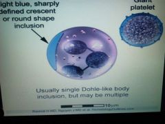
Dohle bodies
|
in neutrophil as irreg. rounded/oval, blue staining; "Pince nez" *
|
|
|
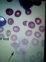
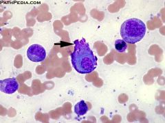
Auer bodies/ Faggot cells
acute myelocytic leukemia |
like twigs, reddish purple in cytoplasm
|
|
|
Alder-Reilly anomaly
|
dense azurophilic granulation in all types of wbc
|
|
|
Czediak Higashi syndrome
partial albinism |
affects all wbc
|
|
|
May-Hegglin anomaly
|
with pale blue, spindled-shape inclusions
|
|
|
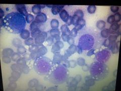
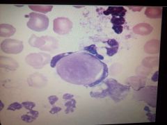
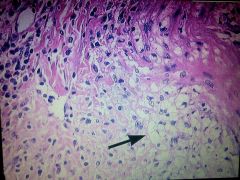
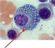
Gaucher's cell
Gaucher's disease |
crinckled appearance
|
|
|
Foam cells
Niemann-Pick disease |
found in BM
|
|
|
Lymphocyte
|
|
|
|
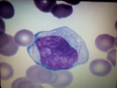
monocyte
|
|
|
|
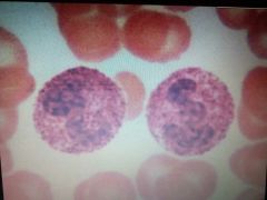
eosinophil
|
|
|
|
basophil
|
|
|
|
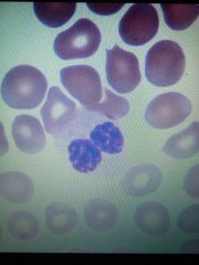
neutrophil
|
|
|
|
stages
|
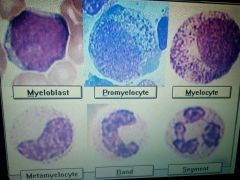
stages of granulopoiesis
|
|
|
Give the normal Values for WBC's |
5,000 - 10,000 uL |
|
|
Give the normal Levels of Hematocrit. |
Female: 37– 46%
Male: 42 –52% |
|
|
Give the normal Levels of Hemoglobin. |
Female: 12.3 – 15.7 g/dL
Male: 14.0 – 17.4 g/dL |
|
|
Give the normal value of RBC's. |
Adult Female: 4.0-5.5 millions/mm3
|
|
|
Give the normal Neutrophil values. |
2,500-8,000 mm3 (55-70%) |
|
|
Give the normal values of Lymphocytes. |
1,000-4,000 mm3 (20-40%) |
|
|
Give the normal values of Monoocytes.
|
150 - 1,000/ul (3-10%) |
|
|
Give the normal values of Basophils.
|
0 - 200/ul (0-2%) |
|
|
Give the normal values of Eosinophils.
|
0 -300/ul (0-3%) |
|
|
Give the normal values of Platelets. |
150,000 - 400,000/ul |
|
|
These cells are increased in: Neutrophil: Lymphocyte: Monocyte: Eosinophil: Basophil: |
Neutrophil: bacterial infections Lymphocyte: viral infections Monocyte: TB, syphilis, malignancies Eosinophil: allergies, parasites, CML Basophil: immediate hypersensitivity, CML |
|
|
Hypersegmented Neutrophil is associated with: |
Pernicious Anemia |
|
|
Hyposegmented Neutrophil is associated with:
|
Pelger Huet Anomaly, Psuedo-Pelger Huet |
|
|
Toxic Granulation and Vacuoles are associated with: |
Bacterial infections, burns, chemotherapy |
|
|
Dohle Bodies (RNA) are associated with: |
Bacterian infections, burns, May-Hegglin |
|
|
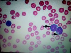
Atypical Lymphs (size increase & basophilia) is associated with: |
Infectious Mononucleosis; other infections |
|
|
Rieder cell |
Has clover leaf like nucleus; found in chronic lymphocity leukemia |
|
|
Flame Cell |
Antibody plasma cell w/ intense eosinophilic cytoplasm; may be seen in IgA myelomas |
|
|
Grape Cell/Motts Cell |
Cytoplasm is filled w/ Russell bodies; seen in multiple myelomas. |
|
|
Tart Cell |
Monocyte which has ingested whole lymph. or nucleus w/ nuclear chromatin |
|
|
Toxic granulations |
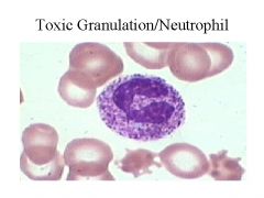
altered primary granules w/c contain myeloperoxidase; seen in chemical poisoning |
|
|
Myeloperoxidase deficiency (MPO) |
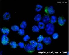
most common antibody |
|
|
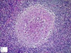
Popcorn Cells / L & H Cells |
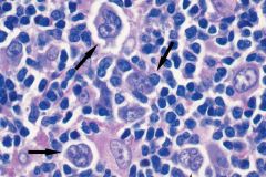
Seen in Nodular Lymph. Predominant Hodgkin's Lymphoma |
|
|
Reed-Sternberg Cells |
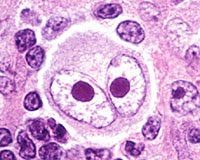
Has an owl's eye appearance; seen in classical Hodgkins Lymphoma |
|
|
Lacunar Cells |
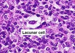
Has beta nucleus; artifactual refraction of cytoplasm; seen in Nodular Sclerosis |
|
|
Sezary Cell |
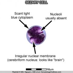
Cells of lymphocytic origin; see in Mycosis Fungoides |
|
|
Lazy Leukocyte Syndrome |
Neutrophils that respond poorty to chemotactic agents; poor directional motiliy |
|
|
Jordan's Anomaly |
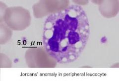
Granulocytes and monocytes w/fat containing vacuoles; seen in ictosis & muscle dystrophy |
|
|
Chronic Granulomatous Disease |
Phagocytes fail to produce super oxide; seen in bacterial and fungal infections |